Joints and Movement Articulations Joints Functional junctions between

Joints and Movement

Articulations = Joints • • • Functional junctions between bones Bind parts of the skeletal system Make bone growth possible Permit skeletal change during childbirth Enable movement in response to muscle contraction • Skeleton Review Video

3 Types of Cartilage

Classification of Joints • Classification based on • Classification the type of tissue that according to degree of binds the bones: movement possible: • Fibrous joints • Cartilaginous joints • Synovial joints • Immovable (synarthrotic) • Slightly movable (amphiarthrotic) • Freely movable (diarthrotic)
 • Dense CT with many collagenous fibers • Lie between bones")
Fibrous Joints (Synarthrotic) • Dense CT with many collagenous fibers • Lie between bones that are in close contact • Suture – only between flat bones of the skull • Gomphosis – joint between tooth and socket; periodontal ligament

Fibrous Joints
 • Consist of hyaline or fibrocartilage • 2 types: • Synchondrosis")
Cartilaginous Joints (Amphiarthrotic) • Consist of hyaline or fibrocartilage • 2 types: • Synchondrosis – bands of hyaline cartilage • Epiphyseal plate • Costal cartilage • Symphysis – pad of fibrocartilage that allows limited movement • Intervertebral disks • Symphysis pubis • Slightly movable (amphiarthrotic)
")
Cartilaginous Joints (amphiarthrotic)
 • Articular cartilage (hyaline cartilage) covers the ends of bones •")
Synovial Joints (Diarthrotic) • Articular cartilage (hyaline cartilage) covers the ends of bones • A fibrous articular capsule encloses joint surfaces • A joint cavity is filled with synovial fluid • Ligaments reinforce the joint • Most joints in the body are synovial
 • Articular cartilage – hyaline cartilage; resists wear and minimizes")
Synovial Joint Structure (Diarthrotic) • Articular cartilage – hyaline cartilage; resists wear and minimizes friction • Subchondral plate – somewhat elastic bone located under the articular cartilage • Absorbs shocks and helps protect joint from stresses • May fracture from excessive stress from obesity or athletic activities

Synovial Joint Structure continued… • Joint capsule – has 2 distinct layers: • Outer layer consists of dense CT whose fibers attach to the periosteum • Flexible enough to allow movement but strong enough to hold the joint together • Ligaments – bundles of strong, tough cartilaginous fibers that reinforce the joint capsule • Synovial membrane – inner layer of the joint capsule • Shiny, vascular lining of loose CT only a few cells thick • Covers all surfaces within the joint capsule except the articular cartilage

Synovial Joint Structure continued… • Synovial cavity – area enclosed by the joint capsule • Synovial fluid – clear fluid secreted by the synovial membrane that moistens and lubricates the joint cavity and supplies nutrients to the cartilage

Synovial Joint Structure continued… • Menisci – fibrocartilage disks that partially or completely divide the joint into compartments between the articular surfaces • Bursae – flattened fibrous sacs lined with synovial membrane and containing a thin film of synovial fluid. Common where ligaments, muscles, skin, tendons, or bones rub together. • Knee Replacement Surgery

Ball-and-Socket Joints • Bone with a globular head articulates with the cup-shaped cavity of another bone • Widest range of motion and rotation • Examples – hip, shoulder

Condyloid Joints • Ovoid condyle fits elliptical cavity • Wide range of motion, but no rotation • Examples – metacarpals and phalanges
 Joints • Nearly flat or slightly curved articulating surfaces • Allow sliding")
Gliding (plane) Joints • Nearly flat or slightly curved articulating surfaces • Allow sliding (back and forth) and twisting movements • Examples – within the wrist and ankle, between vertebrae, sacroiliac joint, between ribs and sternum

Hinge Joints • Convex surface of one bone joins concave surface of another bone • Movement in one plane only • Examples – elbow, phalanges

Pivot Joints • Cylindrical surface of one bone rotates within a ring of bone • Allows rotation only • Examples – proximal radius and ulna, atlas and axis

Saddle Joints • Bones with concave and convex regions on articulating surfaces • Allows movement in 2 planes • Example – trapezium and metacarpal 1 (thumb)
")
Joints Review (8: 11)

Types of Ordinary Body Movements • Flexion • Decreases the angle of the joint • Brings two bones closer together • Typical of hinge joints like knee and elbow • Extension • Opposite of flexion • Increases angle between two bones

Types of Ordinary Body Movements • Rotation • Movement of a bone around its longitudinal axis • Common in ball-andsocket joints • Example is when you move atlas around the dens of axis (shake your head “no”)

Types of Ordinary Body Movements • Abduction • Movement of a limb away from the midline • Adduction • Opposite of abduction • Movement of a limb toward the midline

Types of Ordinary Body Movements • Circumduction • Combination of flexion, extension, abduction, and adduction • Common in ball-andsocket joints

Special Movements • Dorsiflexion • Lifting the foot so that the superior surface approaches the shin • Plantar flexion • Depressing the foot (pointing the toes)

Special Movements • Inversion • Turn sole of foot medially • Eversion • Turn sole of foot laterally

Special Movements • Supination • Forearm rotates laterally so palm faces anteriorly • Pronation • Forearm rotates medially so palm faces posteriorly

Special Movements • Opposition • Move thumb to touch the tips of other fingers on the same hand

Joint Movements • Protraction • moving a part forward • Retraction • moving a part backward • Elevation • raising a part, shrug the shoulder • Depression • lowering a part, droop the shoulder

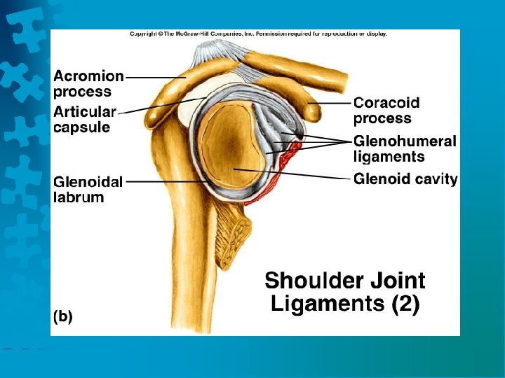
Shoulder Joint • Ball and socket joint made up of the rounded head of the humerus and the glenoid cavity of the scapula. • The joint capsule is loose. Muscles and tendons reinforce the joint. • Shoulder joint is capable of a wide range of movements including flexion, extension, abduction, adduction, rotation, and circumduction.

Shoulder Joint • Ligaments: coracohumeral ligament, glenohumeral ligaments, transverse humeral ligament, and glenoid labrum • Bursae: subscapular, subdeltoid, subacromial, subcorocoid bursae

Coracoacromial Ligament Coracoclavicular Ligament

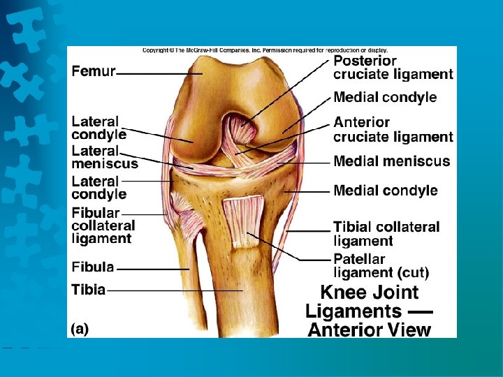
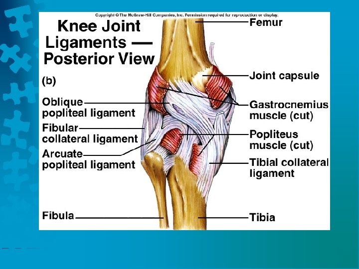
Knee Joint • The knee is the largest and most complex synovial joint. • It consists of the medial and lateral condyles at the proximal end of the tibia. The femur articulates with the patella. • The joint capsule is thin and strengthened by muscles and tendons.

Knee Joint • Ligaments of the knee joint: patella, oblique popliteal, arcuate popliteal, tibial collateral (MCL), fibular collateral ligament (LCL), anterior cruciate ligament (ACL) strengthen the joint capsule. • Cruciate ligaments prevent displacement of articulating surfaces. • Two fibrocartilaginous menisci separate the articulating surfaces.

Life-Span Changes • Joint stiffness occurs due to a change in collagen structure. • Fibrous joints strengthen over a lifetime.

Life-Span Changes • Synchondrosis disappear over time as part of skeletal growth and development. • Symphysis joints may lose water and flexibility may decrease.
- Slides: 39