
Jody Culham Brain and Mind Institute Department of
Jody Culham Brain and Mind Institute Department of Psychology Western University http: //www. fmri 4 newbies. com/ How Neurons Become BOLD Last Update: September 18, 2017 Last Course: Psychology 9223, F 2017, Western University
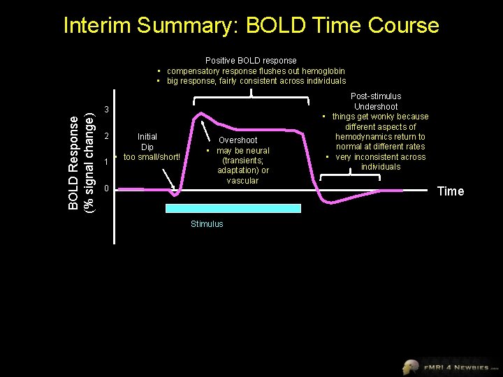
Interim Summary: BOLD Time Course BOLD Response (% signal change) Positive BOLD response • compensatory response flushes out hemoglobin • big response, fairly consistent across individuals 3 2 1 0 Initial Dip • too small/short! Overshoot • may be neural (transients; adaptation) or vascular Stimulus Post-stimulus Undershoot • things get wonky because different aspects of hemodynamics return to normal at different rates • very inconsistent across individuals Time
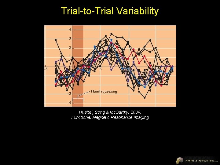
Trial-to-Trial Variability Huettel, Song & Mc. Carthy, 2004, Functional Magnetic Resonance Imaging
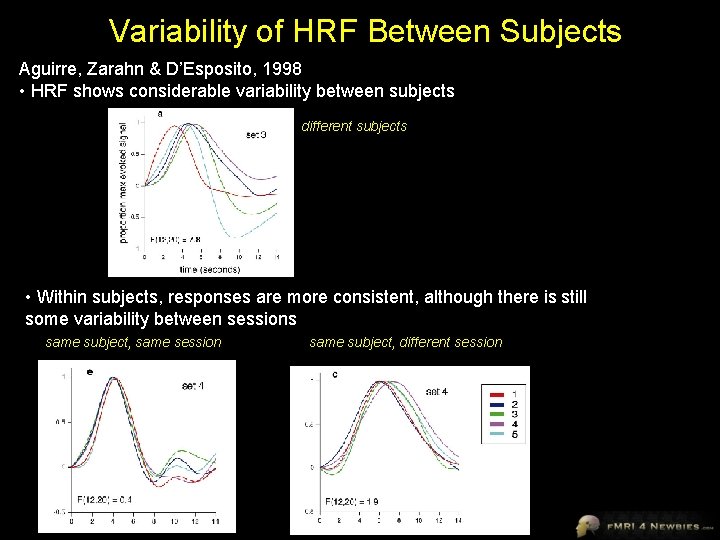
Variability of HRF Between Subjects Aguirre, Zarahn & D’Esposito, 1998 • HRF shows considerable variability between subjects different subjects • Within subjects, responses are more consistent, although there is still some variability between sessions same subject, same session same subject, different session
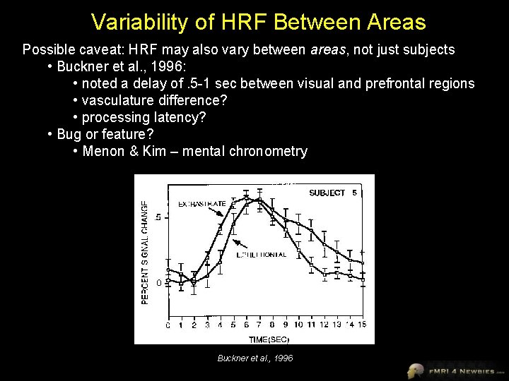
Variability of HRF Between Areas Possible caveat: HRF may also vary between areas, not just subjects • Buckner et al. , 1996: • noted a delay of. 5 -1 sec between visual and prefrontal regions • vasculature difference? • processing latency? • Bug or feature? • Menon & Kim – mental chronometry Buckner et al. , 1996
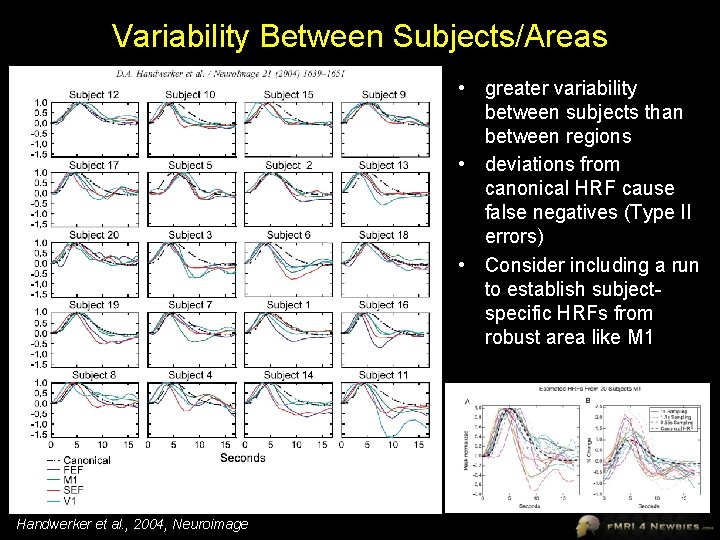
Variability Between Subjects/Areas • greater variability between subjects than between regions • deviations from canonical HRF cause false negatives (Type II errors) • Consider including a run to establish subjectspecific HRFs from robust area like M 1 Handwerker et al. , 2004, Neuroimage
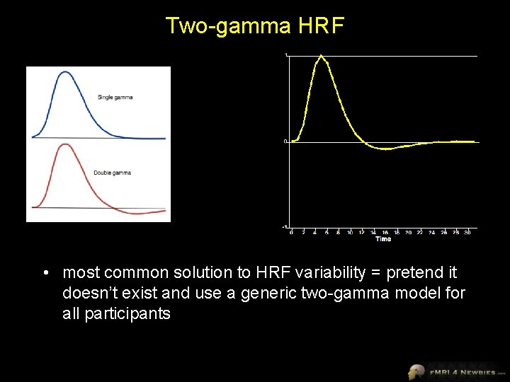
Two-gamma HRF • most common solution to HRF variability = pretend it doesn’t exist and use a generic two-gamma model for all participants
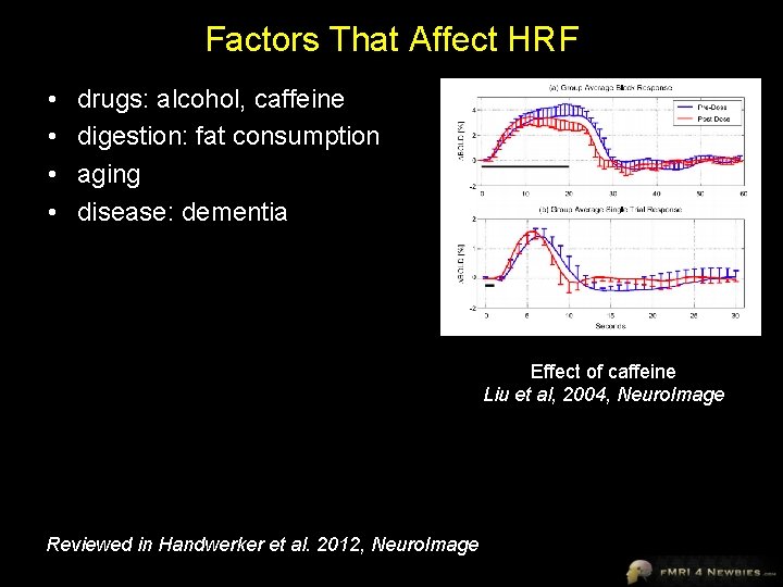
Factors That Affect HRF • • drugs: alcohol, caffeine digestion: fat consumption aging disease: dementia Effect of caffeine Liu et al, 2004, Neuro. Image Reviewed in Handwerker et al. 2012, Neuro. Image
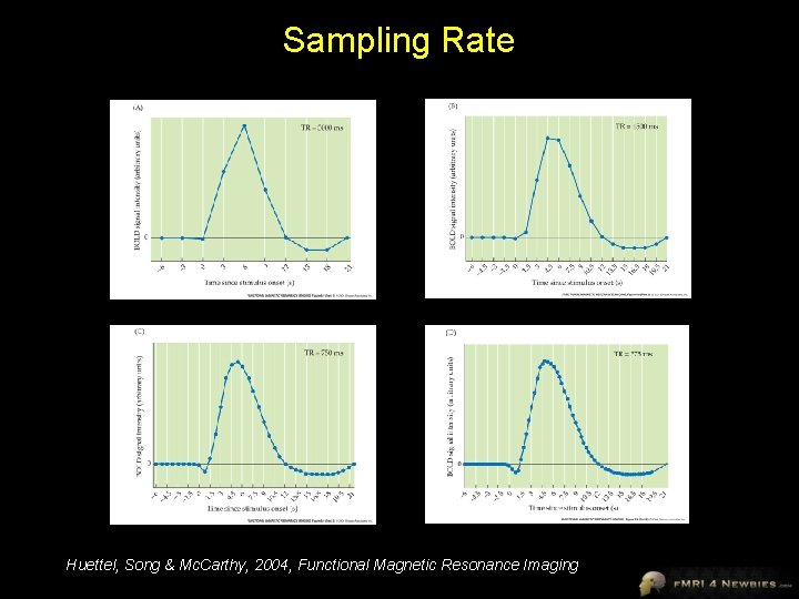
Sampling Rate Huettel, Song & Mc. Carthy, 2004, Functional Magnetic Resonance Imaging
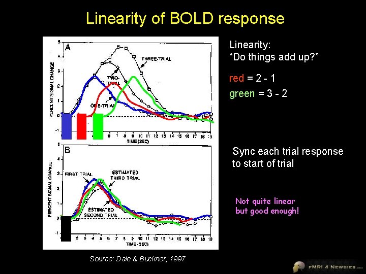
Linearity of BOLD response Linearity: “Do things add up? ” red = 2 - 1 green = 3 - 2 Sync each trial response to start of trial Not quite linear but good enough! Source: Dale & Buckner, 1997
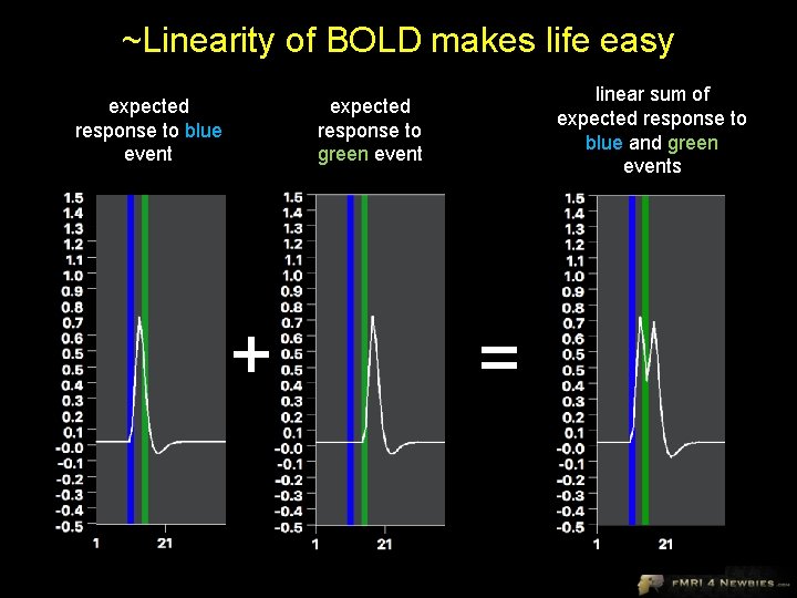
~Linearity of BOLD makes life easy expected response to blue event linear sum of expected response to blue and green events expected response to green event + =
Section 2 From Neurons to BOLD
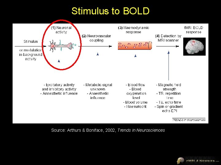
Stimulus to BOLD Source: Arthurs & Boniface, 2002, Trends in Neurosciences
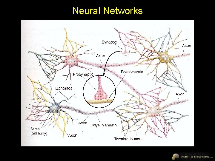
Neural Networks
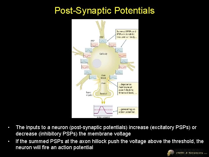
Post-Synaptic Potentials • • The inputs to a neuron (post-synaptic potentials) increase (excitatory PSPs) or decrease (inhibitory PSPs) the membrane voltage If the summed PSPs at the axon hillock push the voltage above threshold, the neuron will fire an action potential
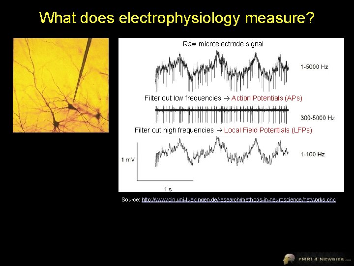
What does electrophysiology measure? Raw microelectrode signal Filter out low frequencies Action Potentials (APs) Filter out high frequencies Local Field Potentials (LFPs) Source: http: //www. cin. uni-tuebingen. de/research/methods-in-neuroscience/networks. php
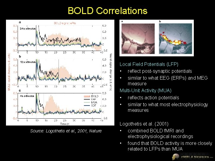
BOLD Correlations 24 s stimulus 12 s stimulus 4 s stimulus Source: Logothetis et al. , 2001, Nature Local Field Potentials (LFP) • reflect post-synaptic potentials • similar to what EEG (ERPs) and MEG measure Multi-Unit Activity (MUA) • reflects action potentials • similar to what most electrophysiology measures Logothetis et al. (2001) • combined BOLD f. MRI and electrophysiological recordings • found that BOLD activity is more closely related to LFPs than MUA
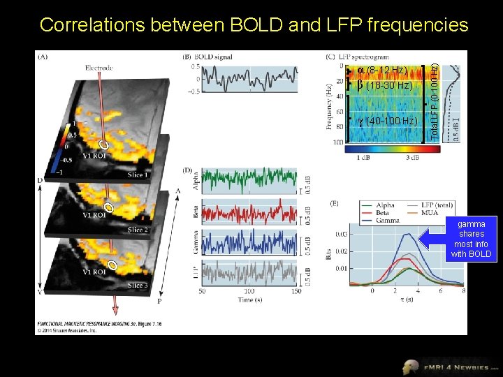
α (8 -12 Hz) β (18 -30 Hz) γ (40 -100 Hz) Total LFP (0 -100 Hz) Correlations between BOLD and LFP frequencies gamma shares most info with BOLD
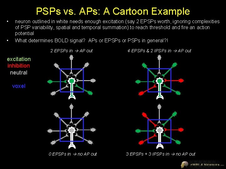
PSPs vs. APs: A Cartoon Example • • neuron outlined in white needs enough excitation (say 2 EPSPs worth, ignoring complexities of PSP variability, spatial and temporal summation) to reach threshold and fire an action potential What determines BOLD signal? APs or EPSPs or PSPs in general? ! 2 EPSPs in AP out 4 EPSPs & 2 IPSPs in AP out 0 EPSPs in no AP out 3 EPSPs + 3 IPSPs in no AP out excitation inhibition neutral voxel
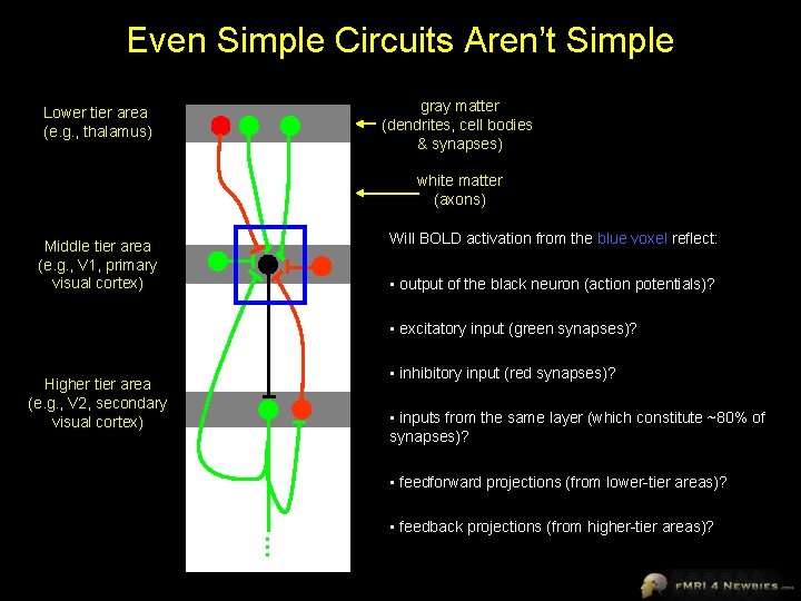
Even Simple Circuits Aren’t Simple gray matter (dendrites, cell bodies & synapses) Lower tier area (e. g. , thalamus) white matter (axons) Will BOLD activation from the blue voxel reflect: Middle tier area (e. g. , V 1, primary visual cortex) • output of the black neuron (action potentials)? • excitatory input (green synapses)? • inhibitory input (red synapses)? Higher tier area (e. g. , V 2, secondary visual cortex) • inputs from the same layer (which constitute ~80% of synapses)? • feedforward projections (from lower-tier areas)? … • feedback projections (from higher-tier areas)?
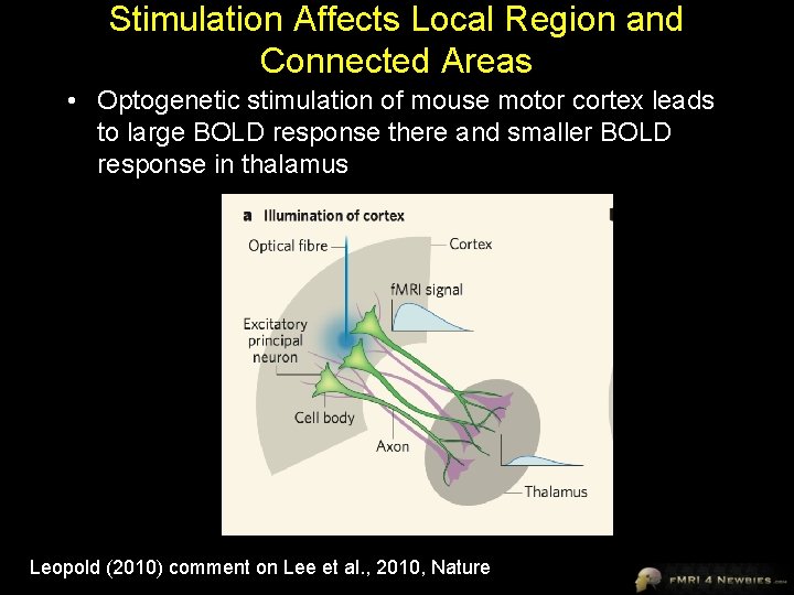
Stimulation Affects Local Region and Connected Areas • Optogenetic stimulation of mouse motor cortex leads to large BOLD response there and smaller BOLD response in thalamus Leopold (2010) comment on Lee et al. , 2010, Nature
What about inhibitory synapses? • GABA = inhibitory neurotransmitter hyperpolarization (IPSP) • less metabolically demanding than excitatory (glutamatergic) activity • GABA can be taken up presynaptically rather than recycled through astrocytes • Therefore, neurotransmission at inhibitory synapses likely influences the BOLD signal less than at excitatory synapses
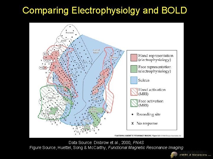
Comparing Electrophysiolgy and BOLD Data Source: Disbrow et al. , 2000, PNAS Figure Source, Huettel, Song & Mc. Carthy, Functional Magnetic Resonance Imaging
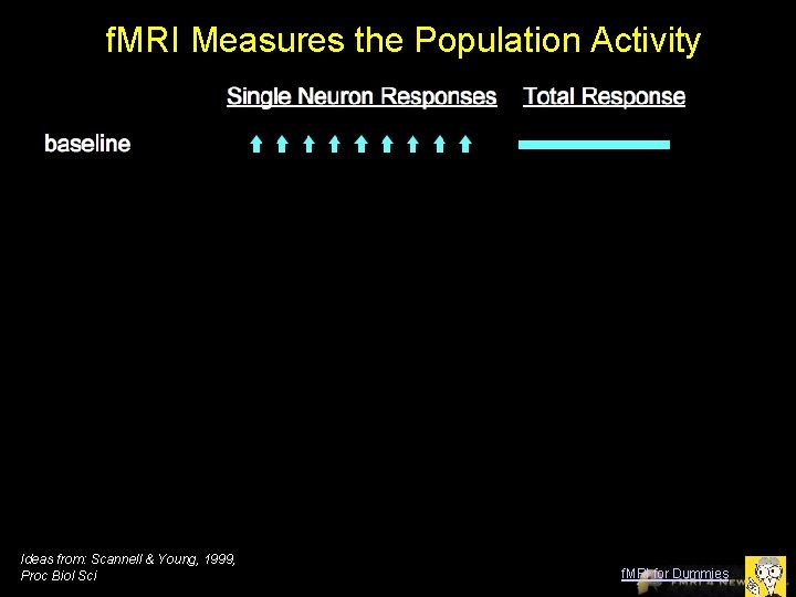
f. MRI Measures the Population Activity Ideas from: Scannell & Young, 1999, Proc Biol Sci f. MRI for Dummies
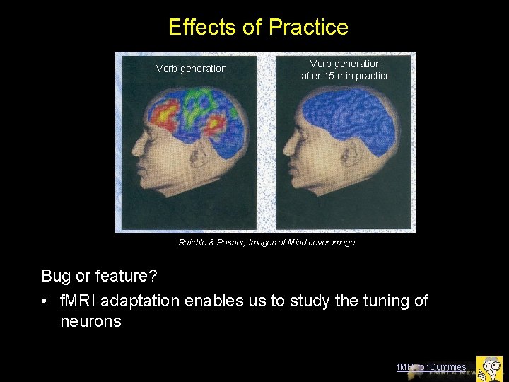
Effects of Practice Verb generation after 15 min practice Raichle & Posner, Images of Mind cover image Bug or feature? • f. MRI adaptation enables us to study the tuning of neurons f. MRI for Dummies
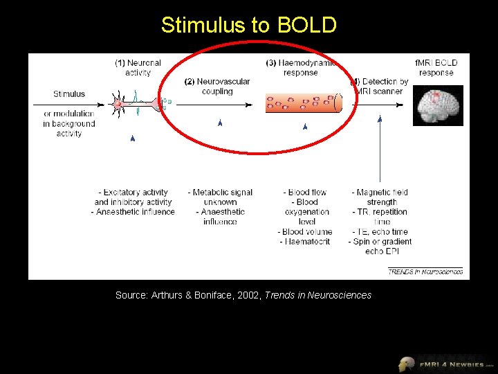
Stimulus to BOLD Source: Arthurs & Boniface, 2002, Trends in Neurosciences
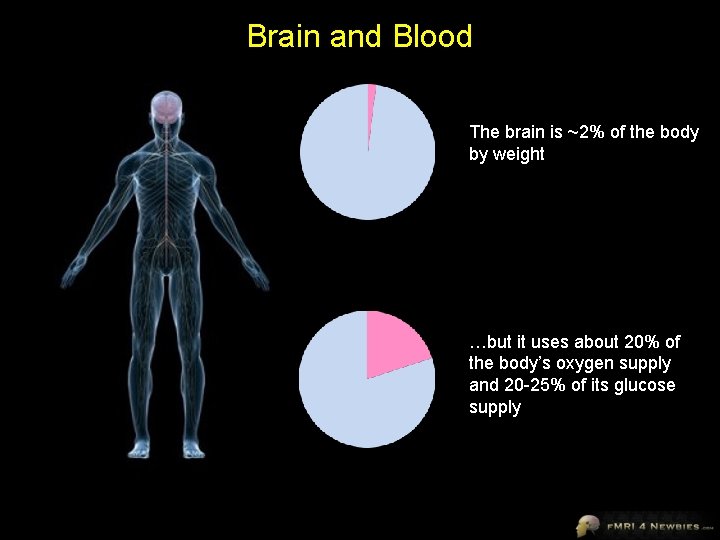
Brain and Blood The brain is ~2% of the body by weight …but it uses about 20% of the body’s oxygen supply and 20 -25% of its glucose supply
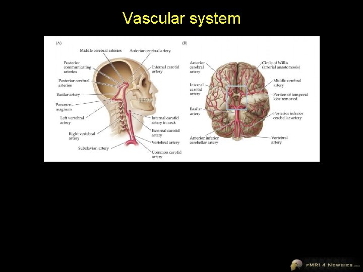
Vascular system
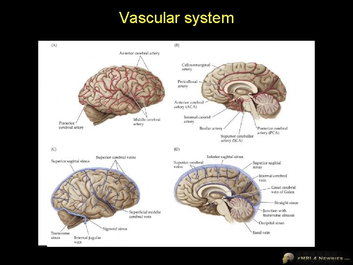
Vascular system
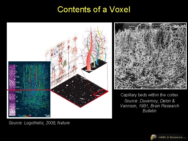
Contents of a Voxel Capillary beds within the cortex Source: Duvernoy, Delon & Vannson, 1981, Brain Research Bulletin Source: Logothetis, 2008, Nature
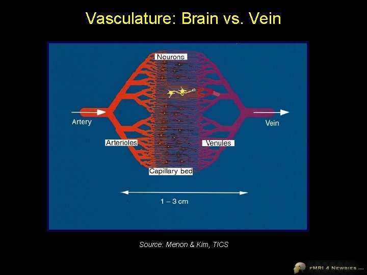
Vasculature: Brain vs. Vein Source: Menon & Kim, TICS
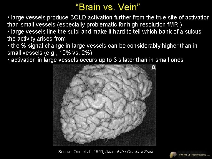
“Brain vs. Vein” • large vessels produce BOLD activation further from the true site of activation than small vessels (especially problematic for high-resolution f. MRI) • large vessels line the sulci and make it hard to tell which bank of a sulcus the activity arises from • the % signal change in large vessels can be considerably higher than in small vessels (e. g. , 10% vs. 2%) • activation in large vessels occurs up to 3 s later than in small ones Source: Ono et al. , 1990, Atlas of the Cerebral Sulci
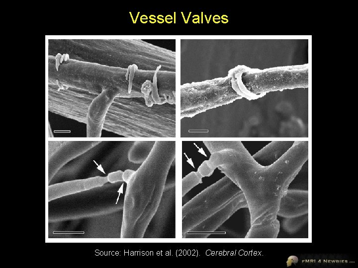
Vessel Valves Source: Harrison et al. (2002). Cerebral Cortex.
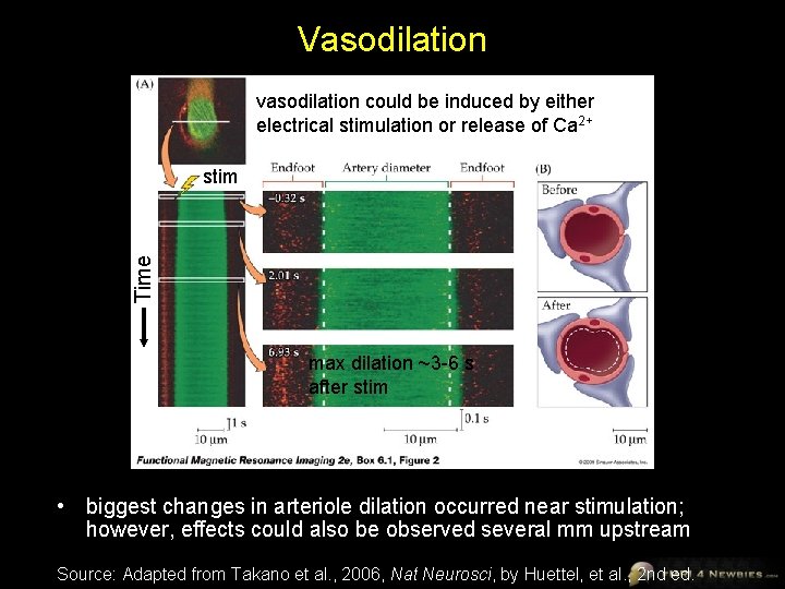
Vasodilation vasodilation could be induced by either electrical stimulation or release of Ca 2+ Time stim max dilation ~3 -6 s after stim • biggest changes in arteriole dilation occurred near stimulation; however, effects could also be observed several mm upstream Source: Adapted from Takano et al. , 2006, Nat Neurosci, by Huettel, et al. , 2 nd ed.
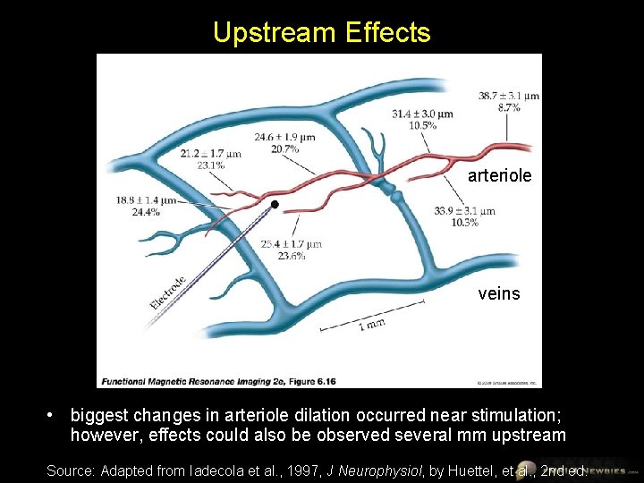
Upstream Effects arteriole veins • biggest changes in arteriole dilation occurred near stimulation; however, effects could also be observed several mm upstream Source: Adapted from Iadecola et al. , 1997, J Neurophysiol, by Huettel, et al. , 2 nd ed.
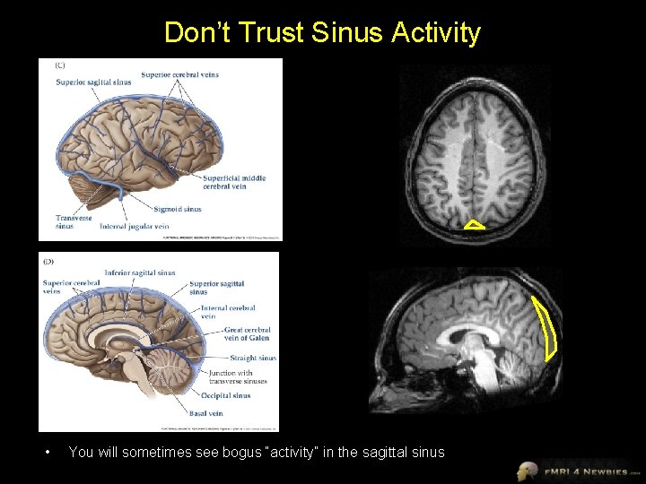
Don’t Trust Sinus Activity • You will sometimes see bogus “activity” in the sagittal sinus
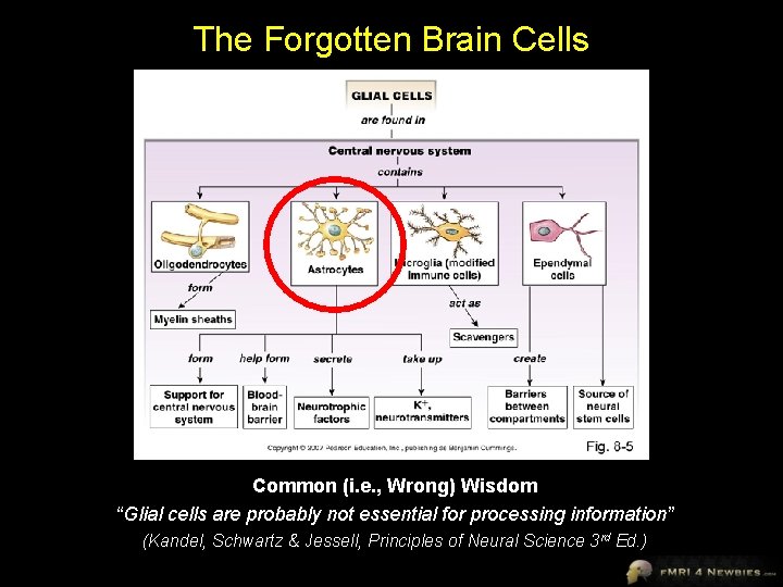
The Forgotten Brain Cells Common (i. e. , Wrong) Wisdom “Glial cells are probably not essential for processing information” (Kandel, Schwartz & Jessell, Principles of Neural Science 3 rd Ed. )
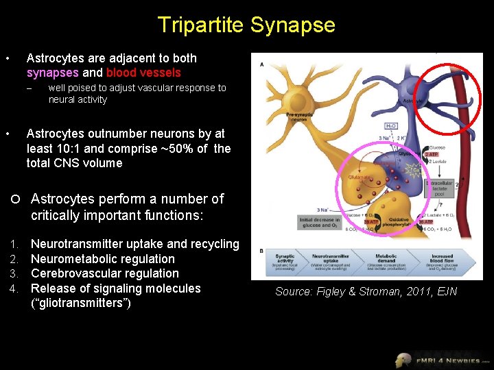
Tripartite Synapse Astrocytes are adjacent to both synapses and blood vessels • – well poised to adjust vascular response to neural activity Astrocytes outnumber neurons by at least 10: 1 and comprise ~50% of the total CNS volume • Astrocytes perform a number of critically important functions: 1. 2. 3. 4. Neurotransmitter uptake and recycling Neurometabolic regulation Cerebrovascular regulation Release of signaling molecules (“gliotransmitters”) Source: Figley & Stroman, 2011, EJN
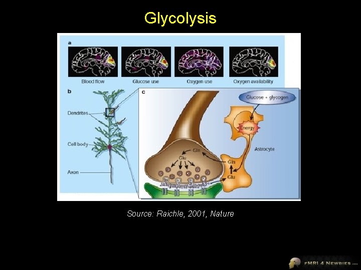
Glycolysis Source: Raichle, 2001, Nature
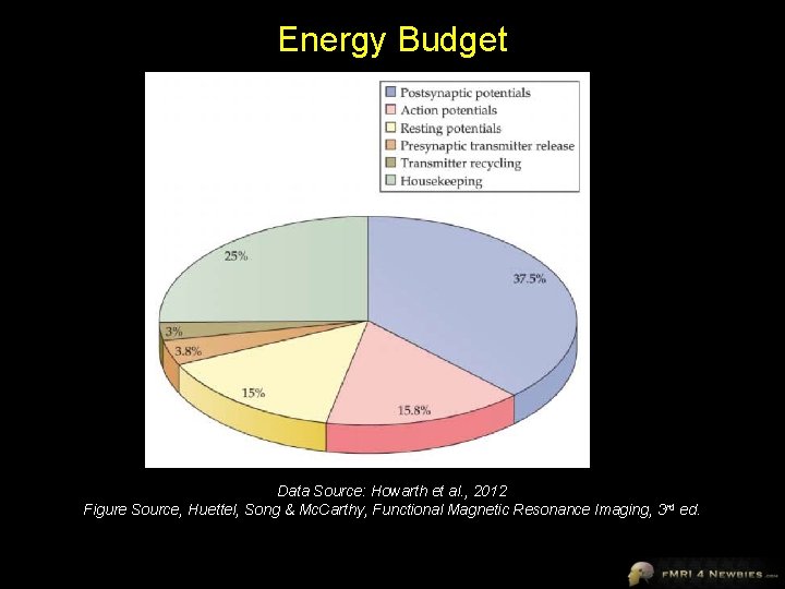
Energy Budget Data Source: Howarth et al. , 2012 Figure Source, Huettel, Song & Mc. Carthy, Functional Magnetic Resonance Imaging, 3 rd ed.
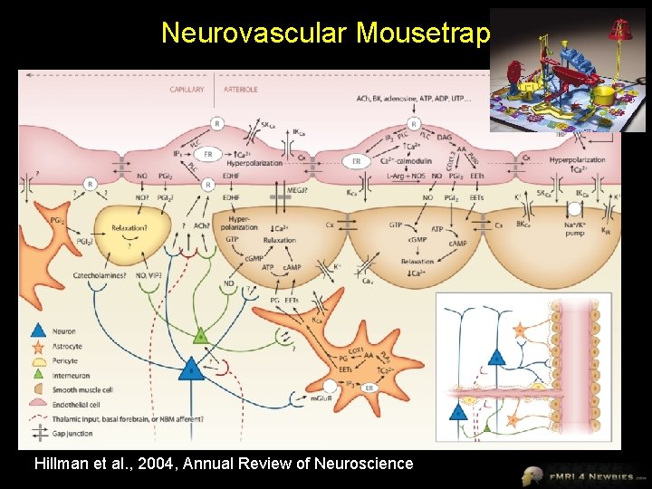
Neurovascular Mousetrap Hillman et al. , 2004, Annual Review of Neuroscience
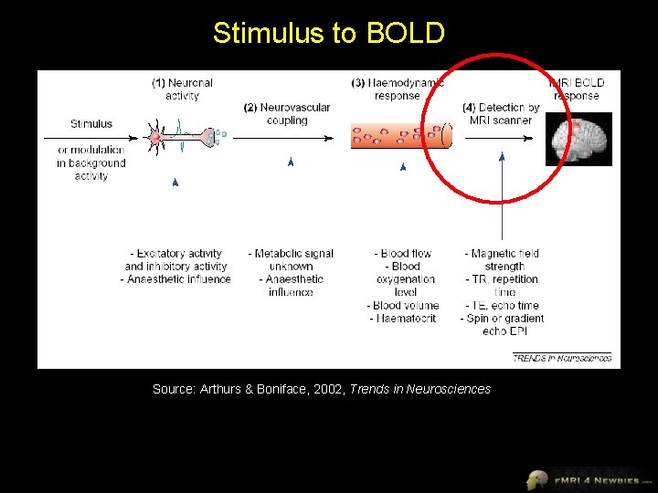
Stimulus to BOLD Source: Arthurs & Boniface, 2002, Trends in Neurosciences
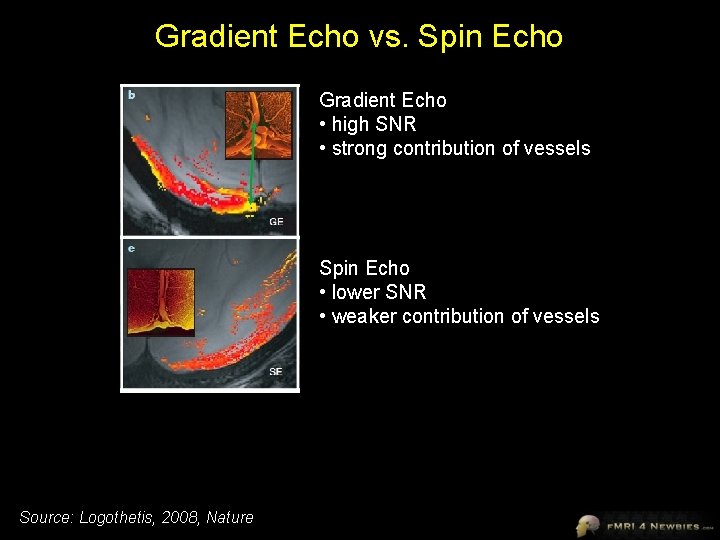
Gradient Echo vs. Spin Echo Gradient Echo • high SNR • strong contribution of vessels Spin Echo • lower SNR • weaker contribution of vessels Source: Logothetis, 2008, Nature
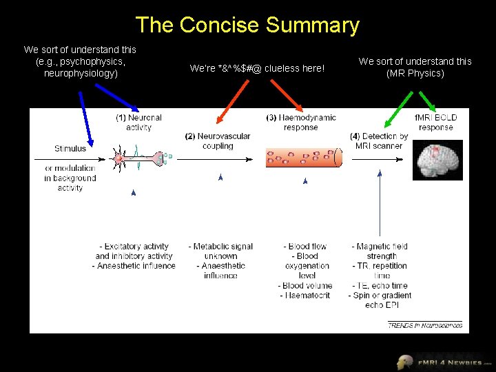
The Concise Summary We sort of understand this (e. g. , psychophysics, neurophysiology) We’re *&^%$#@ clueless here! We sort of understand this (MR Physics)
Summary: BOLD ≠ neural activity • BOLD reflects inputs to neurons (post-synaptic potentials) more than outputs (action potentials) • inputs can come from same brain area or areas in the hierarchy that are earlier (feedforward) or later (feedback) • excitation may have a larger effect on BOLD than inhibition • maps measured by neurophysiology and BOLD agree to a first approximation but not perfectly • f. MRI measures population response not individual neuron responses and requires a different way of thinking about activity – e. g. , a small change to a large number of neurons (e. g. , attention, expectation) can cause a bigger BOLD effect than a large change to a small number of neurons (e. g. , presentation of a stimulus that is optimal for a subset of neurons) • BOLD effects can be seen in downstream vessels (venules, veins, sinuses) and a bit upstream (arterioles)
Is the f. MRI Sky Falling?
Don’t Panic • BOLD imaging is well correlated with results from other methods • BOLD imaging can resolve activation at a fairly small scale (e. g. , retinotopic mapping) • PSPs and action potentials are correlated so either way, it’s getting at something meaningful • even if BOLD activation doesn’t correlate completely with electrophysiology, that doesn’t mean it’s wrong – may be picking up other processing info (e. g. , PSPs, synchrony) – maybe anticipatory changes in blood flow are interesting too
- Slides: 47