jabb modalits LTS Lts Alapveten vizulis alap szlelsnk

Újabb modalitás LÁTÁS

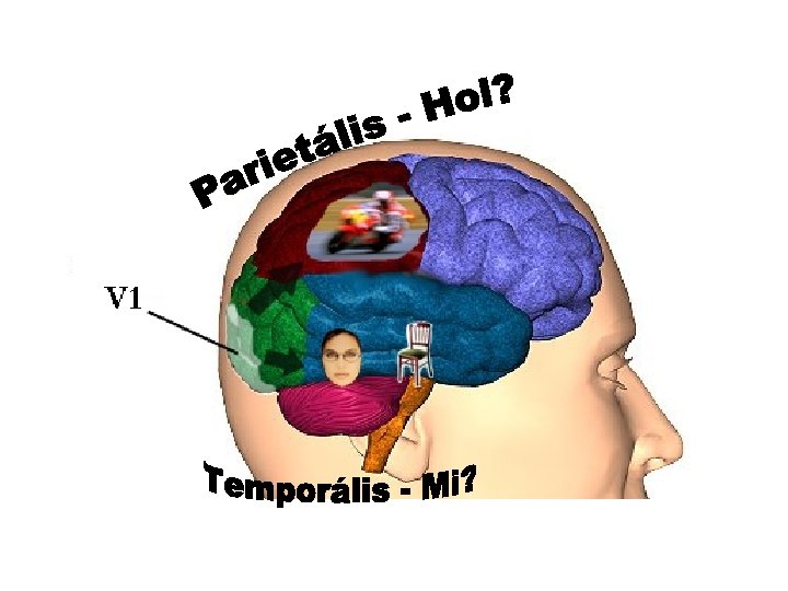
Látás • Alapvetően vizuális alapú észlelésünk • Számos különféle információt dolgozunk fel – Alak, forma – Mozgás – Mélység – Szín • Feldolgozásuk egyidőben, párhuzamosan!!!

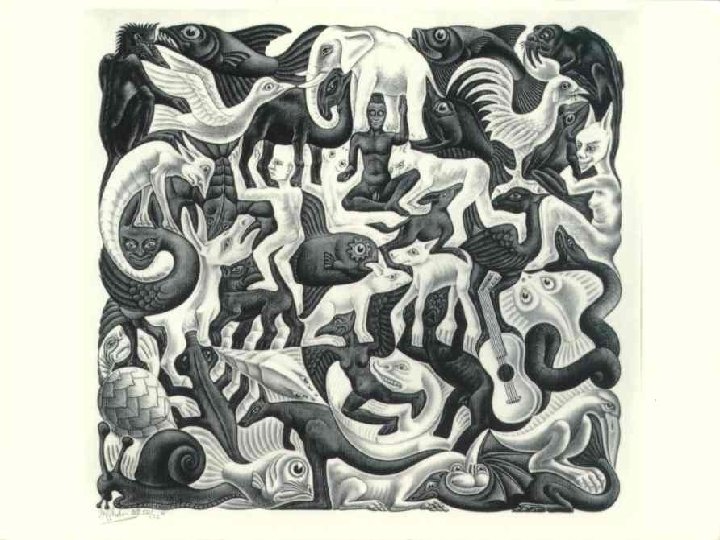
Analógia

DE… • Mi valahogy összerakjuk a 3. dimenziót is! • Ebben még segítenek: kognitív funkciók, pl. : előzetes tudásunk és a bennünk kialakult invarianciák, konstanciák • Azaz: a kamera passzív felvételével szemben látásunk egy AKTÍV FOLYAMAT

Egy kis történet… • Locke és Berkeley: észlelés = olyan egyszerű feldolgozás, melyben az elemi észleletek összeadódnak • Ma: nem atomisztikus, hanem HOLISZTIKUS • Úttörők: Gestalt – Wertheimer, Koffka, Köhler – Központi elv: szerveződés (komputációs elvek alapján az agyban) – Korai: melódia-analógia (interrelationship) – Speciális alapelvek a perceptuális szerveződésre

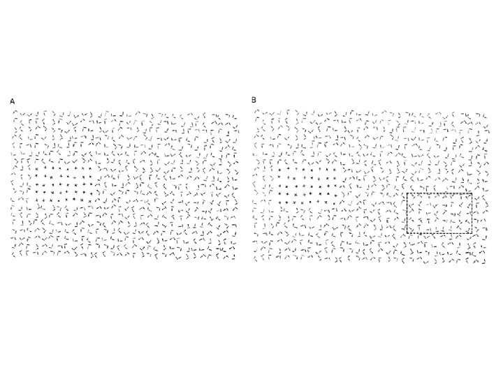
Kétértelmű ábrák I. • Legegyszerűbb: sorok vagy oszlopok?

Kétértelmű ábrák II.

Hasonlóság és közelség

Figura és háttér elkülönülése – a klasszikus …

És mások…

Az agy „félreolvasásai” - illúziók

Néha többet dolgozik – kitöltések Kanizsa ábrák

Segíthet – téri viszonyok

Látjuk vagy nem?

Bezzeg így… összekötöttük!

Konvex - konkáv

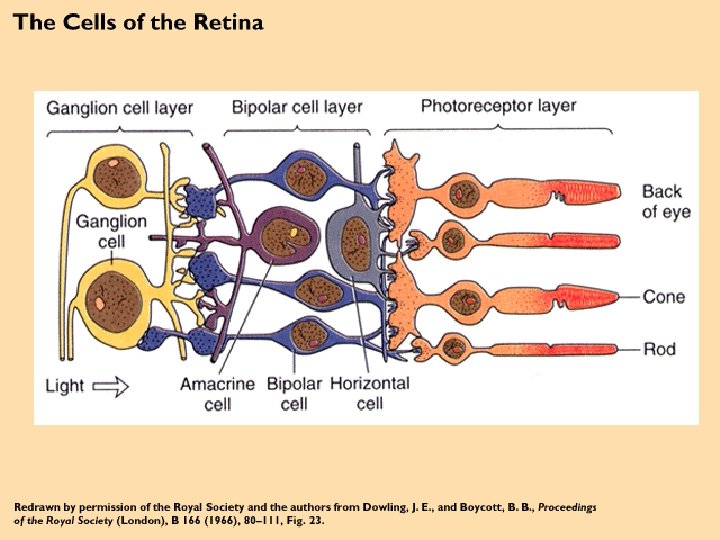
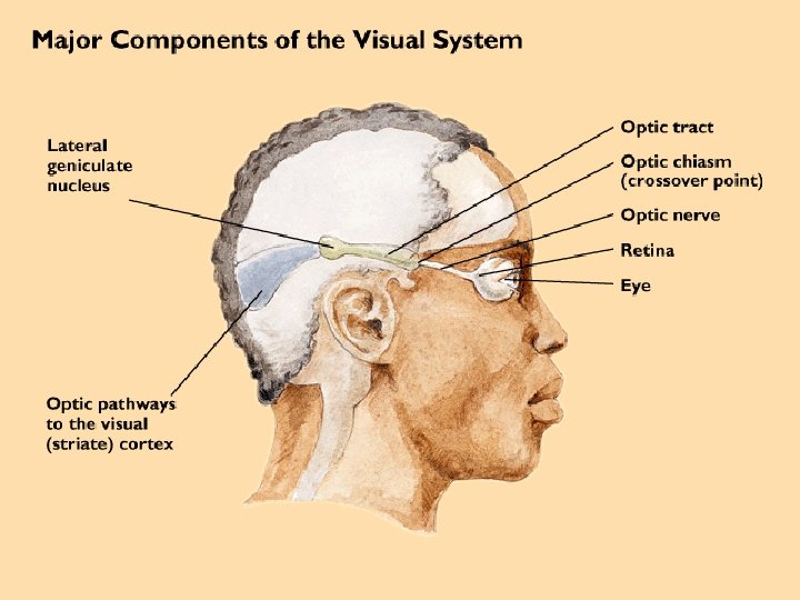
Kezdeti út … • Fotoreceptorok – bipoláris sejtek – ganglionsejtek – axonjaik: nervus opticus – CGL – V 1 (Br 17) • retinotópia

Mára számok, adatok • Retinának legalább 32 reprezentációja az extrastriatális régiókban • A makákó neocortexének több mint fele érintett a látásban • Az egyes régiók eltérő nagyságúak, legnagyobbak: V 1 és V 5, legkisebb: MT (V 5) • Különböző ingertulajdonságokra érzékenyek – Klinikai adatok, képalkotás – Neglect, agnóziák
 (V 1 -től MT-ig)")
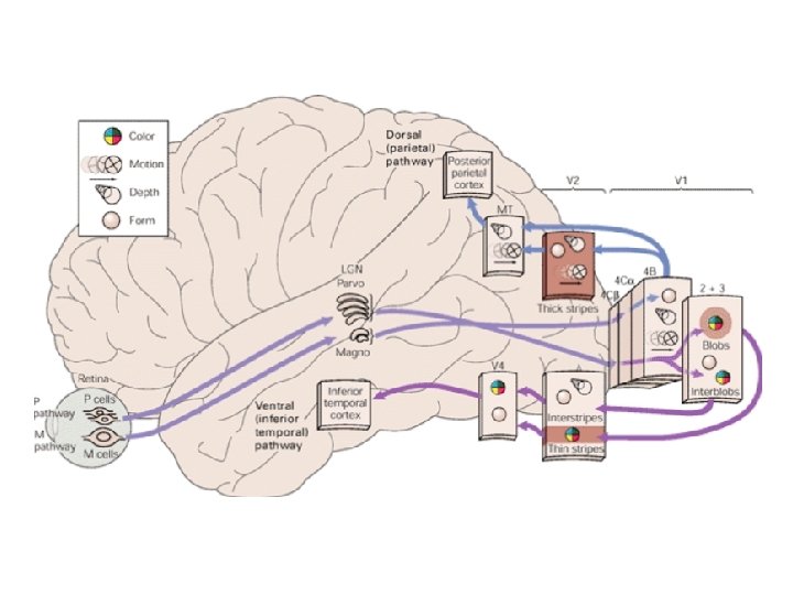
Párhuzamos feldolgozás • Ungerleider és Mishkin – Dorzális (WHERE? ) (V 1 -től MT-ig) és ventrális (WHAT? ) pályarendszer (V 1 -től IT-ig) • Goodale és Milner – Action és Perception pályarendszer

D. F. Kontroll

Út a retinától a kéreg felé • • 2 fő pályarendszer P és M pályák Nagy ganglionsejtek (Magno) Kis ganglionsejtek (Parvo) Más típusú infót szállítanak a Thalamusba M: magnocelluláris rétegekbe P: parvocelluláris rétegekbe Innen V 1 külön rétegébe (M: 4 Cα, P: 4 Cβ)

Kéregben • V 1 -ben és V 2 -ben két fontos alosztály: blobok (V 1) és stripe-ok (V 2) – Szerepükről később • P pálya – blobba és interblobba, onnan rendre stripe vagy interstripe (V 2) – V 4 – ventrális • M pálya – vékony stripes (V 2) – MT (V 5) – dorzális • M ventrálisba is!

Vizuális figyelem • Fő feladat: asszociáció az észlelt vonások között • Treismann és Julesz: ehhez fókuszált figyelem kell! • Mi különül el a háttértől: fényesség, szín, orientáció… – Azonnal kiugró (pop-up) – globális feldolgozás elég, preattentív (bottom-up) – Ha figyelmet igényel – attentív – szeriális feldolgozás – top-down • Treismann: modell


A retina és a pályák

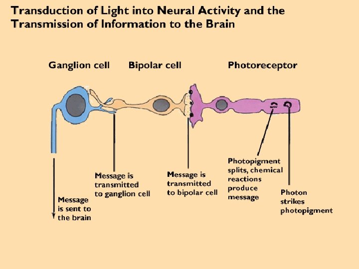
Vizuális percepció kezdete. . • ÚT: fény → cornea → szem belső része • Itt: egy speciális „érzékszerv“ (retina) segítségével elektromos jel generálódik → nervus opticus → magasabb központok

Retina • Amiben más, több: – Alkalmas a szenzoros transzdukció megértésére – nem periférikus, a KIR része • Mindemellett: relatíve egyszerű, 5 fő neuronosztály
 • Más retinális")
Ma. . . • Hogyan lesz a fényből elektromos jel? (receptorok) • Más retinális sejtek hogyan alakítják át ezt a jelet? (út az agyhoz, kapcsolatok) • Központi pályák

Ezek konkrét megtárgyalása előtt • Retina szerveződése • A látás receptorainak alapvető fiziológiai tulajdonságai

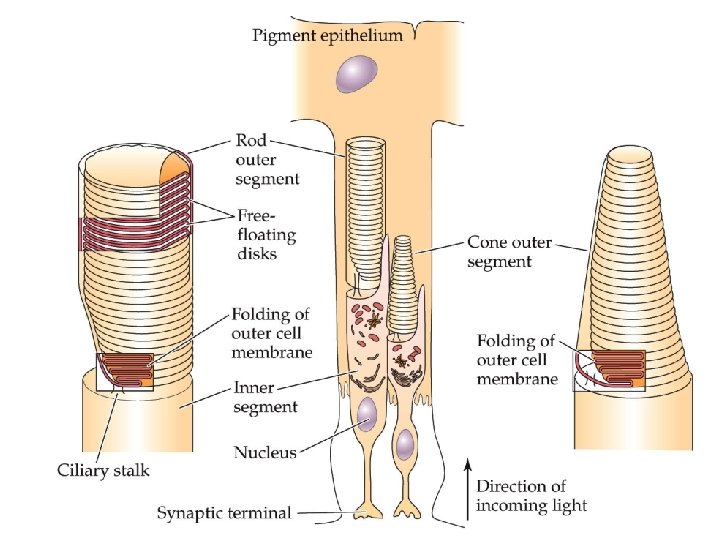
• Cél: a vizuális kép fókuszálása a retinán minimális optikai torzítás/veszteség mellett • Fény fókuszálására: cornea + lencse • Innen az üvegtesten keresztül egyenes az út a fotoreceptorok felé • Pigment epithelium: fekete festékanyaggal (melanin) töltött, elnyeli az összes fényt, mely nem éri el a retinát • Retina: a pigment epitheliummal szemközt a szem belső „borítása“ • Közvetlenül a pigment epitheliummal szemben, a lencséhez legközelebb: retinális sejtek, de axonjaik csupaszak, így ezek a sejtrétegek relatíve transzparensek

A retina KÉT kitüntetett pontja • Fovea: itt a retinális sejtek „kitérnek“, így a fotoreceptorok itt kapják meg a legkevesebb torzítással a vizuális ingert • A legerősebb „kitérés“ a fovea közepénél = foveola • Célunk: a nézett kép mindig ide vetüljön • Vakfolt (optic disc): itt hagyják el a nervus opticus rostjai a retinát, nincsenek fotoreceptorok


Fotoreceptorok PÁLCIKÁK • Magas fényérzékenység, éjszakai látás • Több fotopigment, több fény • Magas erősítés, egyetlen foton detektálása • Alacsony idői felbontás: lassú válasz, hosszú integrációs idő • Érzékenyebb a szétszórt fényre RENDSZER • Foveánál alig, nagy konvergencia a retinális pályán • Akromatikus • Kb. 120 millió CSAPOK • Alacsonyabb érzékenység, nappali látás • Kevesebb fotopigment • Alacsonyabb erősítés • Magas idői felbontás: gyors válasz, rövid integrációs idő • Legérzékenyebb a direkt axiális sugarakra RENDSZER • Foveánál sok, szórt retinális pályák • Kromatikus (3 csaptípus) • Kb. 8 millió Egyik sem generál AP-t, az intenzitást a MP súlyozásával kódolják.
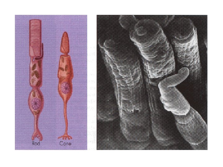

A fotoreceptorok részei • Külső szegmens – Fényelő vizuális pigmentek, melyek egy kisebb fényelő molekulából és egy nagyobb 7 -TM membránproteinből állnak (sok protein – lemez, membránfelszín területét drámaian növelik; csapok esetén a plazma membránjával folytonosak) – Folyamatosan és gyorsan újulnak, régebbiek fagocitózissal távoznak • Belső szegmens – Sejttestek és bioszintetikus rendszer • Szinaptikus végződések (terminálok) – Kapcsolatok

Fototranszdukció • Általánosságok – 3 állomásos kaszkád – A fotoreceptorok pigmentjeinek fényelésével indul a folyamat, fő molekula: c. GMP – c. GMP: pálcikákban second messenger, cytoplazmán át viszi az infót hozzákötve a szabadon lebegő lemezeket a sejt plazmamembránjához; csapoknál: mivel a lemezek folytonosak a plazmamembránnal, itt a c. GMP speciális ioncsatornák nyitásával ionáramokat kontrollál

Röviden • Sötét → c. GMP koncentráció relatíve magas → csatornák nyitva → sejt relatíve depolarizált állapotban → fény aktiválja a vizuális pigmenteket → ezek az aktivált molekulák egy enzimet stimulálnak, mely a citoplazma c. GMP koncentrációját csökkenti → ez bezárja a c. GMP-kapuzott csatornákat → fotoreceptor hiperpolarizációja

1. A fény aktiválja a pigmentmolekulákat a fotoreceptorokban • A vizuális pigment neve: rodopszin – két része van • opszin (protein), beágyazva a lemez membránjába • egy fényelő rész, a retinal (A-vitamin aldehidje), melynek számos izomerje létezik, a két legfontosabb: – nem-aktivált rodopszinban a 11 -cisz izomer, mely az opszin molekula egy kötési helyébe illik bele – Aktiváció esetén (all-)transz, ami már nem illik oda, így az opszin változáson megy át (félstabil állapot) (metarodopszin II. ), de perceken belül szétválik opszinná és (all-)transz retinallá. – Ezután az (all-)transz a pálcikától a pigment epithelium sejtekhez szállítódik, ahol (all-)transz retinollá (ez már az Avitamin) csökken, ami a prekurzor a 11 -cisz szintézisében és ez visszaszállítódik a pálcikákhoz

Kék – opszin Zöld – retinal Forgatás – stabilabb

Csapok esetében • 3 -féle csaptípus – külön fényelésre optimalizált pigmentek • Hasonlóan két részből állnak – Csap opszin (protein) és 11 -cis retinal • Más csap pigment – más opszin izomorf

2. A pigmentmolekulák aktivációja csökkenti a citoplazmás c. GMP koncentrációt • Fény → pigmentmolekula aktivációja → c. GMP citoplazmás koncentrációja csökken (koncentrációját két enzim kontrollálja – GTP-ből szintézis által /ezt egy G-protein, a transzducin GDP-ből állítja elő/ vagy 5´GMP-vé törik le c. GMP-t foszfodiészteráz által – utóbbit a vizuális pigmentek kontrollálják, így fényinfóra aktiválódik a foszfodiészteráz is, így csökken a c. GMP koncentrációja is)

3. A c. GMP koncentráció csökkenése zárja a c. GMPkapuzott ioncsatornákat, így hiperpolarizálja a fotoreceptort • A c. GMP kapuzza ezeket a csatornákat azáltal, hogy direkten kötődik a csatornák citoplazmás oldalához – legalább 3 c. GMP molekula együttműködő kötődése által a csatorna aktiválódik • Fény hiányában a c. GMP-kapuzott ioncsatornák egy belső áramot vezetnek (sötétáram), mely képes depolarizálni a fotoreceptort • E csatornáknak a fény kiváltotta zárása csökkenti ezt az áramot és ezért a sejt hiperpolarizálódik


SÖTÉT glutamát VILÁGOS glutamát

Hogyan tovább? • Ganglion sejtek, de előtte: 3 interneuronosztály – Bipoláris, horizontális, amakrin sejtek – Feladatuk nem csupán jelszállítás, hanem össze is rakják az egyes fotoreceptorokból jövő jelet úgy, hogy a ganglionsejtekben kiváltott elektromos jel döntően a fény pontos téri és idői mintázatától függjön

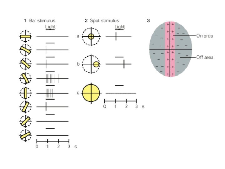
Ganglion sejtek • Van egy alapaktivitásuk, ezt a retinális interneuronokból jövő input modulálja • Input: a környező fotoreceptorokból, a retina egy körülhatárolt területéről (RM) • RM: két fontos vonása – központ (körkörös) és szél (körgyűrű) • Két osztályuk van: ON vagy OFF center • Számuk kb. azonos! Mindegyik fotoreceptor küld kimenetet mindkét típusnak – ez már sugallja a párhuzamos pályákat! • RM mérete a retina egyes részein más – fovea: kicsi, periféria: nagyobb

Ganglionsejtek receptív mezeje

Válaszaik fény jelenlétére

Fő feladatuk • Max válasz, ha központi és széli infó eltér – tehát inkább kontrasztot, mint intenzitást kódol • Miért jó? Mert a transzmissziós hiba minimális lesz. • Szintén növeli a vizuális rendszer teljesítményét, hogy az ON és OFF pályák párhuzamosak

Két funkcionális osztály • M és P osztály – mindkettőben vannak ON és OFF sejtek is • M: nagy RM, fennálló ingerlésre, nagy tárgyakra, gyors változásokra – gyors vonás, mozgás • P: több, kis RM, hullámhosszra specifikus, alak, szín, finom részletek

Interneuronok • Minden típusnak speciális szerepe van • Közülük a legdirektebb a kapcsolat a bipoláris sejtekkel a csapok esetében

Csapoktól a ganglionsjetekig • Két út – Egy ganglionsejt RM-jének közepében lévő csapok – direkt szinaptikus kapcsolat a bipoláris sejtekkel, amelyek direkt kapcsolatban a ganglionsjetekkel (direkt/vertikális pálya) – Szélről: horizontális és amakrin sejteken át indirekten (laterális pálya) • Horizontális: dendritikus fák – feedback a csapok közepének • Amakrin: bipoláris - ganglion

Retina szinaptikus kapcsolatai • Két plexiform réteg – Külső: receptorok, bipoláris, horizontális – Belső: bipoláris, amakrin, ganglion – Azaz: bipoláris mindkettőben – ez a híd a kettő között – De: AP-t még nem tud generálni!!!!

Bipoláris sejtek RM-je • • • Hasonlóan központ/szél szerveződés ON/OFF ON: depolarizáció OFF: hiperpolarizáció A hozzájuk kapcsolódó ganglionsejtosztályokkal serkentő kapcsolatban

És most a pályák. . .

Hálózat • A vizuális rendszer a legkomplexebb neuronális hálózat (kb. 1 millió rost alkotja a nervus opticust) • Út a retinától: középagy – Thalamus – primer látókéreg (V 1/Br 17/strialtális kéreg) • Subcorticalis régiók: pretectális régió, colliculus superior (középagy), CGL (Thalamus) • Ami még: M és P pályák – V 1 – és annak szerveződése

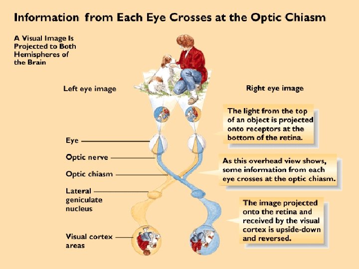
Retinális kép - nomenklatúra • Nazális hemiretina – foveától mediálisan • Temporális hemiretina – foveától laterálisan • Mindkét fele – Dorzális (superior) kvadráns – Ventrális (inferior) kvadráns

Látómező • Az a nézet, amelyet legalább egy szemmel látunk a fej elmozdítása nélkül LVF - RVF

Zónák • Binokuláris – centrális • Monokuláris, ha pl. nagyon temporálisan látható egy tárgy – ekkor csak ipszilaterális nazális
 • Itt lépnek ki a ganglionsejtek axonjai • Itt nincsenek fotoreceptorok")
Vakfolt (optic disc) • Itt lépnek ki a ganglionsejtek axonjai • Itt nincsenek fotoreceptorok = fényre érzéketlen • Mindkét szem esetén foveától nazálisan helyezkedik el, így ha két szemmel nézünk, nem tudatosulhat!!!! • Kísérletesen: monokulárisan megtalálható


• Fontos a VF és a retinális kép közötti szerveződést megérteni, DE: • Két probléma – A lencse megfordítja a vizuális képet, azaz pl. felső VF = inferior (ventrális) retina (sérülésnél erre figyelni kell!!!) – A binokuláris részen megjelenő pont a két retinán máshol található!

Axonok útja • Ganglionsjetek axonjai a vakfolton át kilépnek, myelinizáltak lesznek - nervus opticust alkotják – kereszteződnek a chiasma opticumon át, ahol „újracsomagolódnak“ – tractus opticus – innen 3 subcorticális régió (pretectum, colliculus superior, CGL) • Átkereszteződés szabály: csak a nazális kereszteződik, a temporális marad • Ezáltal a bal oldali retinarészek balra, jobb oldaliak jobbra
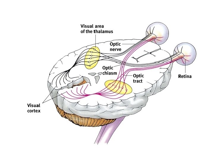

Colliculus superior • Fő feladata: szakkádikus szemmozgás kontrollja • Középagy tetején, sz. á. és f. á. része • Retinális ganglionsejtek direkt projekciója a felső rétegeibe, megalkotja a contralateralis VF térképét – pulvinar (Thal) – kéreg • Jelentős kérgi inputot is kap, felső réteg vizuálisból, alsóbbak más modalitásból is (fütyülő kismadár) • Mélyebb rétegek: erős válasz szakkádikus szemmozgás kezdetekor (középső réteg) – Pl. : bal VFben lévő ingerre válaszoló sejtek balfelé irányuló szakkád kezdete előtt • Input: direkt retinából is DE a legfontosabbak a kéregből

Pretectum • Épp ventrálisan a colliculus superiortól, fő feladat: pupilla reflex • Fény egyik szembe – direkt (abban a szemben) és konszenzuális (másikban) válasz • Reflexet mediálják: retinális ganglionsejtek • Sejtjei: bilaterális Edinger. Westphal nucleusban preganglionáris paraszimpatikus neuronok (III. agyideg mellett) – ciliáris ganglionok beidegzése az okulomotoros idegben • Postganglionárisan: smooth pupilla sphincter izmok, szimpatikusan: radiális izmok dilatatioja

Azért a főnök. . CGL • Retinából kb. 90% axon ide, innen a kéregbe • Blindsight (= vaklátás) • Kontralaterális VF retinotopikus leképeződése • Nem egyforma hangsúlyú reprezentáció (v. ö. : szomatoszenzoros rendszer) – fovea túlreprezentált

CGL felépítése • 6 réteg – közöttük intralamináris rétegek f. á. -nak • M és P retinális ganglionsejtekből – 2 belső: magnocelluláris, 4 dorzális: parvocelluláris – mindegyikben ON és OFF sejtekből is • DE: 1 adott réteg csak az egyik szemből: contralaterális nazális: 1, 4, 6 – ipsilaterális temporális: 2, 3, 5 • RM: központ/szél antagonisztikus szerveződés • Kedvenc inger: kis fényfolt
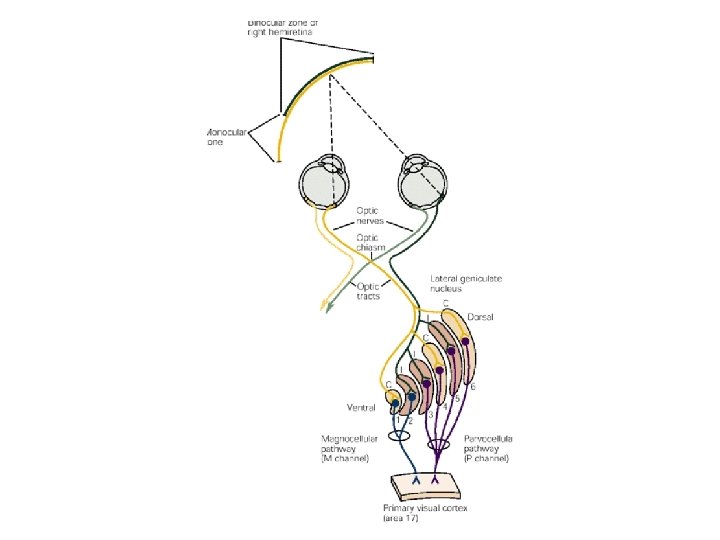

M és P pályák - sejtek • Legfontosabb különbségek a pályák sejtjeiben – táblázat • Szín, luminancia (fényességbeli különbség a 2 szélsőérték között), téri frekvencia (ciklus/fok), idői frekvencia (Hz) • Károsodásuk speciális kiesésekkel

V 1 • Egyszerű retinális inputból a vizuális kép építőkockái!!! • Itt nagyon megváltozik a RM szerveződése, ami marad: ellenoldali VF reprezentációja • Kb. 2 mm vastag, 6 réteg, CGL fő bemeneti rétege: 4 -es – alrétegek: A, B, Cα (M axonjai), Cβ (P axonjai) – szegregáció megmarad • Ezektől függetlenül: intralamináris sejtektől 2, 3 -ba (blobok) • Két fő sejtosztály: piramis sejtek, nem piramis sejtek (kis csillag alakú, tüskés vagy sima dendritekkel) – lokális interneuronok • Tüskés és piramis: serkentő – glutamát vagy aszpartát • Sima: gátló GABA
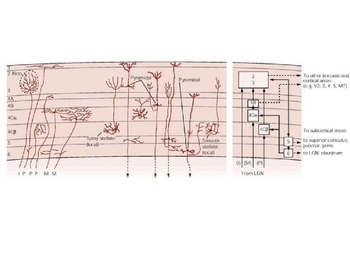

V 1 sejtjei • Egyszerű és komplex sejtek • A vizuális képet apró, különböző orientációjú vonalszegmensekre szedi szét • Hubel és Wiesel – story • Egyszerű sejtek: speciális orientációra, RM szerveződése – serkentő és gátló rész, kialakulás: több ON és OFF összegződése • Komplex: általában nagyobb RM, szintén orientáció, de az inger elhelyezkedése nem annyira döntő
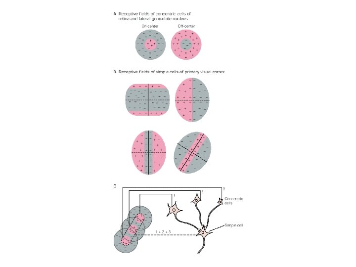

Út visszafelé - avagy erős konvergencia!!! • Bármely komplex sejt egyszerű sejtek csoportjának aktivitását szállítja – bármely egyszerű sejt a CGL sejtcsoportét – bármely CGL a retinális ganglionsjetekét – bármely ganglionsejt a bipoláris sejtekét – azok pedig a receptorsejtekét • Minden szinten minden sejtnek nagyobb absztrakciós kapacitása • A felszálló pálya bármely állomásán egy sejtet aktiváló ingertulajdonság egyre speciálisabb

V 1 – funkcionális modulokba szerveződik • Kolumnáris szerveződés – Orientációs kolumnák, Okuláris dominancia, Hiperkolumnák

Léziók 1. 2. 3. 4. 5. 6. Jobb nervus opticus – totál látásvesztés a jobb szemben Chiasma opticum – bitemporális hemianopsia Jobb tractus opticus – kontralaterális hemianopsia Meyer-loop (rad. opt. ami a temporálisba megy) – felső kontralaterális kvadratikus anopsia Sulcus calcarinus felső partja – másik oldal inferior kvadráns Alsó part – superior kvadráns

Irodalom • Kandel – Schwartz – Jessell: Principles of neural science – 25. – 26. – 27. fejezet
- Slides: 89