
IV Thyroid tumors Clinically the possibility of a
IV. Thyroid tumors
- Clinically , the possibility of a cancer is of major concern in patients who present with thyroid nodules but fortunately, the majority of solitary nodules of the thyroid prove to be either a. Follicular adenomas b. A dominant nodule in multinodular goiter
Note: - Carcinomas of the thyroid, are uncommon, accounting for much less than 10% of solitary thyroid nodules - Several clinical criteria provide a clue to the nature of a given thyroid nodule: a. Solitary nodules, in general, are more likely to be neoplastic than are multiple nodules.
b. Nodules in younger patients are more likely to be neoplastic than are those in older patients. c. Nodules in males are more likely to be neoplastic than are those in females. d. A history of radiation therapy to the head and neck associated with an increased incidence of thyroid cancer mainly papillary carcinoma.
e. Nodules that take up radioactive iodine in imaging studies (hot nodules) are more likely to be benign than malignant • Note: - It is the morphologic evaluation of a given thyroid nodule by pathological study of surgically resected thyroid tissue that provides the most definitive diagnosis
1. Follicular adenomas - Are benign neoplasms - Usually are solitary - Clinically present as painless nodules.
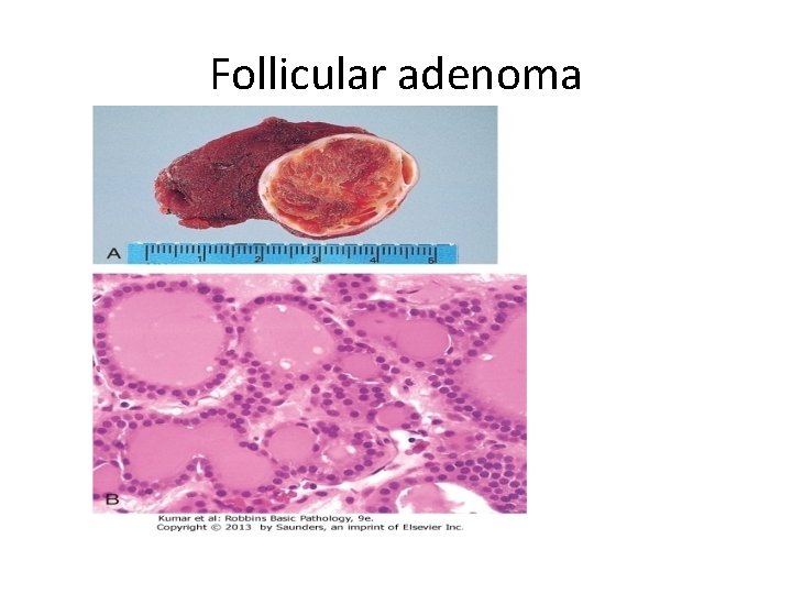
Follicular adenoma
Carcinomas
- The major subtypes of thyroid carcinoma are a. Papillary carcinoma ( for more than 85% of cases) b. Follicular carcinoma (5% to 15% of cases) c. Anaplastic carcinoma (less than 5% of cases)
A. Papillary Carcinoma : - Is most the most common form - Accounts for the majority of thyroid carcinomas associated with previous exposure to ionizing radiation. - May occur at any age and is the most common malignant thyroid tumor in children,
Clinical Features a. Manifest as painless mass in the neck, either within the thyroid or as metastasis in a cervical lymph node b. Are indolent lesions, with 10 -year survival rates of 95%.
c. The presence of isolated cervical nodal metastases does not have a influence on good prognosis of these lesions. d. In a minority of patients, hematogenous metastases are present at the time of diagnosis, most commonly to lung
The bad prognostic factors are: a. Tumors arising in patients older than 40 years b. The presence of extrathyroidal extension c. Presence of distant metastases (stage) -
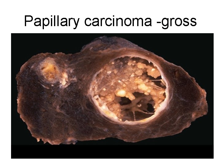
Papillary carcinoma -gross
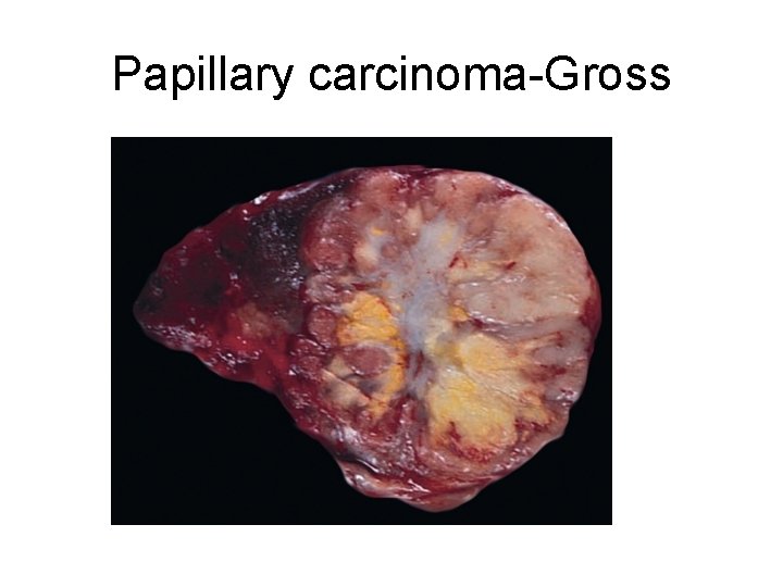
Papillary carcinoma-Gross
B. Follicular Carcinoma : -More common in women and in areas - Common in people with dietary iodine deficiency Clinically manifest as cold thyroid nodules
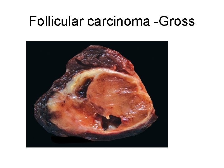
Follicular carcinoma -Gross
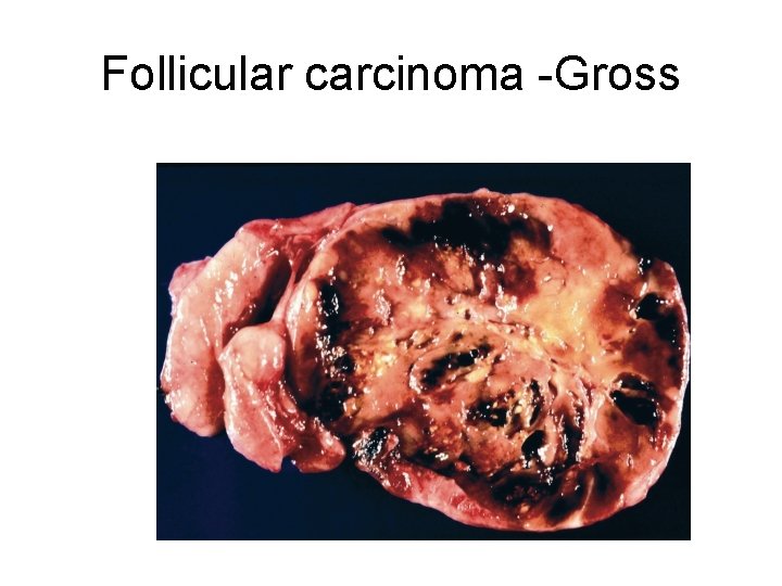
Follicular carcinoma -Gross
- Tends to metastasize through the bloodstream (hematogenous dissemination) to lungs, bone, and liver. - Rgional nodal metastases are uncommon
3. Anaplastic Carcinoma - Affects elderly people - They are aggressive, with a mortality rate of 100%. - Death occurs in less than 1 year as a result of aggressive local growth which compromise of vital structures in the neck.
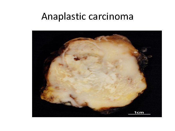
Anaplastic carcinoma
4. Medullary Carcinoma - Are neuroendocrine neoplasms. - Secrete calcitonin, the measurement of which plays an important role in the diagnosis and postoperative follow-up evaluation of patients.
- Are sporadic in about 70% of cases and the remaining 30% are familial cases a. Occurring in the setting of MEN syndrome 2 A or 2 B, b. or Familial medullary thyroid carcinoma without an associated MEN syndrome
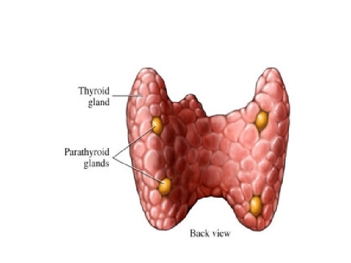
II. Parathyroid gland
I. HYPERPARATHYROIDISM : 3 categories a. Primary type b. Secondary c. Less commonly: tertiary hyperparathyroidism.
a. Primary Hyperparathyroidism Causes 1. Parathyroid adenoma (85% to 95) 2. Primary parathyroid hyperplasia 5% to 10%. 3. Parathyroid carcinoma-(1%)
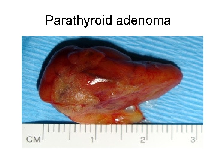
Parathyroid adenoma
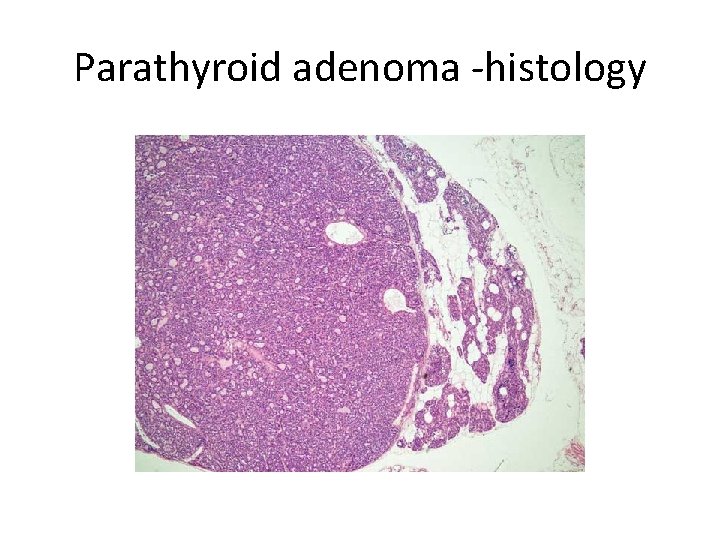
Parathyroid adenoma -histology
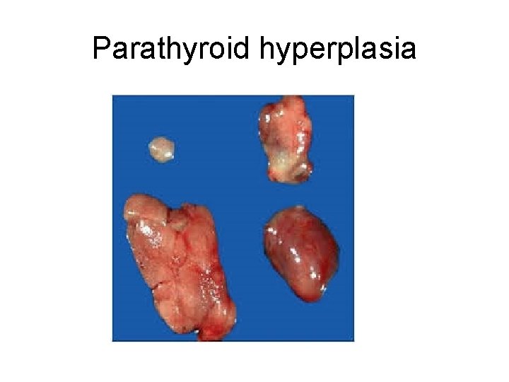
Parathyroid hyperplasia
Morphologic changes in other organs I. Skeletal changes include: a. Osteitis fibrosa cystica) characterized by 1. Increased osteoclastic activity, resulting in erosion of bone, particularly in the metaphyses of long tubular bones. - Bone resorption is accompanied by increased osteoblastic activity and the formation of new bone
. - The cortex is grossly thinned and the marrow contains increased amounts of fibrous tissue accompanied by foci of hemorrhage and cysts b. Brown tumors of hyperparathyroidism - Aggregates of osteoclasts, , and hemorrhage occasionally form masses that may be mistaken for neoplasms like giant cell tumors
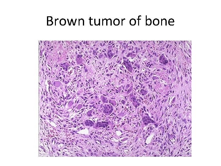
Brown tumor of bone
II. Kidney changes a. PTH-induced hypercalcemia favors the formation of urinary tract stones (nephrolithiasis) s b. Calcification of the renal interstitium (nephrocalcinosis) III. Metastatic calcification may be seen in the stomach, lungs, myocardium, and blood vessels
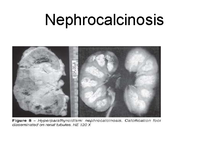
Nephrocalcinosis
Clinical Features - The most common manifestation is an increase in serum calcium and is the most common cause of clinically silent hypercalcemia. NOTE - The most common cause of clinically apparent hypercalcemia in adults is paraneoplastic syndromes
• Clinical Manifestations : 1. Pain is secondary to a. Fractures of bones b. Renal stones 2. Gastrointestinal disturbances, including constipation, peptic ulcers, pancreatitis, and gallstones.
3. CNS alterations, - depression, lethargy, and seizures 4. Neuromuscular abnormalities, weakness and hypotonia
b. Secondary Hyperparathyroidism - Is caused by any condition causing a chronic decreases in the serum calcium level, because low serum calcium leads to compensatory overactivity of the parathyroids. - Renal failure is the most common cause and the mechanisms
1. Chronic renal insufficiency causes decreased phosphate excretion, which in turn results in hyperphosphatemia. and the elevated serum phosphate levels depress serum calcium levels and so stimulate parathyroid gland activity
2. Loss of renal substances reduces the availability of α 1 -hydroxylase enzyme necessary for the synthesis of the active form of vitamin D, which in turn reduces intestinal absorption of calcium
Clinical Features - Are dominated by those related to chronic renal failure
II. HYPOPARATHYROIDISM: - The major causes are: . a. Surgically induced hypoparathyroidism: inadvertent removal of parathyroids during thyroidectomy.
b. Congenital absence: associated with thymic aplasia (Di George syndrome) and cardiac defects c. Autoimmune hypoparathyroidism
Clinical manifestations - Are secondary to hypocalcemia and include: a. Increased neuromuscular irritability (tingling, muscle spasms, , and sustained carpopedal spasm or tetany), b. Cardiac arrhythmias,
- Slides: 45