Isolation and Identification of Viruses By Dr Vikrant

Isolation and Identification of Viruses By Dr. Vikrant P. Wankhade Vidyabharti College of Pharmacy-Amravati

Isolation and Identification of Viruses Definition: Obligate intracellular parasite composed of: Nucleic acid - either DNA or RNA Protein coat Viruses must be grown in living cells. They can't be grown in culture media or on agar plates alone, they must have living cells to support their replication. The easiest viruses to grow are bacteriophages (because the easiest cells to grow in the lab are bacteria). Once viruses have replicated and been harvested the concentration of viral particles (virions) in the viral stock solution must be determined. One of the easiest ways to determine the concentration of a stock solution of bacteriophages is to use the plaque method. Classification: Animal virus Plant virus Bacterial virus (bacteriophage)

Acknowledgement This power point presentation has been adapted from all know and unknown sources and this power point presentation solely used for Teaching purpose only.

Determined by electron microscopy. Ranges from 20 to 14, 000 nm in length. There is also a group of giant viruses, including the giant mimivirus, which is something like 800 nm in diameter and has a genome with 1. 2 Mbp base pairs carrying somewhere in the neighborhood of 1000 genes, 911 of which code for proteins.

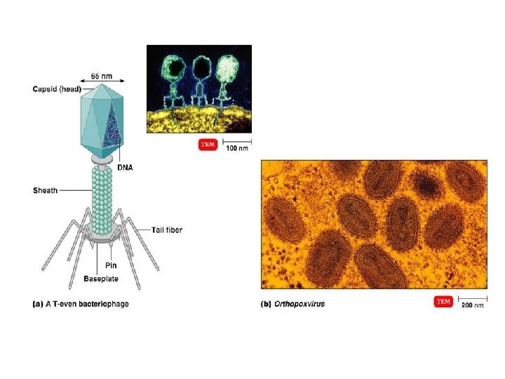
Virus Structure Virions are complete, fully developed viral particles composed of nucleic acid surrounded by a protein coat. Some viruses have an envelope composed of a phospholipid bilayer with viral glycoproteins. 1. Nucleic acid Viral genomes are either DNA or RNA (not both). Nucleic acid may be single- or double-stranded Nucleic acid may be circular or linear or separate molecules. Nucleic acid: protein ranges from about 1% - 50%. 2. Capsid - protein coat Capsomeres are subunits of the capsid Protomeres are capsomere subunits.

3. Envelope – the outer covering of some viruses, the envelope is derived from the host cell plasma membrane when the virus buds out. Some enveloped viruses have spikes, which are viral glycoproteins that project from the envelope. Influenzavirus has two kinds of spikes, H (hemagglutinin) and N (neuraminidase). The H spike allows the virus to attach to host cells (and red blood cells), the N spike is an enzyme that allows the mature viral particles to escape from the host cell Non-enveloped or naked viruses are protected by their capsid alone.

General Morphology Based on capsid architecture, although enveloped viruses end up being approximately spherical. 1. Helical, non-enveloped 2. Helical, enveloped 3. Polyhedral, non-enveloped 4. Polyhedral, enveloped Polyhedral means many sides (most are icosahedral - 20 triangular faces and 12 corners) 5. Complex viruses are, well, complex. See bacteriophages.

The plaque method: Virus, bacteria, and agar mixed, plated and incubated. After replication the virus lyses the bacteria, forming plaques, or clear zones. Each plaque is assumed to come from a single viral particle. The titer (concentration of the stock solution) of the virus is given in plaque forming units.

Growing Animal Viruses In The Laboratory 1. Live animal cultures have to be used for some animal viruses. Simian AIDS and feline AIDS provide models for studying human AIDS. 2. Embryonated eggs can serve as substitutes for some viruses. Can inoculate membrane that best supports specific virus (allantoic, amniotic, chorioallantoic, or yolk sac).

3. Cell culture is a lot cheaper and easier to work with (contamination can be a problem however). Primary cell lines have a short lifespan in culture – a few generations before reaching senescence. Diploid cell lines are derived from embryos and can grow for up to 100 population doublings before senescence. Continuous cell lines are derived from transformed cells and grow indefinitely in culture. Hela cells – 1 st continuous cell line, derived from Helen Lane (fictional name - actually named Henrietta Lacks), a cervical cancer patient who died in 1951. This is the oldest continuous cell line and was first used to culture and identify polio virus.

Viral Identification Serological methods Western blotting Cytopathic effects Diagnostic inclusion bodies are associated with rabies virus, measles virus, vaccinia virus, smallpox virus, herpesvirus, and adenoviruses. Molecular methods include PCR and RFLPs. PCR was used to identify the West Nile virus and the SARS-associated coronavirus

Viruses multiplication Viruses do not contain enzymes for energy production or protein synthesis. For a virus to multiply, it must invade a host cell and direct the host’s metabolic machinery to produce viral enzymes, viral proteins, and copies of its nucleic acid, using the host cell's ATP to power the reactions. Viral particles disappear upon penetration, none are seen during biosynthesis and assembly, and eventually all cells die so no new virions can be produced. The eclipse period is the period when all viral particles are present but before they are assembled. Burst time is the time from phage adsorption to release. Burst size is the number of newly synthesized phages produced from one infected cell.

Multiplication of Bacteriophage The virus may cause lysis or lysogeny. Events of the lytic cycle: Attachment or adsorption Requires a receptor Penetration T-evens release lysozyme to break down a portion of the cell wall. The tail sheath contracts and the tail core is driven through the hole in the wall to the plasma membrane. The viral genome is then injected into the bacterium. Biosynthesis Viral DNA and proteins are synthesized. Host protein synthesis is stopped by degradation of host DNA, interference with transcription, or repression of translation. Maturation During maturation or assembly phage DNA and capsids are assembled into complete viruses. Release occurs when phage lysozyme breaks down the cell wall and newly synthesized phage particles are released.
- Slides: 14