ISCHIOANAL FOSSA DR ABEERA SARFRAZ DR MUHAMMAD ZUBAIR

ISCHIOANAL FOSSA DR ABEERA SARFRAZ/ DR MUHAMMAD ZUBAIR
 on each side of the anal canal are")
INTRODUCTION The ischio-anal fossae (ischiorectal fossae) on each side of the anal canal are large fascia-lined, wedge-shaped spaces between the skin of the anal region and the pelvic diaphragm. The apex of each fossa lies superiorly where the levator ani muscle arises from the obturator fascia. The ischio-anal fossae, wide inferiorly and narrow superiorly, are filled with fat and loose connective tissue. The two ischio-anal fossae communicate by means of the deep postanal space over the anococcygeal ligament (body), a fibrous mass located between the anal canal and the tip of the coccyx. Each ischio-anal fossa is filled with a fat body of the ischio-anal fossa. These fat bodies support the anal canal, but they are readily displaced to permit descent and expansion of the anal canal during the passage of feces. The fat bodies are traversed by tough, fibrous bands, as well as by several neurovascular structures, including the inferior anal/rectal vessels and nerves and two other cutaneous nerves, the perforating branch of S 2 and S 3 and the perineal branch of S 4 nerve
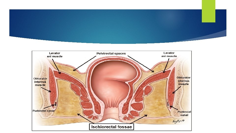

BOUNDRIES Each ischio-anal fossa is bounded as follows: Laterally by the ischium and overlapping inferior part of the obturator internus, covered with obturator fascia. Medially by the external anal sphincter, with a sloping superior medial wall or roof formed by the levator ani as it descends to blend with the sphincter; both structures surround the anal canal. Posteriorly by the sacrotuberous ligament and gluteus maximus. Anteriorly by the bodies of the pubic bones, inferior to the origin of the puborectalis. These parts of the fossae, extending into the UG triangle superior to the perineal membrane (and musculature on its superior surface), are known as the anterior recesses of the ischio-anal fossae.
 is an essentially horizontal passageway within the")
PUDENDAL CANAL The pudendal canal (Alcock canal) is an essentially horizontal passageway within the obturator fascia that covers the medial aspect of the obturator internus muscle and lines the lateral wall of the ischio-anal fossa. The internal pudendal artery and vein, the pudendal nerve, and the nerve to the obturator internus enter the pudendal canal at the lesser sciatic notch, inferior to the ischial spine. The internal pudendal vessels and the pudendal nerve supply and drain blood from and innervate most of the perineum. As the artery and nerve enter the canal, they give rise to the inferior rectal artery and nerve, which pass medially to supply the external anal sphincter and the peri-anal skin. Toward the distal (anterior) end of the pudendal canal, the artery and nerve both bifurcate, giving rise to the perineal nerve and artery, which are distributed mostly to the superficial pouch (inferior to the perineal membrane), and to the dorsal artery and nerve of the penis or clitoris, which run in the deep pouch (superior to the membrane). When the latter structures reach the dorsum of the penis or clitoris, the nerves run distally on the lateral side of the continuation of the internal pudendal artery as they both proceed to the glans penis or glans clitoris.

PUDENDAL NERVE The pudendal nerve is a branch of the sacral plexus (S 2 to 4 anterior rami). It leaves the main pelvic cavity through the greater sciatic foramen, enters the gluteal region of the lower limb, and curls around the attachment of the sacrospinous ligament at the ischial spine. After a brief course in the gluteal region, it passes through the lesser sciatic foramen and enters the posterior aspect of the perineum. The nerve then runs forward in the pudendal canal. Its branches supply the external anal sphincter and the muscles and skin of the perineum. Branches q Inferior rectal nerve: This runs medially across the ischioanal fossa and supplies the external anal sphincter, the mucous membrane of the lower half of the anal canal, and the perianal skin. q Dorsal nerve of the penis (or clitoris): This is distributed to the penis (or clitoris). q Perineal nerve: This supplies the muscles in the urogenital triangle and the skin on the posterior surface of the scrotum (or labia majora).

Internal Pudendal Artery The internal pudendal artery is a branch of the internal iliac artery. It travels with the pudendal nerve and passes from the pelvis through the greater sciatic foramen and enters the perineum through the lesser sciatic foramen. Branches Inferior rectal artery: This supplies the lower half of the anal canal. Branches to the penis in the male and to the labia and clitoris in the female. Internal Pudendal Vein The internal pudendal vein receives tributaries that correspond to the branches of the internal pudendal artery.
 is a potential space")
SUPERFICIAL PERINEAL POUCH The superficial perineal pouch (space or compartment) is a potential space between the perineal fascia and the perineal membrane, bounded laterally by the ischiopubic rami. In males, the superficial perineal pouch contains the root (bulb and crura) of the penis and associated muscles (ischiocavernosus and bulbospongiosus). proximal (bulbous) part of the spongy urethra. superficial transverse perineal muscles. deep perineal branches of the internal pudendal vessels and pudendal nerves.
.")
In females, the superficial perineal pouch contains the clitoris and associated muscle (ischiocavernosus). bulbs of the vestibule and surrounding muscle (bulbospongiosus). greater vestibular glands. superficial transverse perineal muscles. related vessels and nerves (deep perineal branches of the internal pudendal vessels and pudendal nerves).
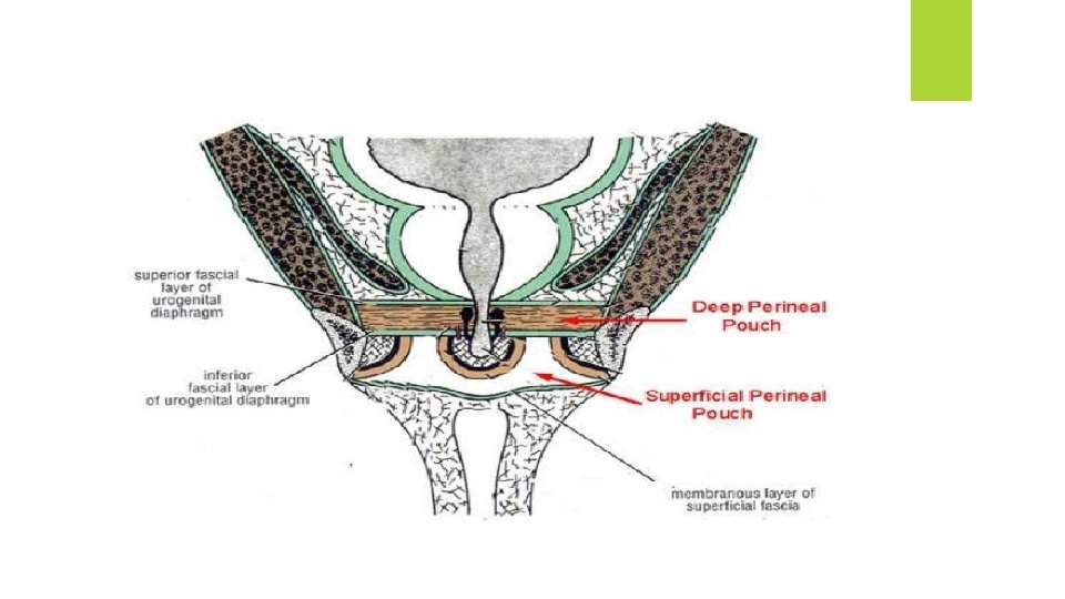
 is bounded inferiorly by the perineal")
DEEP PERINEAL POUCH The deep perineal pouch (space) is bounded inferiorly by the perineal membrane, superiorly by the inferior fascia of the pelvic diaphragm, and laterally by the inferior portion of the obturator fascia (covering the obturator internus muscle). It includes the fat-filled anterior recesses of the ischio-anal fossae. The superior boundary in the region of the urogenital hiatus is indistinct. In both sexes, the deep perineal pouch contains part of the urethra, centrally. the inferior part of the external urethral sphincter muscle, above the center of the perineal membrane, surrounding the urethra. anterior extensions of the ischio-anal fat pads.
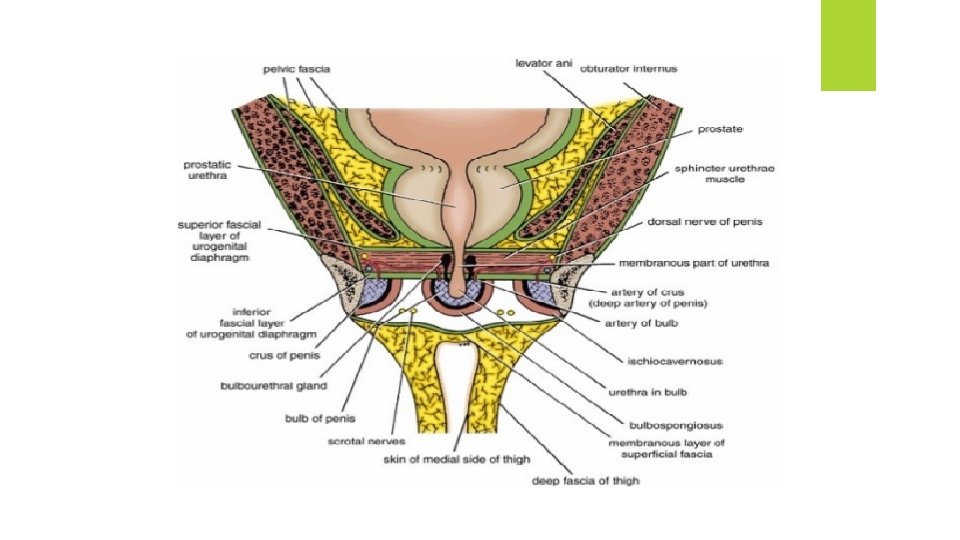

In males, the deep perineal pouch contains the intermediate part of the urethra, the narrowest part of the male urethra. deep transverse perineal muscles, immediately superior to the perineal membrane (on its superior surface), running transversely along its posterior aspect. bulbo-urethral glands, embedded within the deep perineal musculature. dorsal neurovascular structures of the penis. In females, the deep perineal pouch contains the proximal part of the urethra. a mass of smooth muscle in the place of deep transverse perineal muscles on the posterior edge of the perineal membrane, associated with the perineal body. dorsal neurovasculature of the clitoris.

URETHRAL RUPTURE Rupture of the urethra may complicate a severe blow on the perineum. The common site of rupture is within the bulb of the penis, just below the perineal membrane. The urine extravasates into the superficial perineal space and then passes forward over the scrotum beneath the membranous layer of the superficial fascia. If the intermediate (membranous) part of the urethra is ruptured, urine escapes into the deep perineal space and can extravasate upward around the prostate and bladder or downward into the superficial perineal space.
- Slides: 14