Involuntary Movement By Dr M Abbas Involuntary Movement

Involuntary Movement By: Dr. M. Abbas

Involuntary Movement: • An involuntary movement occurs when you move your body in an uncontrollable and unintended way. • You can experience these movements in almost any part of the body, including the neck, face, and limbs.

Reflex Movement • Reflexes are automatic, subconscious response to changes within or outside the body. • Reflexes maintain homeostasis (autonomic reflexes) like heart rate, breathing rate, blood pressure, and digestion. • Reflexes also carry out the automatic action of swallowing, sneezing, coughing, and vomiting.

• Reflexes maintain balance & posture. • ex. Spinal reflexes – control trunk and limb muscles. • Brain reflexes – involve reflex center in brain stem. • ex. Reflexes for eye movement

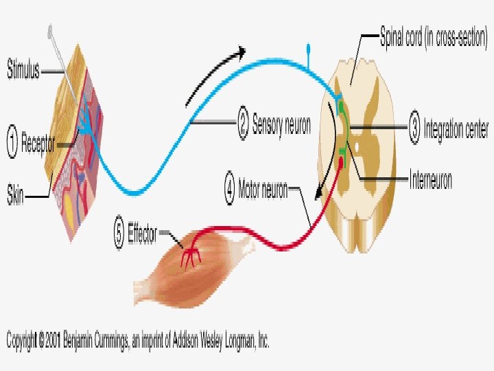
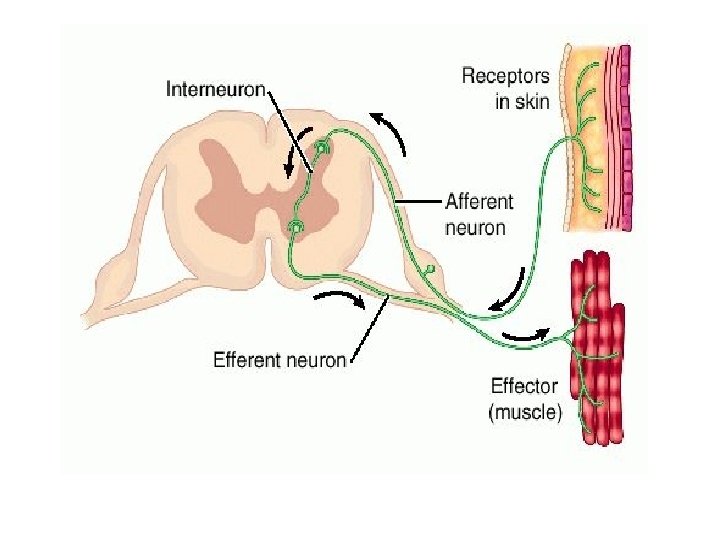
Reflex Arc: • The reflex arc governs the operation of reflexes. Nerve impulses follow nerve pathways as they travel through the nervous system. • The simplest of these pathways, including a few neurons, constitutes a reflex arc. Reflexes whose arc pass through the spinal cord are called spinal reflexes.
 Description: the receptor")
Parts of Reflex Arc 1. Receptor – detects the stimulus a) Description: the receptor end of a particular dendrite or a specialized receptor cell in a sensory organ. b) Function: sensitive to a specific type of internal or external change. .

2. sensory neuron – conveys the sensory info. to brain or spinal cord. a. Description: Dendrite, cell body, and axon of a sensory neuron. b. Function: transmit nerve impulses from the receptor into the brain or spinal cord

3. Interneuron: relay neurons. a. Description: dendrite, cell body, and axon of a neuron within the brain or spinal cord. b. function: serves as processing center, conducts nerve impulses from the sensory neuron to a motor neuron. 4. Motor neuron: conduct motor output to the periphery. a. Description: Dendrite, cell body, and axon of a motor neuron. b. function: transmits nerve impulse from the brain or spinal cord out to an effecter.

5. Effector: a. Description: a muscle or gland. b. function: Response to stimulation by the motor neuron and produces the reflex or behavioral action

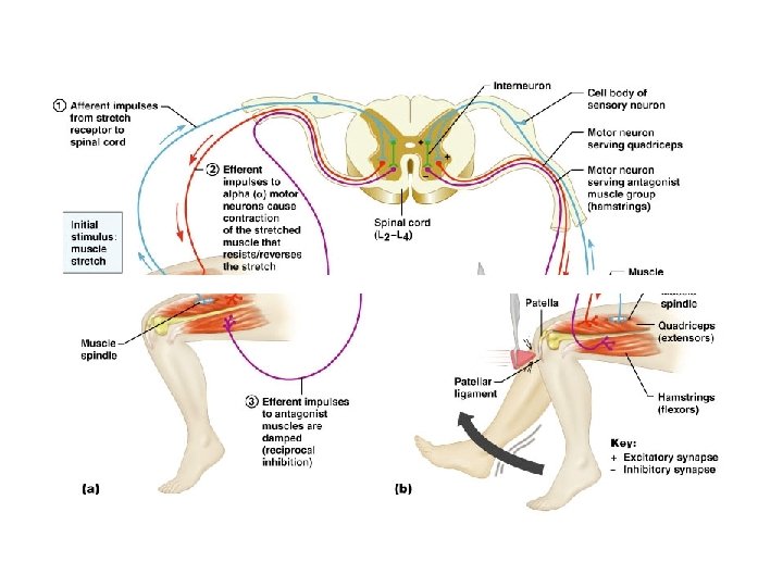
The Stretch Reflex • Simplest spinal reflex • Depends only on the single connection between primary afferent fibres and motor neurons of the same muscle • i. e. Knee-jerk test

Stretch reflex • For skeletal muscles to perform normally: • The Golgi tendon organs (proprioceptors) must constantly inform the brain as to the state of of the muscle. • Stretch reflexes initiated by muscle spindles must maintain healthy muscle tone.

Stretch reflex • This is the most important local static reflex which controls the tone in those extensor muscles which keep the body upright (antigravity muscles)
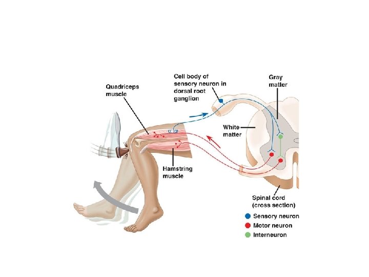

The receptor muscle senses the action of the hammer against the patella ligament through the muscle spindle's sensory neuron The message is transmitted along the afferent (sensory) nerve axon to the spinal cord The afferent neuron synapses with the efferent pathway (motor neuron) of the same muscle An impulse is transmitted along the efferent pathway (motor neuron) to the muscle The motor units contract (knee-jerk to accomodate additional stretch)

Postural Reflexes: • Postural reflexes help to maintain the body in upright and balanced position • They also provide adjustments necessary to maintain a stable posture during voluntary activity

• Afferent Pathway- comes from the eyes, the vestibular apparatus and the proprioceptors • Integrating Centres- are formed by neuronal network in the brain stem and spinal cord • Efferent Pathway- α-motor neurons supplying the various skeletal muscles i. e. the effector organ

Righting reflexes: • Righting reflexes help to correct the position of the body when it goes off balance and falls down • These reflexes consists of a chain of reactions following one another in an orderly sequence

• For example, if an animal is laid on it’s side or back, head rights itself followed by body and animal finally resumes upright posture. The sequence of righting reflexes will be as follows : 1. Head righting reflex or Labyrinthine righting reflex • It is initiated when animal’s head is in lateral position • Impulses arising from the saccules reflexly stimulate the appropriate muscles to bring head back to upright position

Body righting reflex • When the animal lies on the ground, the side in contact with the ground is constantly stimulated while the other side is not • This differential stimulation of the deep structures in the body wall reflexly rights the head 3. Neck righting reflex • The head is righted by above two reflexes but the body still remains in lateral position

• This leads to twisting of neck and this brings thorax and lumbar region successively into upright posture • If the righting of head is prevented , impulses from the body surface may cause righting of the body directly (Body on body righting reflex) 4. Limbs righting reflex • Impulses arising from the limb muscles leads to attainment of appropriate posture of limbs

Optical righting reflexes • Optical impulses also cause righting of the head in animals with intact visual cortex Centers Of Righting Reflexes • Chief center for all righting reflexes, except the optical righting reflex is red nucleus lying in the mid brain • The center for optical righting reflex is in the visual cortex
- Slides: 24