Investigation of Death Autopsy When is an Autopsy


 Toxicology-mass spectroscopy Healthy vs Emphysema Lung tissue (histology Neuropathology: normal")

- Slides: 29
Investigation of Death Autopsy
When is an Autopsy Performed? Whenever the cause of death is unclear or suspicious Generally speaking, the following circumstances require investigation by law: Violent Within crime, suicide, or accident 24 hours of entering a hospital or as a result of surgery A natural death when a doctor is not present or the patient is not under the care of a medical facility Occurs Results in police custody or in a correctional facility from a communicable disease that may pose a threat to public health
In Colorado, counties determine when autopsies are performed. Example from Weld county Where no physician is in attendance, or where though in attendance, the physician is unable (or unwilling) to certify the cause of death. All cases in which the attending physician has not been in actual attendance within 30 days prior to death. All cases in which trauma may be associated with the death (i. e. , falls, traffic accidents industrial accidents. ) Any patient who has sustained a fracture; no matter how long ago Deaths by poison or suspected poisoning, chemicals or bacteria, industrial hazardous materials, or radiation. Known or suspected suicide. Deaths where the deceased has a contagious disease. All operating room deaths and deaths which occurring during a medical procedure. All unexplained deaths due to suspicious circumstances. Deaths which occur within 24 hours of admission to hospital
Definitions Autopsy: “to see one’s self” A post-mortem examination of the body, including dissection of the corpse. Performed by a forensic pathologist (medical doctor)
3 Steps of a Death Investigation 1. Preliminary investigation is conducted at the death scene 2. The body is transported to the morgue where the medical examiner examines the body and performs an autopsy 3. The medical examiner/coroner orders lab tests on biological evidence collected during the autopsy
At the Death Scene The death investigator-Employed by the coroner’s/medical examiner’s office Responsible Initial for; assessment Photographs and sketches of the body on scene Position of the body, face (for identification), underside of the body (for lividity, blood, and trace evidence) Document Collect signs of trauma information regarding livor and rigor mortis, degree of decomposition to help establish time of death
At the Death Scene Investigators look for scene markers; any non-biological evidence that provides clues about time of death, (unopened mail, newspaper near the body, etc) Any evidence collected is properly stored and a chain of custody is established Once victim is identified, medical records are gathered and witnesses and the victim’s family are interviewed Paper bags are placed over the victims hands to protect trace evidence from being lost or preventing cross contamination
Medical Examination The medical examination is to determine the manner, cause and mechanism of death 2 stages: 1. External Examination 2. Internal Examination/Autopsy
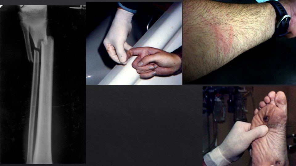
External Examination What is examined? Clothing, boots/shoes, belongings in pockets should be examined and removed carefully for storage/packaging Surface of body Signs of trauma/x-ray Fingernail Hair scrapings samples Fingerprints Biological evidence collection Hairs, blood, plant debris, etc. Non-biological evidence collection Glass, soil, artificial fibers, etc
Internal Examination/Autopsy Estimation of time of death: Algor, Livor, and Rigor Mortis Stomach contents Insects (from death scene investigation) Stages of Decomposition Fresh Bloat Active Decay Advanced Decay Dry/remains
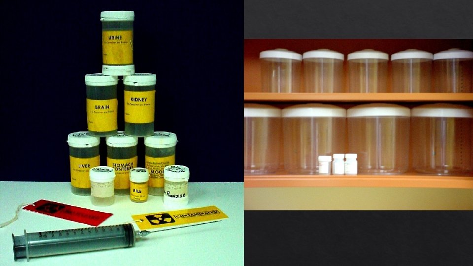
Fluids Collected Blood Collected from femoral artery Used to Collect Vitreous Urine Collected Vitreous from bladder Humor Collected from inside of eye Cerebro-Spinal Fluid Stomach/Intestinal
Stages of Decomposition 1. Fresh: Livor, algor, rigor mortis, autolysis, stoppage, blowflies arrive 2. Bloat: accumulation of gases from microbes, hemoglobin breaks down to form other colored pigments (marbling), maggots hatch, distinctive odors 3. Active Decay: loss of mass (maggots feeding/purging of fluids, liquefaction and disintegration of tissues, strong odors 4. Advanced Decay: reduced insect activity, death of surrounding vegetation 5. Dry/remains: resurgence of plant growth, remains=dry skin, cartilage, and bones
. Fresh Bloat Active Decay https: //youtu. be/B 4 y. Di_w. WMVk Advanced Decay Dry/Remains
Types of Autopsy Medico-Legal Autopsy/Forensic Autopsy: determine cause and manner of death and identify the decedent Clinical/Pathological Autopsy: diagnose a particular disease or for research. Can clarify or confirm medical diagnoses Anatomical/Academic Autopsy: performed by students of anatomy for study Virtual Autopsy: performed using MRI’s and CT scans
Autopsy Trunk dissection; Y-shaped incision From the shoulders to the pelvic bone This incision is deep
Opening the Chest Skin & muscle, are pulled from the chest wall Chest Plate is extracted
Removal and Dissection of the Organs Many methods of removal serve different purposes Rokitansky method is an in-situ and en bloc examination of organs intact (still connected to one another) Virchow method is an organ by organ removal. Not great forensic autopsy-connections are lost between organs Letulle method is the En Masse removal of all the viscera (thoracic, cervical, abdominal, pelvic organs) then dissected in organ blocks Preserves Gohn vascular supply and connections between organs. method is “En Bloc” removal of organs that are physiologically connected to another, (thoracic, coeliac, urogenital)
After Organ Removal Upon removal each organ is: Weighed & measured Examined Sliced in cross sections Sampled for microscopic & chemical analysis
“Running the Gut” The contents of the stomach, intestines, and bowels must be bsapp. com
Removing the Brain The Scalp is cut ear to ear across the crown of the head bsapp. com
Exposing the Skull Next the scalp is pulled forward and back to expose the skull bsapp. com
Exposing the Brain Two methods of cutting the skull cap bsapp. com
Removal of the Brain n Spinal Cord is cut n The soft brain is removed n Brain is so soft it must be placed in formaldehyde for about a week before an in depth examination bsapp. com
Close Up Skull cap is replaced Skin pulled back in place Body Organs may or may not be replaced Incisions are sown up with the use of a baseball stitch bsapp. com
Laboratory Analysis Toxicology: the science related to the detection of drugs, alcohol, and poisons using bodily fluids such as blood, vitreous humor, and urine. Histology: the study of tissues. Slides are made of organ tissue to analyze using a microscope. Disease/abnormalities can be detected. Neuropathology: the study of disease and trauma associated with the nervous system Serology: the study of blood, semen, and other body fluids with reference to legal matters
Malaria in Blood (Serology) Toxicology-mass spectroscopy Healthy vs Emphysema Lung tissue (histology Neuropathology: normal brain vs. Alzheimer’s Disease
Parts: The Autopsy Report Heading Case number, victim info, date/time of death, etc External Full Examination description of body & clothing, evidence of disease/trauma Evidence of Injury Description Internal of any injuries and record of all affected organs Examination Weights and descriptions of all major organ systems and abnormalities, findings from toxicology/histology Medical Cause Examiner’s findings and opinion and manner or death, results and outcomes of tests and examinations