INTV 3 PERCUTANEOUS TREATMENT OF FACET JOINT SYNOVIAL


(4): � It’s a manifestation of progressive posterior facet arthrosis :")


(2)(3)(4): � CT-arthrography can identified synovial cysts communicating with the adjacent")


, in his study of 30 Patients, found that")

 Another alternative of treatment is")


- Slides: 28
INTV 3 PERCUTANEOUS TREATMENT OF FACET JOINT SYNOVIAL CYST DEVELOPPED IN INTRA DUCTAL S. KOUKI , W. AMORRI, M. LANDOULSI , S. BOUGUERRA , Y. AROUS , H. BOUJEMAA , N. BEN ABDALLAH Military Hospital of Tunis
OBJECTIVE: To study the results of facet joint intraarticular steroid injections in a patient with symptomatic lumbar facet joint synovial cysts developped in intra ductal.
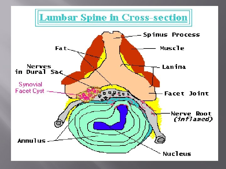
INTRODUCTION üFacet joint synovial cyst is an Expansion of the joint capsule and synovium into the spinal canal üBy definition it communicates with the adjacent joint üThe üIt‘s average when it occured is 60 years a rare cause of radicular pain üClinical signs are unilateral nerve root or radicular claudication bilateral lower üIs easily diagnosed by new medical imaging modalities üImage-guided percutaneous steroid injections presents often an effective alternative to surgery
CASE REPORT üThe patient is a 59 -years woman üWithout individual medical history outside of an overweight ücomplaining of low back sciatica type left L 5, associated with a left cruralgia, medical treatment refractory to
EXPLORATION BY IMAGING üRadiographs of the lumbosacral spine : a degenerative spinal disco, more advanced at L 4 -L 5 segment, associated with a degenerative Lowgrade isthmic spondylolisthesis. üThe CT scan : an intra ductal synovial cyst, next to the left posterior facet joint L 4 -L 5, measuring 2 cm long axis, which causes a conflict with the L 5 root at its emergence, and L 4 ipsilateral root.
THERAPEUTIC MANAGEMENT üA well conducted medical treatment with rest did not lead to a favorable outcome. ü A surgical treatment proposed refused by the patient üShe was entrusted to us for a percutaneous treatment
PERCUTANEOUS TREATMENT UNDER SCANNER 1/ INSTALLATION OF THE PATIENT, AND TRACKING: üThe patient is prone positioned. üThe procedure is performed in the interventional scanner room üWe conducted a helix centered on the lumbar spine to identify the left facet joint L 4 -L 5. üThe CT features of the facet joint synovial cyst is a Rounded picture of homogeneous fluid density intra ductal with hyper dense fibrous shell.
2/ PROGRESS OF INTERVENTIONAL GESTURE: üAfter local anesthesia and surgical skin disinfection üJoint aspiration and injection of 1 ml of iodinated contrast in facet joint, opacified both the joint and the cyst intra canal, objectifying the communication between them. üfluid üThen content was aspirated we have inject a bulb of a prolonged action corticosteroid (Altim®) combined with 1 cc of Xylocaine® under pressure until rupture of the cyst, as evidenced by a loss of strength and opacification of the epidural space on the acquisition of control.
FIG 1: AXIAL CT SCAN OF L 4 IN BONE WINDOW SHOWING THE AVERAGE LOAD OF INTRA DUCTAL CYST WITH MASS EFFECT ON THE DURAL SHEATH
FIG 2: AXIAL CT SCAN OF L 4 IN BONE WINDOW SHOWING THE COMPLETE FILLING OF THE CYST WITH EARLY EXTRA VASATION OF CONTRAST
FIG 3 : AXIAL CT SCAN OF L 4 IN BONE WINDOW SHOWING THE COMPLETE FILLING OF THE CYST WITH CLEAR EXTRAVASATION OF CONTRAST MATERIAL BY CRACKING CYSTIC
FIG 4 : SAGITTAL RECONSTRUCTIONS SHOWING OPACIFICATION AND SIGNS OF INTRA DUCTAL CYST
3/ RESULT AND EVOLUTION: üImmediately, the patient describes an exaggeration of pain followed by a relief ü This is likely due to the effect of Xylocaine® and the reduction of pressure in the cyst after its cracks. üThis cracking is a cure of this cyst, it is evidenced by the extravasation of contrast outside the cyst. üThe decline in two years was marked by a favorable clinical course, especially since the patient has lost weight and always wore a lumbar corset.
DISCUSSION 1/ PATHOPHYSIOLOGY (1)(4): � It’s a manifestation of progressive posterior facet arthrosis : during outbreaks of effusion, the normal joint recess become diverticula, synovial recesses would enlarge with progressive fibrous thickening and inflammation of their walls.
� By definition, intraspinal synovial cysts communicate with the adjacent facet joint. � They are characterized by the presence of synovial lining and clear or xanthochromic content � Opposed to ganglion cysts that do not communicate with the facet joint, have a fibrous wall, and contain gelatinous myxoid material
Both entities often are described as juxtaarticular or synovial cysts. Synovial cysts would be a manifestation of facet degeneration: The L 4 -5 level is most commonly involved because it corresponds to the level of maximal mechanical stress and motion.
2/ IMAGING STUDY (1)(2)(3)(4): � CT-arthrography can identified synovial cysts communicating with the adjacent facet joint with marked degeneration and a spondylolisthesis � Diagnosis at non contrast CT is based on the detection of a cystic structure next to a degenerated facet joint, such as in our case. The cyst may sometimes extend into the lateral recess. � The presence of bony erosions or remodeling suggest the possibility of Tarlov cyst, arachnoid cyst, or cystic nerve sheath tumor, but these changes have also been described in patients with synovial cysts. � Facet joint injection demonstrating communication of the facet joint with the cyst is pathognomonic for the presence of a synovial cyst.
� In the MRI signal is variable: * Hypo. T 1, hyper. T 2: type fluid * Hyper. T 1, hypo. T 2: type haem * Hypo. T 1, hypo. T 2: gas, calcification, hemosiderin * Hyper. T 1, hyper. T 2: blood, fat � The differential diagnosis includes ganglion cysts, posterior longitudinal ligament cysts, and ligamentum flavum cysts; however, these cysts do not communicate with the facet joint and are not lined with epithelium. � The cysts often are of fluid density, they rarely contain blood products, calcium, or gas (gas in the facet joint). � The presence of increased wall density improves diagnosis and narrows the differential diagnosis.
3/ Type of therapeutic management : � At the time of imaging, our patient had already undergone medical management, combining rest and NSAIDs, with support device. � The detection of a symptomatic synovial cyst may require percutaneous steroid injection or surgery. � Surgery, performed initially, allows resection of the cyst and treatment of other potential abnormalities: disk herniation, spinal stenosis, narrowing of the lateral recess, spondylolisthesis.
Long-term follow-up for surgical excision of symptomatic juxtafacet cysts without spinal fusion revealed excellent to good results in 92% of the patients, with a satisfaction rate of 80%, in the study of El Shazly AA. (3). Common surgical risks include spinal instability, dural tear, neurologic injury, epidural hemorrhage and hematoma, seroma, and cyst recurrence While surgery is the gold standard for the treatment for symptomatic facet joint cysts, conservative options include bed rest, physical therapy, acupuncture, oral analgesics and anti-inflammatories, and percutaneous injection and aspiration
� Arthrography-infiltration is a good alternative in case of cons-indication to surgery or refusal � Percutaneous interventions are usually indicated in elderly or high-risk patients (1)(2)(3). � Under image-guided assistance, transforaminal or interlaminar epidural corticosteroid anesthetic injection can be performed pre-emptively or concurrently to reduce the risk of procedure-related pain (1)(2)(3).
� In long-term follow, C Parlier-Cuau(6), in his study of 30 Patients, found that One-third had long-lasting acceptable benefit, and Bureau NJ(5) objective that among his 12 patients, 75% experienced complete resolution of their radiculopathy and 50% of patients, long-term follow-up imaging demonstrated complete regression of the lumbar facet synovial cyst. � Although results are variable and the significant failure rate, this gesture can usually pass a course of acute pain. In most cases, the improvement made possible the resumption of professional activity or at least allows to establish the normal posture (1)(4).
� In our case, CT-guided percutaneous infiltration, has enabled us to confirm the diagnosis, and treat the cyst, which allowed an immediate relief of pain without recurrence after a decline of three years. � J. F. Martha et al. (1) Have a large series of 101 injections with rupture of the cyst showed an immediate analgesic effect in 80% of cases and stressed that the infiltration allowed to postpone surgery in half of cases and follow up to 3 years showed an analgesic effect the same on both therapeutic. � Complications of facet infiltrations in the lumbar spine are rare, shared with corticosteroid infiltrations to other sites such as risks of infection or local hematoma(1)(4).
� In the study by Allen et al. (2) Another alternative of treatment is the under fluoroscopics percutaneous contrast distention, and rupture of the lumbar Z-joint cyst, it can expect about a 70% chance of a successful long-term outcome. Recurrence rate is high (37. 5%) and usually occurs in the first 3 months. However, patients still have a 45% chance of a successful outcome after the second cyst rupture. � The advantage of CT over fluoroscopy is the direct treatment of synovial cysts as well as ganglion, posterior longitudinal ligament, and ligamentum flavum cysts that do not communicate with the facet joint, therefore allowing direct, safe, and reliable puncture of the cyst without dural violation
CONCLUSION Arthrography of the facet joint, supplemented by intra-articular injection of corticosteroids, is the last step of medical management, it’s simple to perform, useful to confirm the diagnosis, may provides complete or significant regression of radicular symptoms, and may be an alternative to surgical excision of the cyst.
REFERENCES 1. Martha JF, Swaim B, Wang DA, Kim. DH, Hill J, Bode R, et al. Outcome of percutaneous rupture of lumbar synovial cysts: a case series of 101 patients. Spine J 2009; 9: 899 -904. 2. Allen TL, Tatli Y, Lutz GE. Fluoroscopic percutaneous lumbar zygapophyseal joint cyst rupture: a clinical outcome study. Spine J 2009; 9: 387 -95. 3. El Shazly AA, Khattab MF. Surgical excision of a Juxtafacet cyst in the lumbar spine: A report of thirteen cases with long-term follow up. Asian J Neurosurg 2011; 6: 78 -82 4. Anthony Chang. Percutaneous CT-Guided Treatment of Lumbar Facet Joint Synovial Cysts. HSS Journal 5: 2, 165 -168. 5. Bureau NJ, Kaplan PA, Dussault RG. Lumbar Facet Joint Synovial Cyst: Percutaneous Treatment with Steroid Injections and Distention-Clinical and Imaging Follow-up in 12 Patients. Radiology 2001; 221: 179 -185. 6. C Parlier-Cuau; M Wybier; R Nizard; P Champsaur; P Le Hir; J D Laredo. Symptomatic lumbar facet joint synovial cysts: clinical assessment of facet joint steroid injection after 1 and 6 months and long-term follow-up in 30 patients. Radiology 1999; 210(2): 509 -13.
THANX