INTRODUCTION Ultrasound scans are high frequency sound waves


 imaging US scanning Ultrasonography")


 v Pelvic ultrasound")


 v first trimester ultrasound v obstetrical ultrasound")


, which")


: polyps, fibroids, cancer, congenital anomaly,")

, fibrocystic breast condition")


. Arcuate ligament. Vagina. Bladder mucosa. Urethral mucosa. Anterior edge")


 Prepubien. Symphysis pubis. Arcuate ligament. Anterior edge of the urethra. Urethral mucosa.")


- Slides: 66
INTRODUCTION Ultrasound scans are high frequency sound waves too high for humans to hear. After the Titanic hit an iceberg and sank in 1912, scientists researched ways to find underwater icebergs. During this time, SONAR (sound navigation and ranging), which uses ultrasound, was developed. Ultrasound waves sent to the part of the body being examined are reflected, refracted, or absorbed at the interfaces inside the body. Echoes that return in this way carry information about the size, distance, and uniform of internal organs. This is displayed on a monitor to create an ultrasound image.
HISTORY Ultrasonic energy was first applied to the human body for medical purposes by Dr. George D. Ludwig at the Naval Medical Research Institute, Bethesda, Maryland in the late 1940 s. Medical ultrasonography was used 1953 at Lund University by cardiologist Inge Edler and Carl Hellmuth Hertz, the son of Gustav Ludwig Hertz, who was a graduate student at the department of nuclear physics.
OTHER NAMES Ultrasound (US) imaging US scanning Ultrasonography
DEFINITION An ultrasound is something like an x-ray. But it uses sound waves rather than radiation to make black-and white pictures from inside the body. A hand-held device called a transducer sends highfrequency sound waves through the body. The sound waves echo off of body structures. A computer converts the echoes into visual images. An ultrasound allows a healthcare provider to view a pregnant woman's organs and the growing baby.
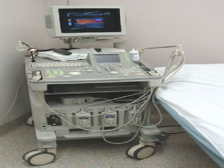
INSTRUMENTATION
FROM SOUND TO IMAGE The creation of an image from sound is done in three steps producing a SOUND WAVES receiving ECHOS interpreting those ECHOS
PRODUCING A SOUND WAVE In medical ultrasonography, a sound wave is typically produced by creating short, strong pulses of sound transducers (usually a type of ceramic). The electrical wiring and transducers are encased in a probe. The electrical pulses vibrate the ceramic to create a series of sound pulses from each. The frequencies present in this sound wave can be anywhere between 2 and 13 MHz. Any frequency above the capabilities of the human ear is referred to as 'ultrasound'. The goal is to produce a single focused arc-shaped sound wave from the sum of all the individual pulses emitted by the transducer.
PRODUCING A SOUND WAVE
RECEIVING THE ECHOS The return of the sound wave to the transducer results in the same process that it took to send the sound wave, just in reverse. The return sound wave vibrates the transducer's elements and turns that vibration into electrical pulses that are sent from the probe to ultrasound scanner where they are processed and transformed into a digital image.
FORMING THE IMAGE The ultrasound scanner must determine three things from each received echo: 1. ) Which transducer elements received the echo (there are multiple elements on a transducer). 2. ) How strong was the echo. 3. ) How long did it take the echo to be received from when the sound was transmitted. Once the ultrasound scanner determines these three things, it can locate which pixel in the image to light up and to what brightness. Transforming the received signal into a digital image can be best explained by using a blank spreadsheet as an analogy.
Types of ultrasound v Abdominal ultrasound v Obstetric ultrasound (pregnancy ultrasound) v Pelvic ultrasound v Transvaginal ultrasound v Hysterosonography v Musculoskeletal ultrasound v Thyroid ultrasound v Testicle ultrasound v Vascular ultrasound v Doppler ultrasound of extremities v Carotid duplex v Breast ultrasound v Neurosonography v Transrectal ultrasound
USES Evaluate a fetus. Diagnose gallbladder disease. Evaluate flow in blood vessels. Guide a needle biopsy. Guide the biopsy and treatment of a tumor. Check your thyroid gland. Study the heart. Diagnose some forms of infection. Diagnose some forms of cancer. Reveal abnormalities in scrotum and prostate.
PRINCIPLE Ultrasound is based on the same principles as sonar — a technology used to detect underwater objects. During an ultrasound, a technician trained in ultrasound imaging (sonographer) presses a small hand-held device (transducer), about the size of a bar of soap, against your skin. The transducer generates and receives high frequency sound waves that can't be heard by the human ear. As the sonographer places the transducer on your skin, crystals inside the transducer emit pulses of sound waves that travel into your body.
CONT… Our tissues, bones and body fluids reflect the sound waves and bounce them back to the transducer. The transducer then sends this information to a computer, which composes detailed images based on the patterns created by the sound waves.
PREPARATIONS FOR THE PROCEDURE Wear comfortable, loose-fitting clothing. Take off your clothes in the examination area and put on a hospital gown. For an abdominal ultrasound, you shouldn't eat or drink for as many as 12 hours before your examination. For pelvic ultrasounds, you may need to drink six glasses of water one to two hours prior to your examination and avoid urinating. For transvaginal ultrasound, you need to urinate before your examination.
Cont… For thyroid and breast sonography, you will be asked to remove all jewellery. No special preparation is needed in other ultrasound exams. In an emergency, exams can be done without special preparations. We have to tell the sonographer, sonologist, or physician sonologist conducting the examination about pain, bleeding, discharge, fever or any other symptoms you might have. Also, telling the examiner about past ultrasounds and surgeries is helpful, and sometimes it is crucial information. Examinations usually take 10 -30 minutes
HOW IT IS PERFORMED During ultrasonography, a hand-held device called a "transducer" is placed on the area being examined and moved around. This transducer generates ultrasound and sends it through the body. It also detects the returning echoes and transmits them as electrical signals. Because one transducer continuously generates many ultrasound waves while detecting echoes, a real time image can be produced on a viewing monitor. These images can be recorded on videotape, or images can be frozen and recorded on to film.
Cont… During ultrasound, lubricating gel is applied to the skin so that the transducer can be moved around to produce real time images. Ultrasound is similar to audible sound in that it can pass through water and human organs easily, but it can't pass through air or bone. So gel is applied between the transducer and the skin to bridge the gap, and effectively send the ultrasound waves. Because US images are real time images, blood flow, blood vessels, bowel movement, and the movement of internal organs from breathing can be seen
PREGNANCY ULTRASOUND
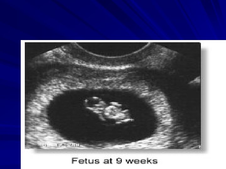
PREGNANCY ULTRASOUND v An ultrasound is used in women who are pregnant, or who might be pregnant. An ultrasound might be done more than once during a pregnancy, depending on the health of the baby or mother. v Used to study fetal development.
OTHER NAMES v transvaginal ultrasound ( TVUS) v first trimester ultrasound v obstetrical ultrasound v pelvic ultrasonography in pregnancy v obstetric sonogram v obstetric ultrasonography
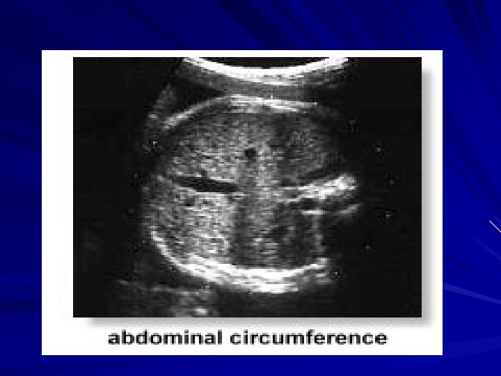
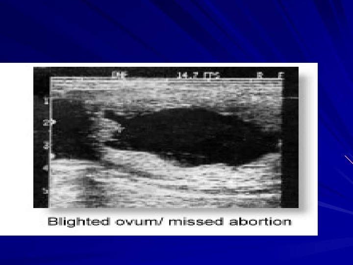
USES OF PREGNANCY ULTRASOUND confirm the expected date of a baby's birth look for size or placement problems with the placenta evaluate causes of vaginal bleeding, such as a blighted ovum, which is a fertilized egg that has stopped growing rule out ectopic pregnancy, a condition in which the fertilized egg grows outside of the uterus check for intrauterine growth retardation, which occurs when the baby grows too slowly evaluate the volume of amniotic fluid assess enlarged ovaries
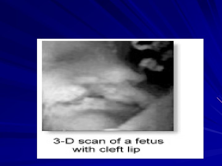
Cont… diagnose multiple pregnancies, such as twins or triplets rule out molar pregnancy, a situation in which the fetus itself becomes a tumor check for problems with the baby, including spina bifida, where the spine fails to close during development, or cleft palate, which is abnormal closure of the lip and roof of the mouth determine if the baby is alive and healthy, with good movement, heart function, and placement in the uterus
TRANSVAGINAL TRANSDUCER A transvaginal transducer, also known as an endovaginal transducer or probe, uses a small electronic array as the scanhead and is inserted directly into the vagina for the scan. This is used as an alternative to an ultrasound examination done via an abdominal scanner. This technique produces a much sharper image, not only because of the scanhead's closer proximity to the uterus, but also because the transducer operates at a higher frequency and hence has a higher resolving power. Transvaginal examinationis are usually done for assessment of very early pregnancies
TRANSVAGINAL TRANSDUCER A sterile condom is slipped over the handheld transducer (or probe), which is then covered with lubricating gel and placed in the vagina. The probe rests up against the cervix and is being moved in several directions by the sonographer. The rest of the test proceeds the same way as transabdominal ultrasound.
TRANSVAGINAL TRANSDUCER
TRANSVAGINAL TRANSDUCER
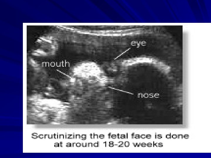
DIAGNOSIS OF FETAL MALFORMATIONS Many structural abnormalities in the fetus can be reliably diagnosed by an ultrasound scan, and these can usually be made before 20 weeks. Common examples include hydrocephalus, anencephaly, myelomeningocoele, achondroplasia and other dwarfism, spina bifida, exomphalos, Gastroschisis, duodenal atresia and fetal hydrops. With more recent equipment, conditions such as cleft lips/ palate and congenital cardiac abnormalities are more readily diagnosed and at an earlier gestational age.
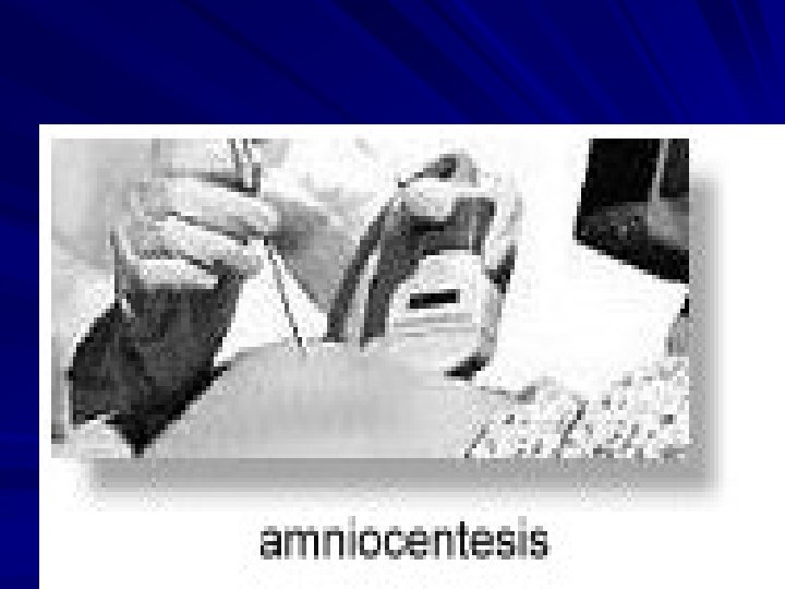
Cont… First trimester ultrasonic 'soft' markers for chromosomal abnormalities such as the absence of fetal nasal bone, an increased fetal nuchal translucency (the area at the back of the neck) are now in common use to enable detection of Down syndrome fetuses. Ultrasound can also assist in other diagnostic procedures in prenatal diagnosis such as amniocentesis, chorionic villus sampling, cordocentesis (percutaneous umbilical blood sampling) and in fetal therapy.
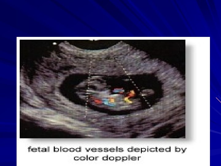
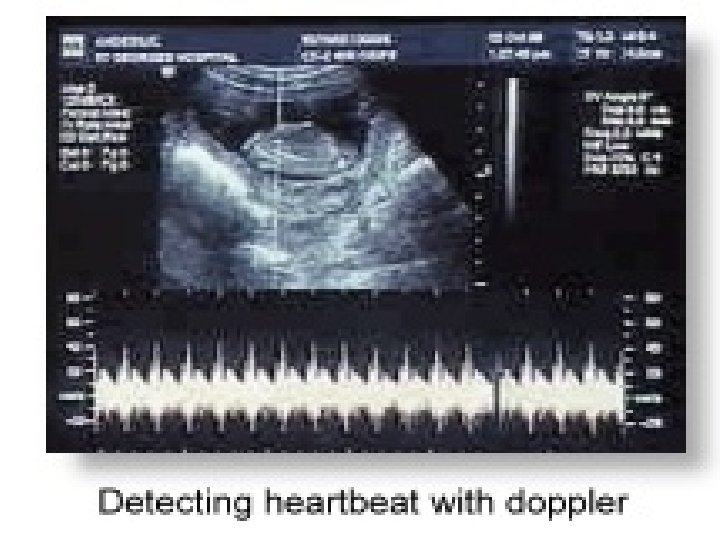
DOPPLER SONOGRAPHY Ultrasonography can be enhanced with Doppler measurements, which employ the Doppler effect to assess whether structures (usually blood) are moving towards or away from the probe, and its relative velocity. By calculating the frequency shift of a particular sample volume, for example a jet of blood flow over a heart valve, its speed and direction can be determined and visualised. This is particularly useful in cardiovascular studies (ultrasonography of the vasculature and heart) and essential in many areas such as determining reverse blood flow in the liver vasculature in portal hypertension.
Cont… The Doppler information is displayed graphically using spectral Doppler, or as an image using colour Doppler or power Doppler. It is often presented audibly using stereo speakers: this produces a very distinctive, although synthetic, sound.
SPECTRAL DOPPLER OF COMMON CAROTID ARTERY
COLOUR DOPPLER OF COMMON CAROTID ARTERY
ADVANTAGES OF THE PROCEDURE Safe, painless, easy, fast, and widely available No radiation Real time imaging -- ultrasonography can be used to guide invasive procedures such as biopsy, and to visualize bowel movement and blood flow. In case of an emergency, bedside sonography can be done without particular patient preparations.
Cont… During pregnancy -- to evaluate fetus, uterus To determine the causes of pain, swelling, bleeding, or infection -- stones, tumor, cyst, inflammations, injury, congenital anomalies To examine blood flow and discover blockages -- atherosclerotic plaque, blood clots in the arteries and veins of extremities, abdomen, pelvis, and neck To guide invasive procedures -- needle biopsy (i. e. , breast cancer), sampling or withdrawal of fluid (i. e. , amniocentesis, amoebic liver abscess), insertion of tubes, catheters, stents, wires
APPLICATIONS Abdominal ultrasound: gallstones, kidney stones, appendicitis, enlarged organs (liver, spleen, kidneys, lymph nodes, etc. ) Obstetric (pregnancy) ultrasound: abnormalities in the unborn child (fetus), uterus, and placenta (such as placenta previa), monitoring fetal development Pelvic ultrasound: cysts, fibroid (uterine myoma), ovarian cancers, uterine anomalies, abscesses, ectopic pregnancy, abortion, prostate cancer/hyperplasia, bladder tumor Transvaginal ultrasound: ectopic pregnancy, fetal death, hydatidiform mole, cysts, tumor of uterus/ovaries
Cont… Hysterosonography (especially in patients with abnormal uterine bleeding): polyps, fibroids, cancer, congenital anomaly, endometrial scars, evaluating the patency of fallopian tubes Musculoskeletal ultrasound: tendon tears (i. e. , tears of Achilles tendon), tears of muscles, masses or tumors, bleeding or other fluid collections within the muscles, bursae, and joints Thyroid ultrasound: cysts, goiter (enlarged thyroid), tumor/cancer Testicle ultrasound: cysts, tumor, fluid collection, torsion, inflammation (epididymitis, tuberculosis, etc. )
Cont… Vascular ultrasound: blood clots, atherosclerotic plaque, injury, congenital malformation, monitoring of vascular graft or bypass blood vessels Doppler ultrasound of the extremities: venous occlusion, deep vein thrombosis, arterial occlusion, arteriosclerosis, trauma to the arteries/veins, monitoring vascular graft/stent/other reconstruction, vascular masses (such as, aneurysm, pseudoaneurysm), measurement of blood flow Carotid duplex: carotid stenosis (narrowing), occlusion, thrombosis
Cont… Breast ultrasound: breast cancer, cyst, benign tumor (such as fibroadenoma), fibrocystic breast condition Neurosonography: prenatal hypoxic brain damage, congenital anomaly Transrectal ultrasound: prostate cancer, benign prostatic hyperplasia Others: pleural effusion, superficial mass EUS can find lesions that are less than 1 to 2 cm, it can find metastatic tumors that cannot be seen on other imaging tests such as CT or MRI.
3 -D ULTRASOUND
YAWNING FOETUS!
FOETUS POSITION
Pubo-rectal muscle: Transvaginal para-sagittal section
Urethro-vesical area "Prepubien" (bulbocavernosus muscle). Arcuate ligament. Vagina. Bladder mucosa. Urethral mucosa. Anterior edge of the urethra.
Doppler of the Urethra Pubic bone. Vascular plexus of the urethra. Bladder neck.
The Urethra Symphysis pubis. Pubic bone. Urethra. Arcuate ligament
The"Prepubien(bulbo-cavernosus muscles) Prepubien. Symphysis pubis. Arcuate ligament. Anterior edge of the urethra. Urethral mucosa.
The anal sphincter Vagina. External sphincter. Internal sphincter.
The anal canal and the levator plate External anal sphincter. Internal anal sphincter. Levator plate.
LIMITATIONS Ultrasound does not penetrate air or bone. So if an abnormality is behind bowel gas, ribs, or calcified rib cartilage, it may not be discovered. Because ultrasound is absorbed and reflected inside the body, only some of the waves reach deep places farthest from the transducer. There's a limitation to ultrasound's ability to look deep into the body. Of two tumors of equal size, the tumor closest to the transducer will be discovered more readily than the more distant one. Examinations, consequently, are not as productive for obese, tall patients as they are for thin or petite ones.
Cont… Ultrasonography is an operator-dependent, subjective test. The more experience the operator has, the better the patient listens to his instructions (hold your breath, do not eat, repress the urge to urinate), the better the results. Further, the more the operator knows about the patient's past medical history, current medical history, and the results of other radiological and laboratory tests, the better the examination. For best results, before taking ultrasonography, ask if the practice where the scan is being performed is accredited either by the American Institute of Ultrasound in Medicine or the American College of Radiology.
Cont… Ultrasonography can't discover all abnormalities. If there's an ulcer, small polyps, or diverticuli, or infection/inflammation in the stomach, intestine, or colon, ultrasound will not be effective in finding them. In such cases, endoscopy, barium enema, or an upper GI series must be done. Ultrasound is an incomplete examination, and a CAT scan or MRI will have to be done for more accurate diagnosis.
Thank you