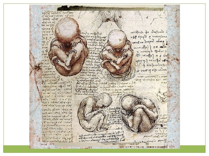
Introduction to the Human Body Chapter One Locate

Introduction to the Human Body Chapter One

Locate these Structures… 1. Kidney 2. Medulla Oblongata 3. Liver 4. Carpels 5. Pyloric Sphincter Valve 6. Femur 7. Achilles Tendon 8. Biceps brachii 9. Patella 10. Iris 11. Atrium 12. Gall Bladder 13. Sternum 14. Ulna 15. Pancreas 16. Cochlea 17. Cornea 18. Mammary Gland 19. Coccyx 20. Mandible

Understanding the Human Body �Curiosity Illnesses, injuries, death � Obtained knowledge regarding the human body as science and medicine advanced

Anatomy vs. Physiology �Anatomy: examines the structures, or morphology, or body parts – their forms, organization -tomy: (Greek) cutting up Anatomists rely on examination �Physiology: considers the functions of body parts – what they do and how they do it Also Greek origin; relationship to nature Physiologists rely on experimentation

A few definitions… �Regional anatomy: study of anatomy based on regions or divisions of the body; emphasis on relations between the structures of that region �Systemic anatomy: study of anatomy based on body systems and their functioning throughout the body �Gross anatomy: study of anatomy that is visible to the naked eye; macroscopic �Microscopic anatomy: study of anatomy at the microscopic level; cells

�The human body is composed of parts within parts, which vary in complexity

Maintenance of Life �Requirements of Organisms Water metabolic processes; transport of substances Food/Nutrients energy Oxygen release energy from food substances Heat regulation of metabolic reaction rates Pressure breathing (atmospheric); blood circulation (hydrostatic) �Although we require the aforementioned, these factors alone are not enough to ensure survival; quality and quantity matter.

Homeostasis �Equilibrium of the body’s internal environment produced by the interaction of organ systems and regulatory processes (feedback systems). Homeostasis is a dynamic condition in response to changing conditions. So important that it requires most of our metabolic energy

�Body maintains homeostasis through a number of selfregulating control systems – homoeostatic mechanisms Receptors: provide information about the stimuli in the internal environment Control center: includes a set point, tells what a particular value should be (body temperature) Effectors: elicit responses that alter conditions in the internal environment ; muscles or glands

Feedback Mechanisms � Negative feedback: almost all homeostatic control mechanisms are negative feedback mechanisms. These mechanisms reduce the variable back to its original state or “ideal value”. Just remember that positive Blood sugar receptors sense the change pancreas (control center) secretes insulin blood sugar levels feedback mechanisms reduce � Positive feedback: the output enhances the enhance the original stimulus. Child birth: oxytocin is released that intensifies and negative feedback speeds up contractions. Blood clotting: vessel is damaged, platelets start to mechanisms inhibit it. cling to the injured site and release chemicals that attract more platelets. The platelets continue to pile up and release chemicals until a clot is formed.

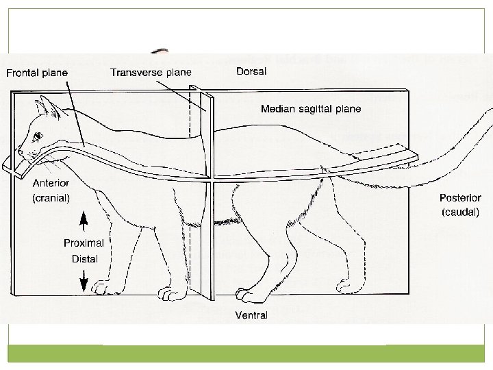
Body Planes and Terminology Day Two – Chapter One

�Standing straight, body erect �Feet slightly apart �Palms facing forward �Thumbs point away from body

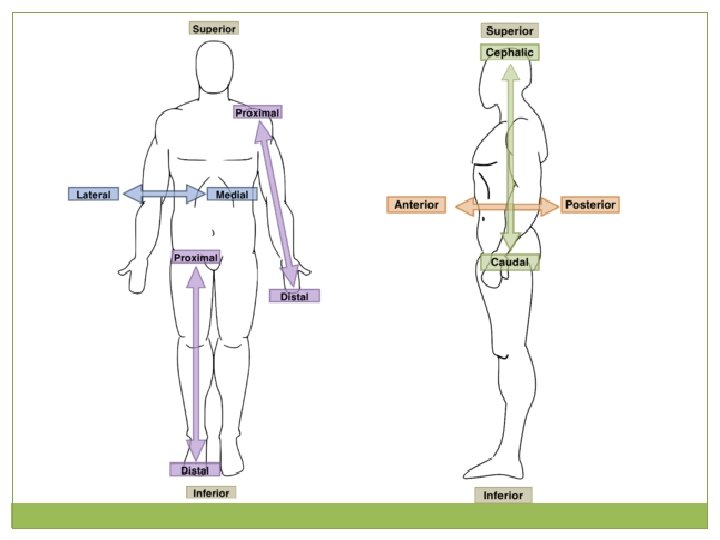
�Sagittal: divides the body into right and left parts �Frontal/coronal: divides the body into anterior and posterior parts �Transverse/horizontal: divides the body into superior and inferior parts

S/I 1. The head is _______ to the belly button 2. The hip is ________ to the shoulder P/D 3. The wrist is ___________ to the elbow 4. The ankle is _________ to the toes M/L 5. The ears are _________ to the nose 6. The sternum/breast bone is ______ to the shoulders A/P 7. The gluteus maximus is _________ to patella. 8. The belly button is _________ heel


Body Cavities and Terminology Day Three – Chapter One

�Dorsal cavity: protects the nervous system, Any fluid filled space in a and is divided into two subdivisions multicellular organism Cranial cavity: within the skull; encases the brain Vertebral cavity: runs within the vertebral column; encases the spinal cord �Ventral cavity: houses the internal organs (viscera), and is divided into two subdivisions Thoracic Abdominopelvic

�Thoracic cavity is subdivided into two pleural cavities, the mediastinum, and the pericardial cavity Right & Left Pleural cavities: each houses a lung Mediastinum: central compartment surrounded by loose connective tissue �Contains all the organs of the thoracic cavity aside from the lungs • Superior portion (above the heart) • Inferior portion that is further subdivided into the anterior, middle, and posterior portions �Pericardial cavity: encloses the heart, contains pericardial fluid

�The abdominopelvic cavity is separated from the superior thoracic cavity by the domeshaped diaphragm �Composed of two subdivisions Abdominal cavity– contains the stomach, intestines, spleen, liver, gall bladder Pelvic cavity– lies within the pelvis and contains the bladder, reproductive organs, and rectum
 Pleural cavity")
Cranial cavity Vertebral cavity Superior mediastinum Thoracic cavity (contains heart and lungs) Pleural cavity Pericardial Cavity within the mediastinum Key: Dorsal body cavity Ventral body cavity (b) Anterior view Figure 1. 9 b

Body Cavities Diaphragm Key: Dorsal body cavity Ventral body cavity Abdominal cavity (contains digestive viscera) Abdominopelvic cavity Pelvic cavity (contains bladder, reproductive organs, and rectum) Figure 1. 9 b
 Thoracic cavity Dorsal body cavity (contains heart and lungs) Vertebral")
Cranial cavity (contains brain) Thoracic cavity Dorsal body cavity (contains heart and lungs) Vertebral cavity (contains spinal cord) Key: Dorsal body cavity Ventral body cavity (a) Lateral view Diaphragm Abdominal cavity (contains digestive viscera) Pelvic cavity (contains bladder, reproductive organs, and rectum) Ventral body cavity (thoracic and abdominopelvic cavities)

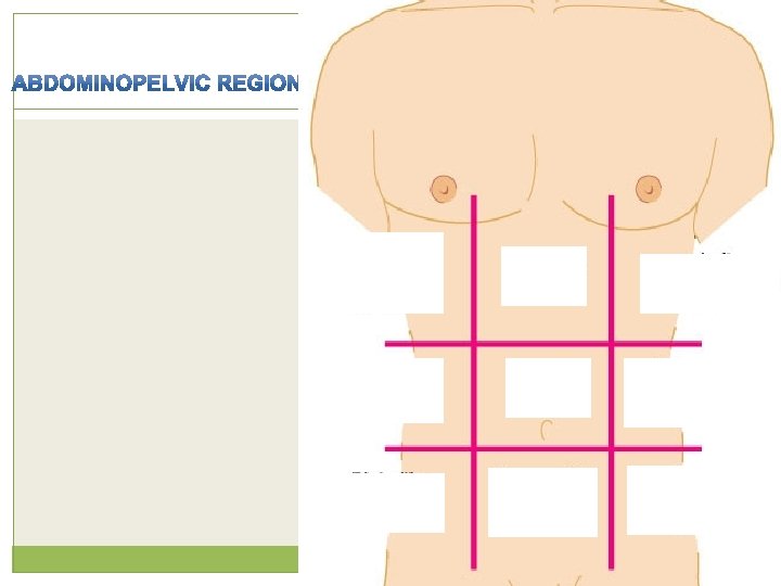
�Right upper Why abdominal divisions? • Theoretical divisions used by clinicians to help localize, �Left upper identify and diagnose a patient’s symptoms. • Two forms of categorization �Right lower • Four quadrants �Left • lower Nine regions • Either categorization is internationally recognized and can be used on a daily basis during clinical practice. Figure 1. 12

� Oral: the space in the mouth inside the teeth and gums and is filled with the tongue when it is relaxed. � Nasal: located within and posterior to the nose � Orbital: house the eyes � Middle ear: contains bones (ossicles) that transmit sound vibrations � Synovial: are inside the joint capsules that surround freely moving joints (such as the hip, knee, elbow, and shoulder)
- Slides: 28