Introduction to the Cell 2015 Pearson Education Inc


- Slides: 44
Introduction to the Cell © 2015 Pearson Education, Inc.
4. 1 Microscopes reveal the world of the cell • Using microscopes, scientists studied • microorganisms, • animal and plant cells, and • some structures within cells. • In the 1800 s, these studies led to cell theory, which states that Light microscope Scanning Electron Microscope • all living things are composed of cells and • all cells come from other cells. Transmission Electron Micrograph © 2015 Pearson Education, Inc.
10 m Length of some nerve and muscle cells Chicken egg 10 mm (1 cm) 1 mm 100 μm 100 nm 1 nm 0. 1 nm © 2015 Pearson Education, Inc. Frog egg Paramecium Human egg Most plant and animal cells Nucleus Most bacteria Mitochondrion Smallest bacteria Viruses Ribosome Proteins Lipids Small molecules Atoms Electron microscope 100 mm (10 cm) Human height Light microscope 1 m Unaided eye Figure 4. 1 e-0
4. 2 The small size of cells relates to the need to exchange materials across the plasma membrane • Cell size must • be large enough to house DNA, proteins, and structures needed to survive and reproduce, but • remain small enough to allow for a surface-tovolume ratio that will allow adequate exchange with the environment. © 2015 Pearson Education, Inc.
4. 2 The small size of cells relates to the need to exchange materials across the plasma membrane • The plasma membrane forms a flexible semipermeable boundary between the living cell and its surroundings. • Phospholipids form a two-layer sheet called a phospholipid bilayer. © 2015 Pearson Education, Inc.
4. 2 The small size of cells relates to the need to exchange materials across the plasma membrane • Membrane proteins are embedded in the lipid bilayer. • Some are channels that shield ions and other hydrophilic molecules • Other proteins serve as pumps, moving molecules into or out of the cell. © 2015 Pearson Education, Inc.
4. 3 Prokaryotic cells vs. eukaryotic cells Prokaryotic cells are small and simpler in structure. • Bacteria and archaea are prokaryotic cells. • Eukaryotic cells have • a membrane-enclosed nucleus and • many membrane-enclosed organelles that perform specific functions. • All other forms of life are composed of eukaryotic cells. © 2015 Pearson Education, Inc.
4. 3 Prokaryotic cells vs. eukaryotic cells • Both prokaryotic and eukaryotic cells have: • a plasma membrane, • an interior filled with a thick, jellylike fluid called the cytosol, • one or more chromosomes, which carry genes made of DNA, • ribosomes, tiny structures that make proteins according to instructions from the genes. • cytoplasm, the interior environment of a cell © 2015 Pearson Education, Inc.
4. 3 Prokaryotic cells • In a prokaryotic cell, • the DNA is coiled into a nucleoid • no membrane surrounds the DNA. • outside the plasma membrane of most prokaryotes is a fairly rigid, chemically complex cell wall • There may be surface projections © 2015 Pearson Education, Inc.
4. 4 Eukaryotic cells are partitioned into functional compartments • A eukaryotic cell contains • a membrane-enclosed nucleus and • various other organelles (“little organs”), which perform specific functions in the cell. • Eukaryotic organelles can be grouped on the basis of four functions: 1. genetic control, 2. manufacturing, distribution, and breakdown of materials, 3. energy processing, and 4. structural support, movement, and intercellular communication. © 2015 Pearson Education, Inc.
4. 4 Eukaryotic cells are partitioned into functional compartments • Almost all of the organelles and other structures of animals cells are present in plant cells. • A few exceptions exist. • Lysosomes and centrosomes containing centrioles are not found in plant cells. • Only the sperm cells of a few plant species have flagella. © 2015 Pearson Education, Inc.
4. 4 Eukaryotic cells are partitioned into functional compartments • Plant but not animal cells have • a rigid cell wall that contains cellulose, • plasmodesmata, cytoplasmic channels through cell walls that connect adjacent cells, • chloroplasts, where photosynthesis occurs, and • a central vacuole, a compartment that stores water and a variety of chemicals. © 2015 Pearson Education, Inc.
Figure 4. 4 a Rough endoplasmic reticulum NUCLEUS Nuclear envelope Nucleolus Chromatin Ribosomes CYTOSKELETON Microtubule Microfilament Intermediate filament Peroxisome Smooth endoplasmic reticulum Plasma membrane Golgi apparatus Lysosome Mitochondrion © 2015 Pearson Education, Inc. Centrosome with pair of centrioles
Figure 4. 4 b Smooth endoplasmic reticulum NUCLEUS Nuclear envelope Nucleolus Chromatin Rough endoplasmic reticulum Mitochondrion CYTOSKELETON Microfilament Microtubule Central vacuole Ribosomes Chloroplast Cell wall Plasmodesma Cell wall of adjacent cell Golgi apparatus Peroxisome Plasma membrane © 2015 Pearson Education, Inc.
© 2015 Pearson Education, Inc.
THE NUCLEUS AND RIBOSOMES © 2015 Pearson Education, Inc.
4. 5 The nucleus contains the cell’s genetic instructions • The nucleus • contains the cell’s DNA and • controls the cell’s activities by directing protein synthesis by making messenger RNA (m. RNA). • DNA is associated with many proteins and is organized into structures called chromosomes. • When a cell is not dividing, this complex of proteins and DNA, called chromatin, appears as a scattered mass within the nucleus. © 2015 Pearson Education, Inc.
4. 5 The nucleus contains the cell’s genetic instructions • The double membrane surrounding the nucleus is called the nuclear envelope • has pores that regulate the entry and exit of large molecules • connects with the cell’s network of membranes called the endoplasmic reticulum. © 2015 Pearson Education, Inc.
4. 5 The nucleus contains the cell’s genetic instructions • The nucleolus is • the site of ribosomal RNA (r. RNA) synthesis. © 2015 Pearson Education, Inc.
4. 6 Ribosomes make proteins for use in the cell and for export • Ribosomes are involved in the cell’s protein synthesis. • Ribosomes are the cellular components that use instructions from the nucleus, written in m. RNA, to build proteins. © 2015 Pearson Education, Inc.
4. 6 Ribosomes make proteins for use in the cell and for export • Some ribosomes are free ribosomes; others are bound. • Free ribosomes are suspended in the cytosol. • Bound ribosomes are attached to the outside of the endoplasmic reticulum or nuclear envelope. © 2015 Pearson Education, Inc.
THE ENDOMEMBRANE SYSTEM © 2015 Pearson Education, Inc.
4. 7 Many organelles are connected in the endomembrane system • Many of the membranes within a eukaryotic cell are part of the endomembrane system. • Some of these membranes are physically connected, and others are linked when tiny vesicles (sacs made of membrane) transfer membrane segments between them. © 2015 Pearson Education, Inc.
4. 7 Many organelles are connected in the endomembrane system • Many of these organelles interact in the synthesis, distribution, storage, and export of molecules. • The endomembrane system includes the • • • nuclear envelope, endoplasmic reticulum (ER), Golgi apparatus, lysosomes, vacuoles, and plasma membrane. © 2015 Pearson Education, Inc.
4. 7 Many organelles are connected in the endomembrane system • The largest component of the endomembrane system is the endoplasmic reticulum (ER), an extensive network of flattened sacs and tubules. • There are two kinds of endoplasmic reticulum, which differ in structure and function. 1. Smooth ER lacks attached ribosomes. 2. Rough ER has bound ribosomes that stud the outer surface of the membrane. © 2015 Pearson Education, Inc.
© 2015 Pearson Education, Inc.
4. 8 The endoplasmic reticulum is a biosynthetic workshop • Smooth ER is involved in a variety of metabolic processes • the production of enzymes important in the synthesis of lipids, oils, phospholipids, and steroids, • the production of enzymes that help process drugs, alcohol, and other potentially harmful substances, and • the storage of calcium ions. • Rough ER makes • additional membrane for itself and • secretory proteins. © 2015 Pearson Education, Inc.
4. 9 The Golgi apparatus modifies, sorts, and ships cell products • The Golgi apparatus serves as a molecular warehouse and processing station for products manufactured by the ER. 1. Products travel in transport vesicles from the ER to the Golgi apparatus. 2. One side of the Golgi stack acts as a receiving dock. 3. Products of the ER are modified in the Golgi stacks. 4. The “shipping” side of the Golgi functions as a depot, where products in vesicles bud off and travel to other sites. © 2015 Pearson Education, Inc.
Figure 4. 9 “Receiving” side of Golgi apparatus Transport vesicle 1 from the ER 2 3 Golgi apparatus 4 Transport vesicle from the Golgi “Shipping” side of Golgi apparatus © 2015 Pearson Education, Inc.
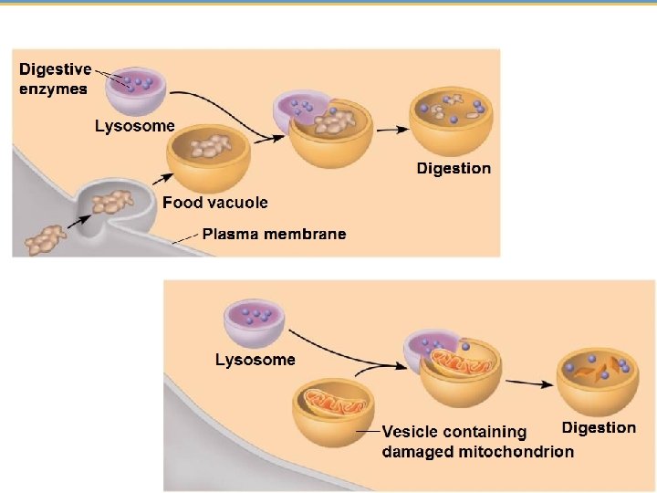
4. 10 Lysosomes are digestive compartments within a cell • A lysosome is a membrane-enclosed sac of digestive enzymes • fuse with food vacuoles and digest food, • destroy bacteria engulfed by white blood cells, or • fuse with other vesicles containing damaged organelles or other materials to be recycled within a cell. © 2015 Pearson Education, Inc.
4. 11 Vacuoles function in the general maintenance of the cell • Vacuoles are large vesicles that have a variety of functions. • Some protists have contractile vacuoles, which help to eliminate water from the protist. • In plants, vacuoles may • • have digestive functions, store water, nutrients contain pigments, or contain poisons that protect the plant. © 2015 Pearson Education, Inc.
The following figure summarizes the relationships among the major organelles of the endomembrane system. Nucleus Smooth ER Nuclear envelope Rough ER Golgi apparatus Transport vesicle Lysosome © 2015 Pearson Education, Inc. Plasma membrane Transport vesicle
4. 12 A review of the structures involved in manufacturing and breakdown • Peroxisomes are metabolic compartments that do not originate from the endomembrane system. – How they are related to other organelles is still unknown. – Some peroxisomes break down fatty acids to be used as cellular fuel. © 2015 Pearson Education, Inc.
ENERGY-CONVERTING ORGANELLES © 2015 Pearson Education, Inc.
4. 13 Mitochondria harvest chemical energy from food • Mitochondria are organelles that carry out cellular respiration eukaryotic cells. • converts the chemical energy in foods to chemical energy in ATP. © 2015 Pearson Education, Inc.
4. 14 Chloroplasts convert solar energy to chemical energy • Chloroplasts are the photosynthesizing organelles of plants and algae. • conversion of light energy from the sun to the chemical energy of sugar molecules.
4. 15 EVOLUTION CONNECTION: Mitochondria and chloroplasts evolved by endosymbiosis • The endosymbiosis theory states that • mitochondria and chloroplasts were formerly small prokaryotes • they began living within larger cells. • they both contain DNA and ribosomes
THE CYTOSKELETON AND CELL SURFACES © 2015 Pearson Education, Inc.
4. 16 The cell’s internal skeleton • Cells contain a network of protein fibers, called the cytoskeleton. • Microtubules: straight, hallow tubes made of tubulin, shape and support the cell, act as tracks which help organelles move • Intermediate filaments: found in most animal cells, reinforce cell shape and anchor some organelles • Microfilaments (actin filaments): support the cell’s shape and are involved in motility
4. 18 Cilia and flagella move when microtubules bend • A flagellum propels a cell by an undulating, whiplike motion. • Cilia work more like the oars of a boat. • Both have a common structure and mechanism of movement. • Both are composed of microtubules wrapped in an extension of the plasma membrane.
4. 19 The extracellular matrix of animal cells functions in support and regulation • Animal cells synthesize and secrete an elaborate extracellular matrix (ECM) • helps hold cells together in tissues • protects and supports the plasma membrane.
4. 20 Three types of cell junctions are found in animal tissues • Adjacent cells adhere, interact, and communicate through specialized junctions between them. • Tight junctions prevent leakage of fluid across a layer of epithelial cells. • Anchoring junctions fasten cells together into sheets. • Gap junctions are channels that allow small molecules to flow through protein-lined pores between cells.
4. 21 Cell walls enclose and support plant cells • A plant cell has a rigid cell wall that • protects and provides skeletal support that helps keep the plant upright and • is primarily composed of cellulose. • Plant cells have cell junctions called plasmodesmata that allow plants tissues to share water, nourishment, and chemical messages.