
Introduction to Special Stains Dr Vivien Rolfe De
- Slides: 26
Introduction to Special Stains Dr Vivien Rolfe De Montfort University This is an Open Educational Resource (OER) that is globally available on the web Creative Commons BY SA
Summary �Why are special stains still important and relevant today? �What are some of the chemical principles behind these stains? �Some common examples that can be prepared in student laboratory teaching.
What are “special stains”? � H&E was first introduced in the 1870’s and the term special stain came to refer to any technique other than H&E used in the clinical environment. � Whilst the H&E stain is the most common staining method in hospital and research laboratories, it isn’t without its limitations. • H&E cannot visualize micro-organisms. • H&E is not good for distinguishing connective tissue and nerve tissue. • H&E cannot distinguish molecular basis of disease and immunohistochemistry might be preferred.
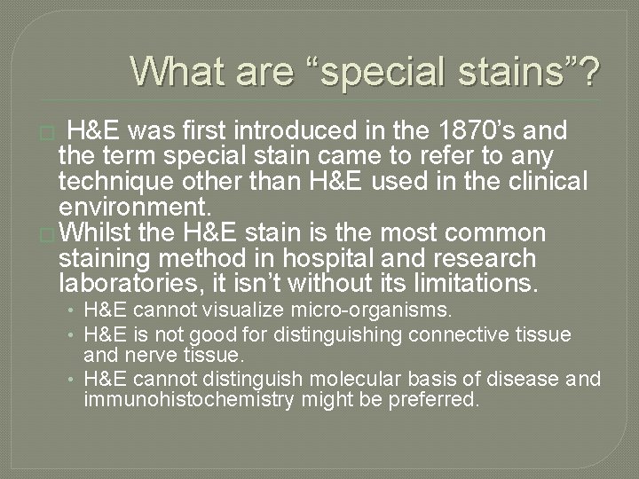
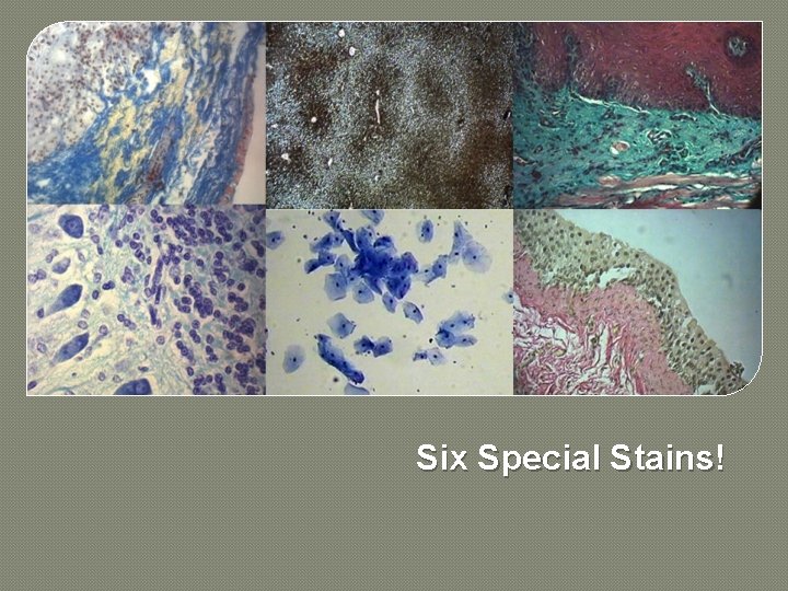
Six Special Stains!
1) Toluidine Blue �Tolonium Chloride �Useful blue cationic dye �Cheap and simple application
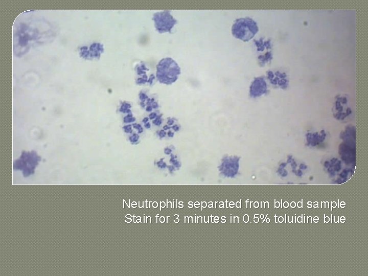
Neutrophils separated from blood sample Stain for 3 minutes in 0. 5% toluidine blue
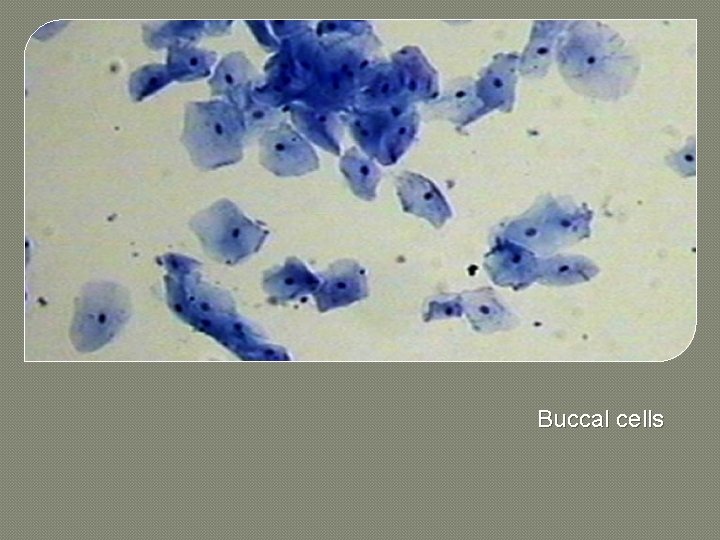
Buccal cells
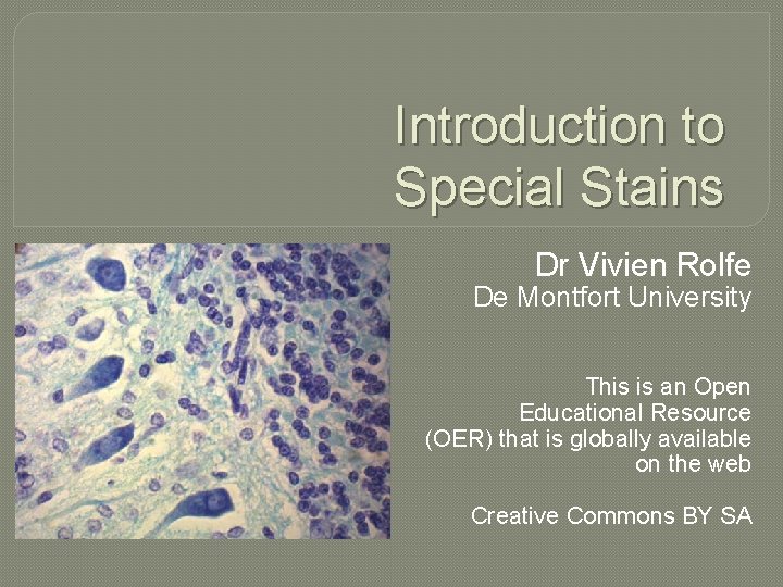
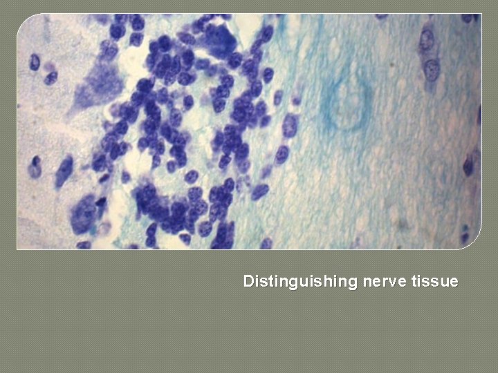
Distinguishing nerve tissue
2) Luxol Fast Blue �Immunohistochemical methods have advanced but are costly and reagents deteriorate quickly. �Special stains include silver methods (such as Gordon and Sweet’s), gold, or Luxol fast blues to stain myelin. �Other special stains identify nerve cells. The techniques are important for looking and neurodegeneration.
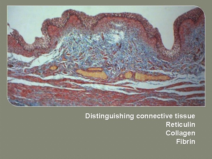
Distinguishing connective tissue Reticulin Collagen Fibrin
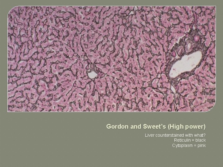
3) Gordon and Sweet’s �One of several silver methods for staining reticulin. �Tissue treated with potassium permanganate to enable the silver to bind. �Uses an ammoniacal silver solution. �What is reticulin? �Why is it important?
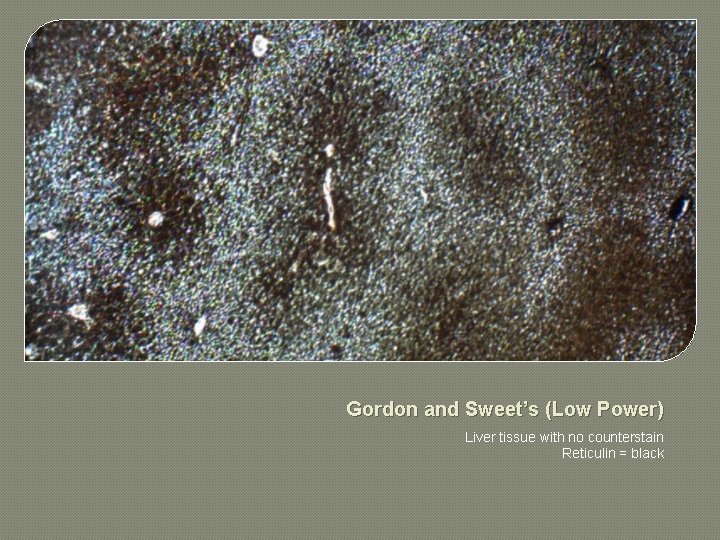
Gordon and Sweet’s (Low Power) Liver tissue with no counterstain Reticulin = black
Gordon and Sweet’s (High power) Liver counterstained with what? Reticulin = black Cytoplasm = pink
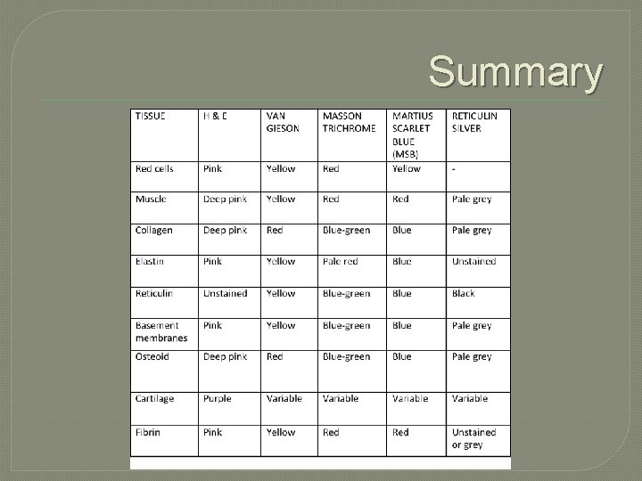
4) Van Gieson �Trichrome stain – producing 3 colours. �Anionic dye and a cationic counterstain. �Nuclear stain applied first such as Weigert’s haematoxlin. �Collagen stains red with acid fuchsine. �Cytoplasm including muscle stains yellow. �Washing in acidified water differentiates tissue producing two colours.
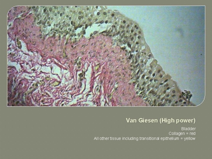
Van Giesen (High power) Bladder Collagen = red All other tissue including transitional epithelium = yellow
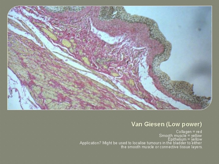
Van Giesen (Low power) Collagen = red Smooth muscle = yellow Epithelium = yellow Application? Might be used to localise tumours in the bladder to either the smooth muscle or connective tissue layers.
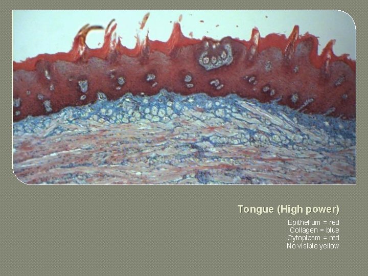
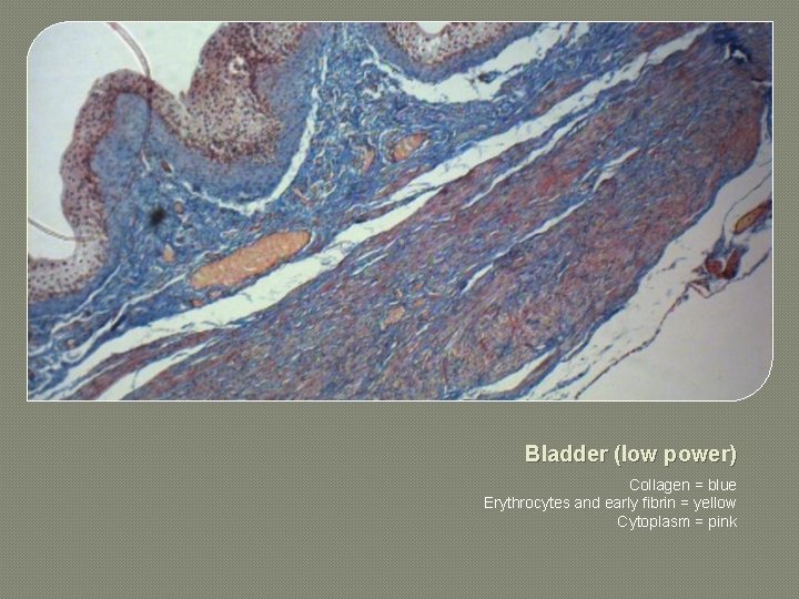
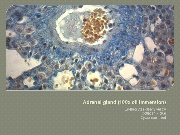
5) Martius scarlet blue stain �Trichrome stain. �Martius yellow and phosphotungstic acid. �Brilliant crystal scarlet. �Methyl blue. �What do the dyes stain?
Tongue (High power) Epithelium = red Collagen = blue Cytoplasm = red No visible yellow
Bladder (low power) Collagen = blue Erythrocytes and early fibrin = yellow Cytoplasm = pink
Adrenal gland (100 x oil immersion) Erythrocytes clearly yellow Collagen = blue Cytoplasm = red
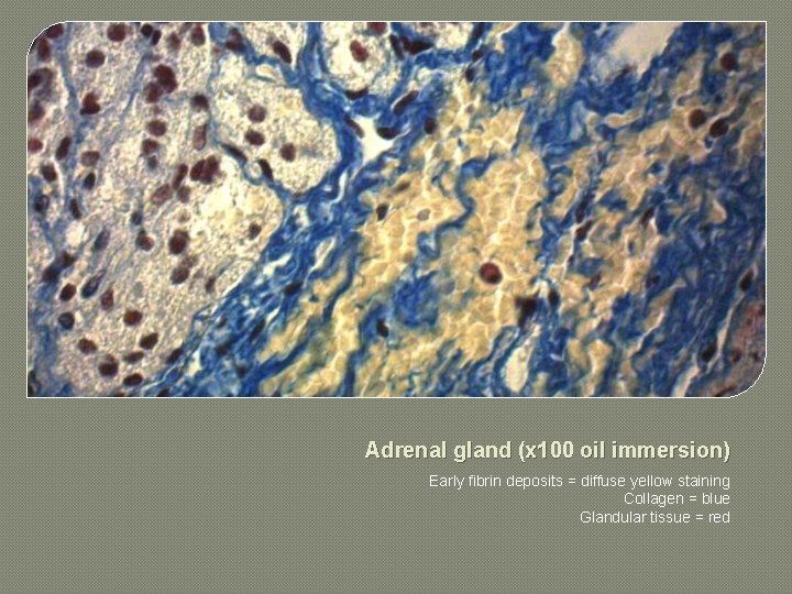
Adrenal gland (x 100 oil immersion) Early fibrin deposits = diffuse yellow staining Collagen = blue Glandular tissue = red
6) Masson’s Trichrome �Trichrome stain. �Iron-haematoxylin plus two anionic dyes. �MSB is a variation of this. �Iron-haematoxylin. �Scarlet-acid fuchsine. �Light green (more of a turquoise stain).
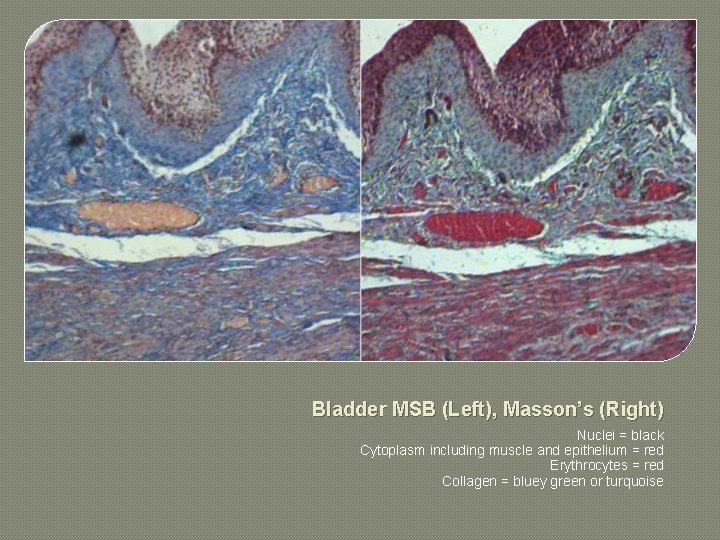
Bladder MSB (Left), Masson’s (Right) Nuclei = black Cytoplasm including muscle and epithelium = red Erythrocytes = red Collagen = bluey green or turquoise
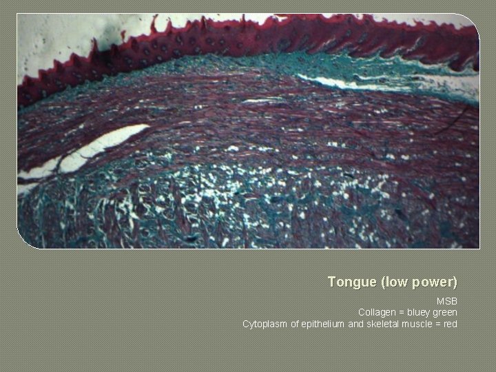
Tongue (low power) MSB Collagen = bluey green Cytoplasm of epithelium and skeletal muscle = red
Summary
Further Resources and OERs � Histological and Histochemical Methods. 4 th Edition. By JA Kiernan. 2007. Scion publishing. Available: http: //www. scionpublishing. com � Histopathology: Fundamentals of Biomedical Science. By G Orchard and B Nation. 2012. Oxford University Press. Available: http: //www. oup. com/uk/orc/bin/fbs/ � Laboratory skills open educational resources. De Montfort University. Available: https: //www. youtube. com/user/biologycourses