
Introduction to Proteomics Dr Ahmed A Mirza MLT


- Slides: 61
Introduction to Proteomics Dr. Ahmed A. Mirza MLT, FAM, KAU
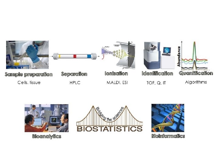
Objectives • Protein expression review • What is proteomics • Basic methods in protein biochemistry • Protein extraction – Physical force – enzymatic • Separation – Chromotography • • Size exclusion Ion exchange Affinity hydrophobicity Analysis Semi-Quantification Gel electrophoresis 2 D gel electrophoresis Differential expression Mass Spectrometry Separation Ionization Identification Quantification microarrays Proteomics Definition Application
What is Proteomics? ?
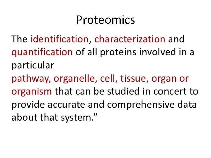
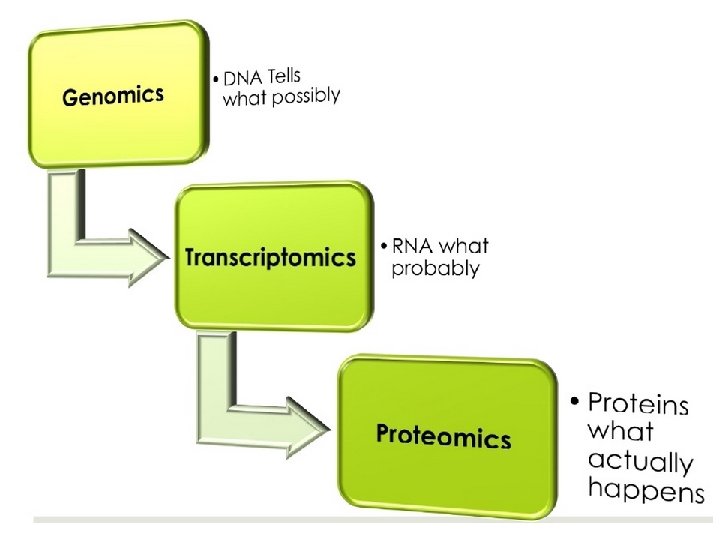
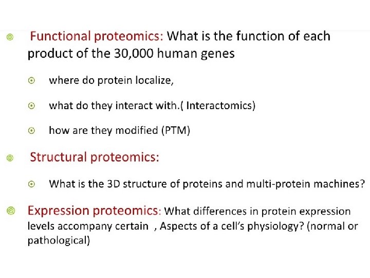
Proteomics
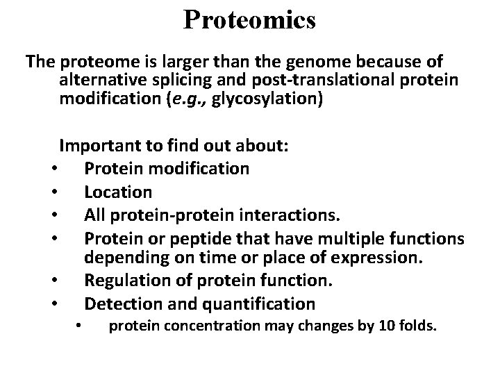
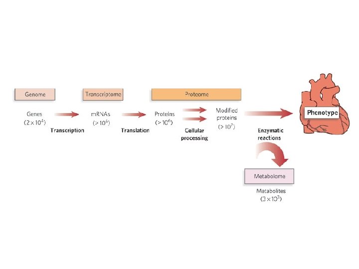
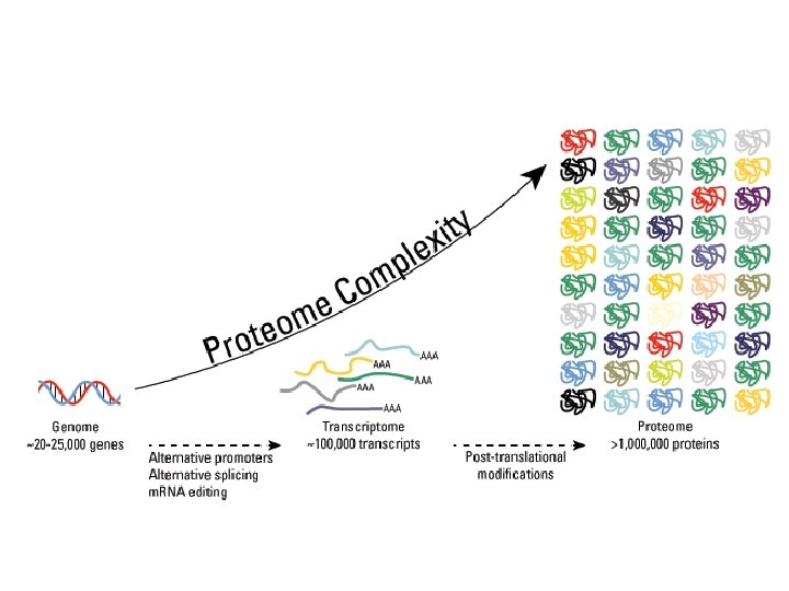
Proteomics The proteome is larger than the genome because of alternative splicing and post-translational protein modification (e. g. , glycosylation) Important to find out about: • Protein modification • Location • All protein-protein interactions. • Protein or peptide that have multiple functions depending on time or place of expression. • Regulation of protein function. • Detection and quantification • protein concentration may changes by 10 folds.
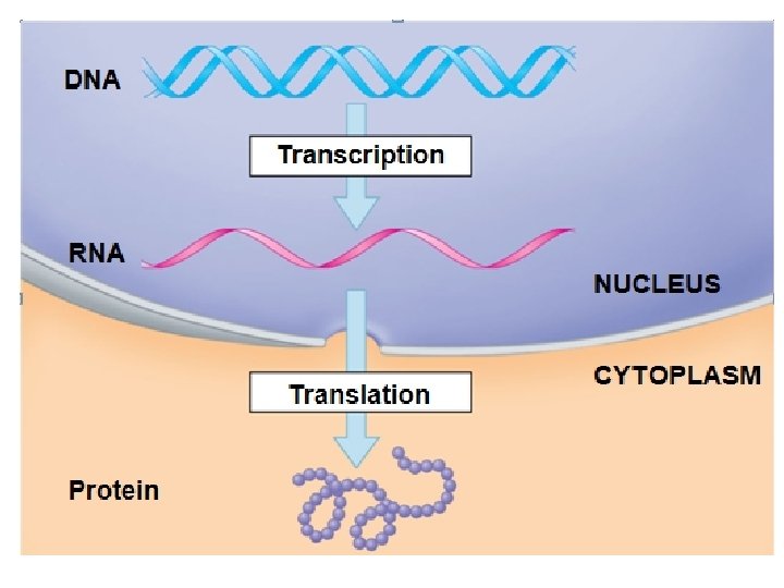
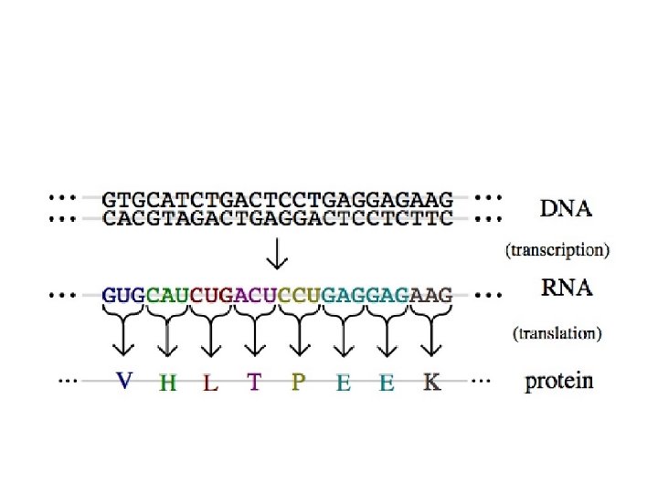
Gene expression… review
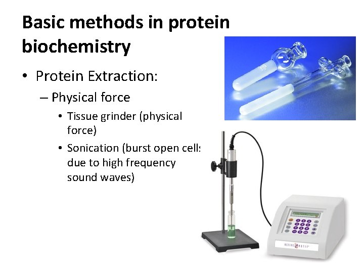
Basic methods in protein biochemistry • Protein Extraction: – Physical force • Tissue grinder (physical force) • Sonication (burst open cells due to high frequency sound waves)
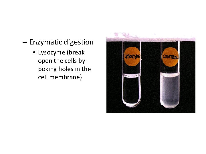
– Enzymatic digestion • Lysozyme (break open the cells by poking holes in the cell membrane)
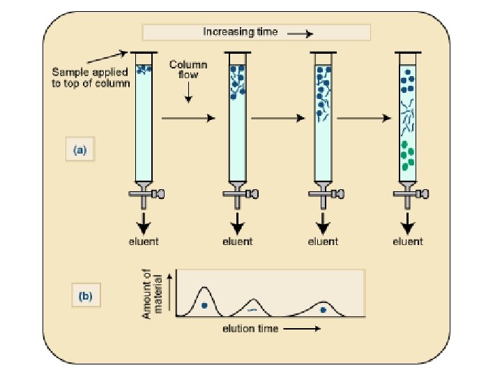
Protein separation – More than one level • Fractionate by organelle (mitochondria, Golgi, etc) (using centrifugation) • Fractionate by protein (size, charge, affinity) using chromatography: – – Size exclusion Ion exchange Affinity Hydrophobic interaction
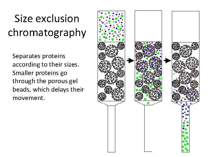
Size exclusion chromatography Separates proteins according to their sizes. Smaller proteins go through the porous gel beads, which delays their movement.
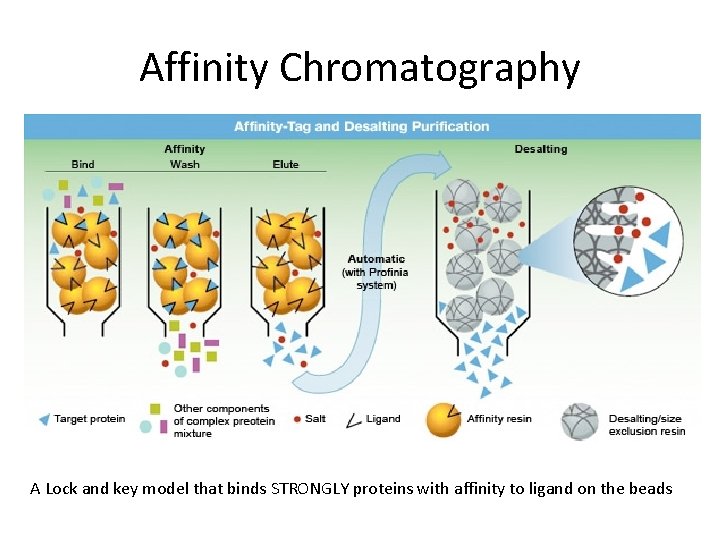
Affinity Chromatography A Lock and key model that binds STRONGLY proteins with affinity to ligand on the beads
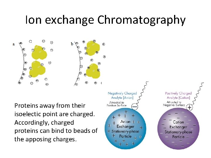
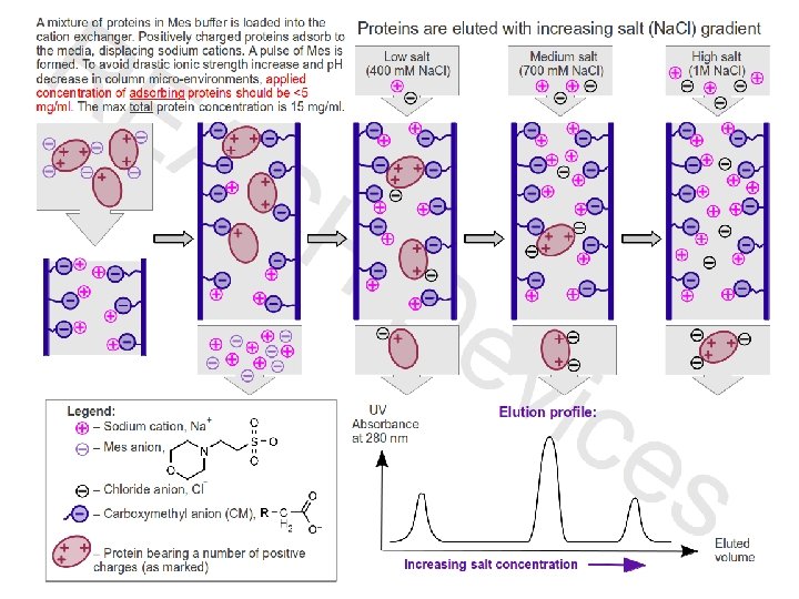
Ion exchange Chromatography Proteins away from their isoelectic point are charged. Accordingly, charged proteins can bind to beads of the apposing charges.
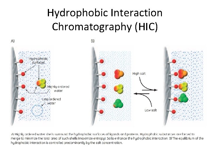
Hydrophobic Interaction Chromatography (HIC)
Analysis
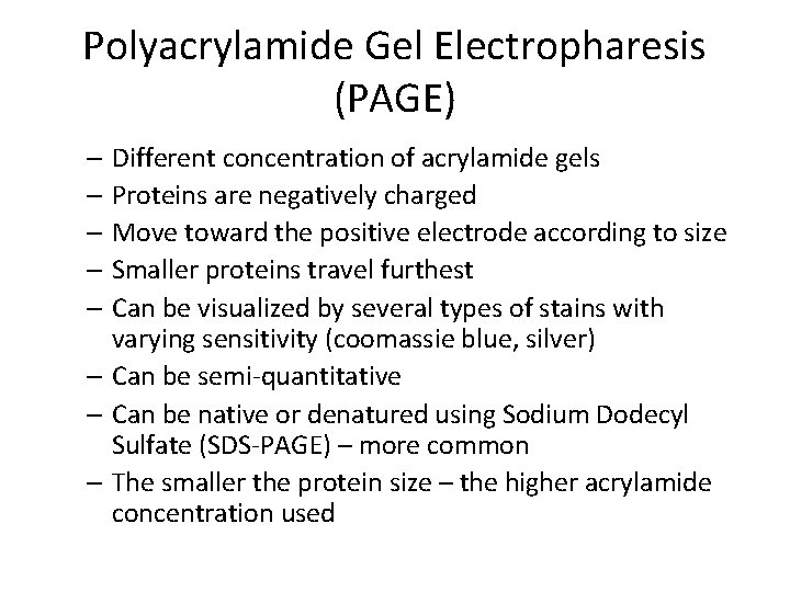
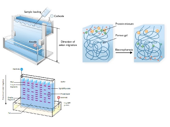
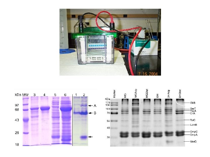
Polyacrylamide Gel Electropharesis (PAGE) – Different concentration of acrylamide gels – Proteins are negatively charged – Move toward the positive electrode according to size – Smaller proteins travel furthest – Can be visualized by several types of stains with varying sensitivity (coomassie blue, silver) – Can be semi-quantitative – Can be native or denatured using Sodium Dodecyl Sulfate (SDS-PAGE) – more common – The smaller the protein size – the higher acrylamide concentration used
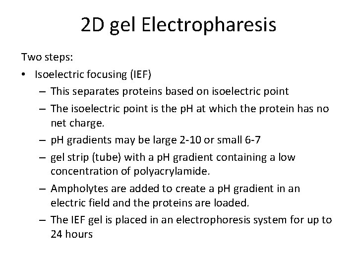
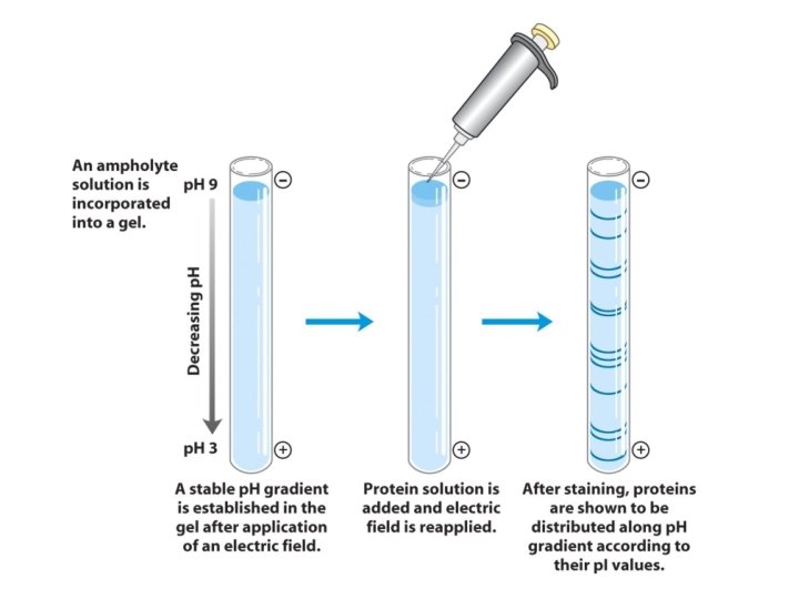
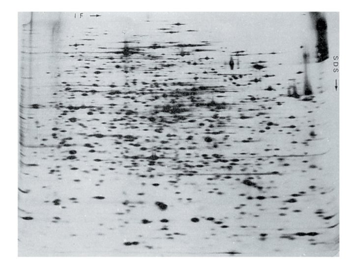
2 D gel Electropharesis Two steps: • Isoelectric focusing (IEF) – This separates proteins based on isoelectric point – The isoelectric point is the p. H at which the protein has no net charge. – p. H gradients may be large 2 -10 or small 6 -7 – gel strip (tube) with a p. H gradient containing a low concentration of polyacrylamide. – Ampholytes are added to create a p. H gradient in an electric field and the proteins are loaded. – The IEF gel is placed in an electrophoresis system for up to 24 hours
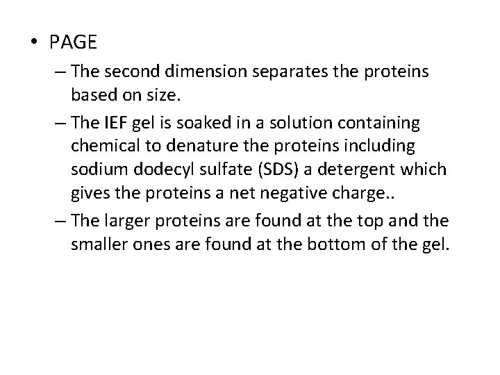
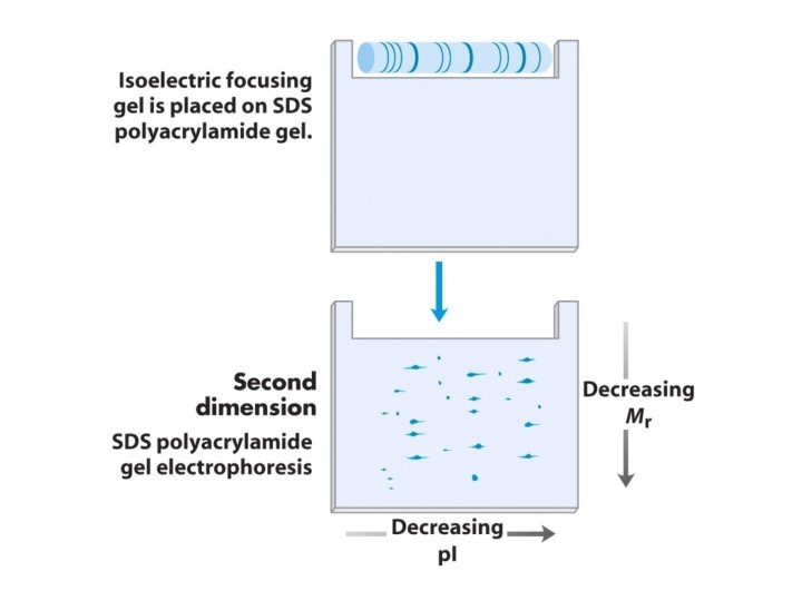
• PAGE – The second dimension separates the proteins based on size. – The IEF gel is soaked in a solution containing chemical to denature the proteins including sodium dodecyl sulfate (SDS) a detergent which gives the proteins a net negative charge. . – The larger proteins are found at the top and the smaller ones are found at the bottom of the gel.
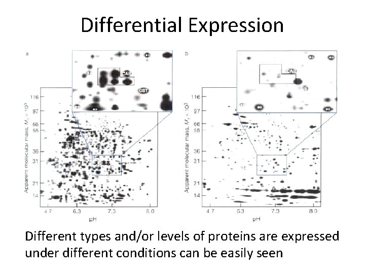
Differential Expression Different types and/or levels of proteins are expressed under different conditions can be easily seen
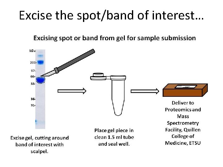
Excise the spot/band of interest…
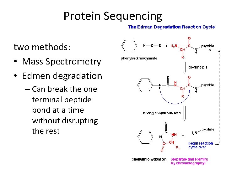
Protein Sequencing two methods: • Mass Spectrometry • Edmen degradation – Can break the one terminal peptide bond at a time without disrupting the rest
Mass Spectrometry http: //www. youtube. com/watch? v=t. OGM 2 g. OHKPc http: //www. youtube. com/watch? v=J-wao 0 O 0_q. M
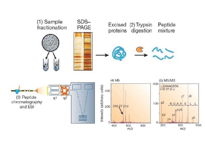
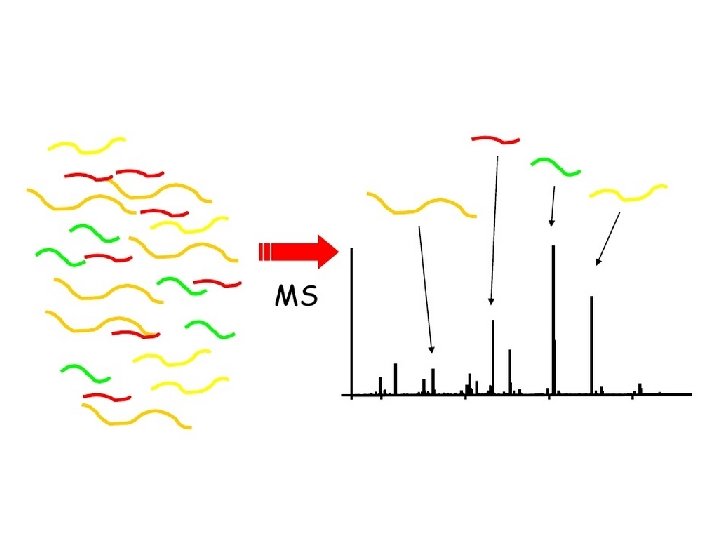
• Mass Spectrometry is a technique for the detection and resolution of a sample of ions by their mass-to-charge ratio represented by m/z where m is the mass in Daltons and z is the charge. • Samples are isolated, digested or not, then is analyzed to determine exact AA sequence MW.
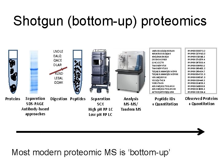
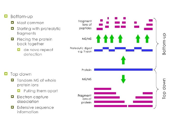
Shotgun (bottom-up) proteomics LNDLE EALQ QACE DLAR N KLND LEEAL QQAK Proteins Separation Digestion Peptides Separation SDS-PAGE SCX Antibody-based High p. H RP LC approaches Low p. H RP LC LNDLEEALQQAKEDLAR NKLNDLEEALQQAK NVQDAIADAEQR SKEEAEALYHSK SLVGLGGTK TAAENDFVTLKK TSQNSELNNMQDLVEDYKK VDLLNQEIEFLK YEELQVTVGR YLDGLTAER ADLEMQIESLTEELAYLKK AETECQNTEYQQLLDIK Analysis MS-MS/ Tandem MS Peptide IDs + Quantitation Most modern proteomic MS is ‘bottom-up’ IPI: IPI 00000073. 2 IPI: IPI 00217963. 3 IPI: IPI 00031065. 1 IPI: IPI 00376379. 4 IPI: IPI 00397801. 4 IPI: IPI 00009950. 1 IPI: IPI 00395488. 2 IPI: IPI 00295414. 7 IPI: IPI 00554711. 3 IPI: IPI 00009867. 3 IPI: IPI 00019449. 1 IPI: IPI 00016915. 1 IPI: IPI 00060800. 5 IPI: IPI 00013885. 1 IPI: IPI 00221224. 6 Observed Proteins + Quantitation
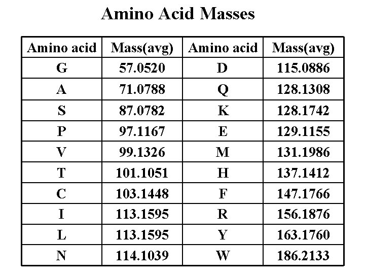
Amino Acid Masses Amino acid G Mass(avg) 57. 0520 Amino acid D Mass(avg) 115. 0886 A 71. 0788 Q 128. 1308 S P V T C I L N 87. 0782 97. 1167 99. 1326 101. 1051 103. 1448 113. 1595 114. 1039 K E M H F R Y W 128. 1742 129. 1155 131. 1986 137. 1412 147. 1766 156. 1876 163. 1760 186. 2133
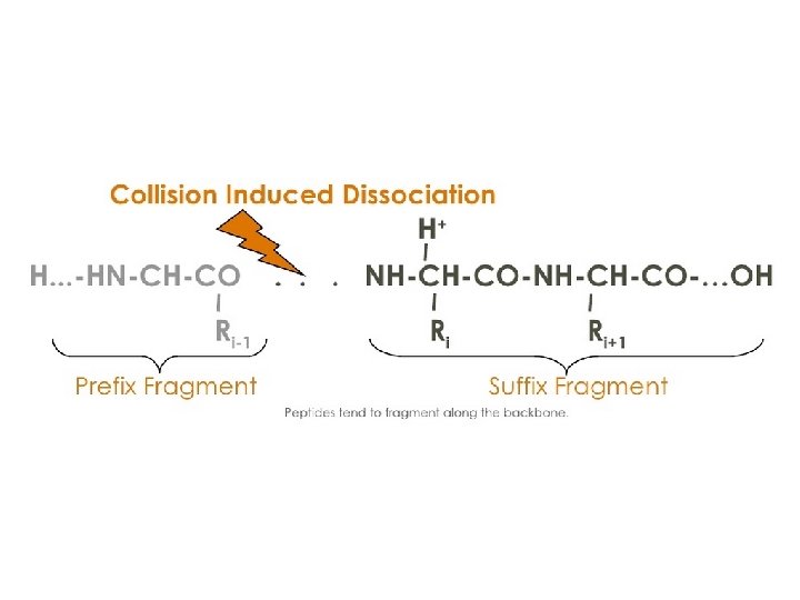
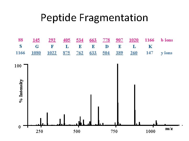
Peptide Fragmentation 88 S 1166 145 G 1080 292 F 1022 405 L 875 534 E 762 663 E 633 778 D 504 907 E 389 1020 L 260 1166 K 147 b ions y ions % Intensity 100 0 250 500 750 1000 m/z
Peptide mass fingerprinting • is an analytical technique for protein identification in which the unknown protein of interest. • The peptide masses are compared to either a database containing known protein sequences or even the genome. • The results are statistically analyzed to find the best match.
Microarrays
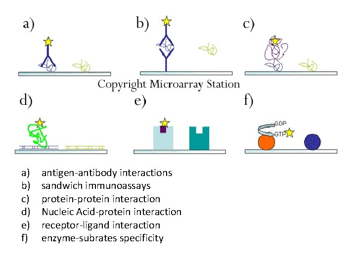
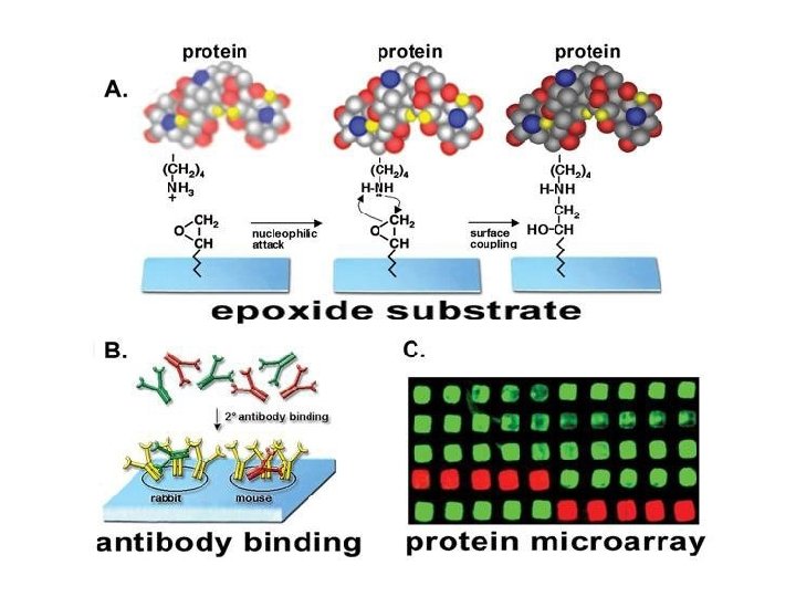
a) b) c) d) e) f) antigen-antibody interactions sandwich immunoassays protein-protein interaction Nucleic Acid-protein interaction receptor-ligand interaction enzyme-subrates specificity
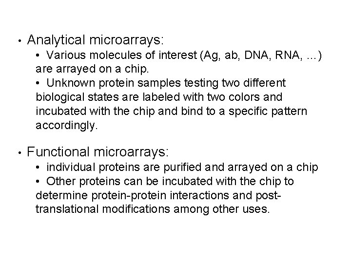
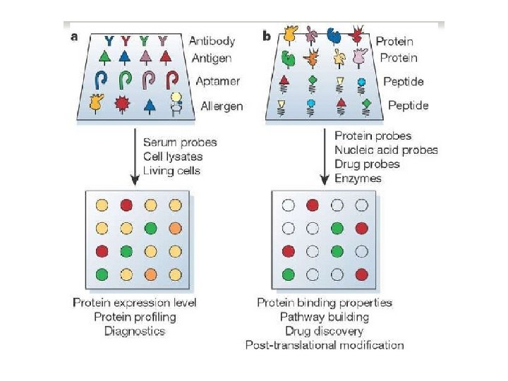
• Analytical microarrays: • Various molecules of interest (Ag, ab, DNA, RNA, …) are arrayed on a chip. • Unknown protein samples testing two different biological states are labeled with two colors and incubated with the chip and bind to a specific pattern accordingly. • Functional microarrays: • individual proteins are purified and arrayed on a chip • Other proteins can be incubated with the chip to determine protein-protein interactions and posttranslational modifications among other uses.
Why should we care?
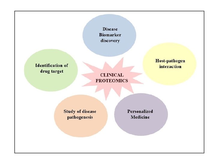
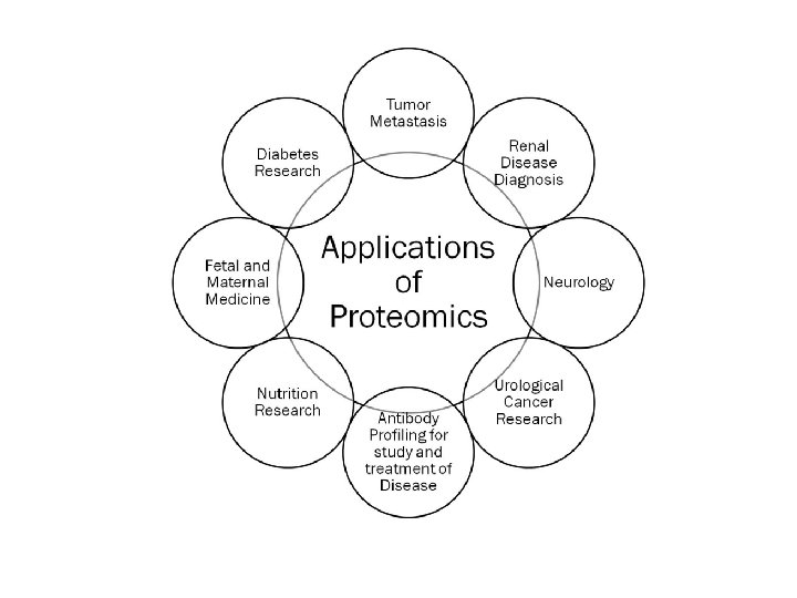
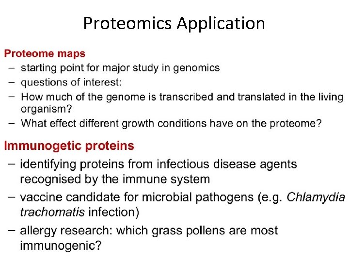
Proteomics Application
Assignment • Write one full page description of two clinical uses of Proteomics.
References All resources and links below were last accessed on 12/09/2014 • Mass Spectroscopy, Bhavana Vedantam. http: //www. slideshare. net/mnmnmknmjjk/mass-spectroscopy-28595894? related=2 • Proteomics, Dimitri Raptis. http: //www. slideshare. net/dimitriraptis/proteomics 6887316 • Proteomics 101, http: //www. slideshare. net/many 87/proteomics-101? related=1 • Application of Proteomics Workflows to Biomedical Research. Karen Jonscher. http: //www. ucdenver. edu/academics/colleges/medicalschool/programs/bioanalytic s/education/Documents/Lecture%201. pdf • Proteomics, Ruchi Yadav. http: //www. slideshare. net/ruchibioinfo/proteomics? related=2 • Proteomics Processes and Applications. Khalid Hakeem. http: //www. slideshare. net/khalidhakeem/proteomics-processes-andapplications? related=3 • Introduction to Proteomics. Phil Charles. http: //www. well. ox. ac. uk/dtc/bioinformatics_course/Introduction. To. Proteo mics. pptx • Proteomics PPT. http: //www. ndsu. edu/pubweb/~mcclean/plsc 411/proteomics%20 lecture%202 b. ppt