Introduction to Proteins Amino acids Proteins 1 o


 is defined as the four")


. •")
 • Many proteins or portions of proteins within the cell")
- Slides: 55
Introduction to Proteins • Amino acids • Proteins 1 o, 2 o, 3 o and 4 o structure • Proteins: Structural Parameters
Found in nature Lehninger ch. 3 Not used in nature
Generally found in the interior of proteins. G A V L M I P
F Interior Y W Surface/Interior
Generally found on the exterior of proteins. Note: I have moved proline from this group to the non-polar group. S T N C Q
Generally found on the exterior of proteins. Sometimes in the interior but then always combined with a negatively charged aa in order to form a salt bridge. H K R
Generally found on the exterior of proteins. Sometimes in the interior but then always combined with a positively charged aa in order to form a salt bridge. D E
Interior of the cell is reducing so cysteine is usually in the sulfhydryl form.
An example of a disulfide containing protein, the peptide hormone insulin. The active form of insulin is initially made from a single polypeptide (proinsulin) and cleaved into the final form. Upon cleavage it is excreted into the extracellular millieu where the cysteines are oxidized to form disulfide bridges.
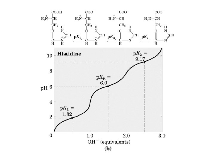
The ionization state of aa’s. Some aa’s can be protonated/deprotonated depending on the p. H. The midpoint of this process is called the p. K. You can ignore p. K 1 and p. K 2 since they don’t occur in proteins (except for the N and C termini respectively).
d a g b a d b g Proline has 2 conformers that are related by rotation about peptide bond. This occurs because the e-’s of the N form part of a s bond with the d carbon. The rotation is slow so that to distinct populations can be found in proteins. Trans is usually 90 -95%.
Formation of the peptide bond. Condensation reaction forms the bond and releases water. Resonance structures indicate a partial p structure for the N-C bond and therefore there is no rotation.
Y V S G A
Due to the partial double bond character of the N-C bond, peptide bonds are planar!
Electron distribution in a peptide bond. Calculated Determined from 0. 54Å structure.
Structure of the peptide bond Electron density of a high resolution xtal structure. The backbone can readily be seen. At right, the location of the backbone atoms in a peptide bond from the structure above. N, C, O, HN.
The levels of protein structure Molten globule
The dihedral angles of the protein backbone. Phi (f) is defined as the four atoms C(i-1) - >N(i) - Ca(i) - C(i). Psi (y) is composed of the four atoms N(i) - Ca(i) - C(i) >- N(i+1). The blue rectangles indicate the plane of the peptide bond.
The range of the dihedral angles is limited by steric clash.
Glycine Only right handed ahelices are observed in proteins. The Ramachandran plot
The Elements of Secondary Structure I. The a-Helix
C-terminus N-terminus Left vs Right Handed Helices in proteins are right handed.
2 In a-helices, the CO of residue I H -bonds to the HN of residues i+3 and i+4. Because all the HN’s and all the CO’s point in the same direction, an a-helix has a net dipole (electric charge). 1 3 5 4 H-bond
Helices have a straight axis. Here you are looking down the axis of a helix. Clearly the sidechains (represented by the purple balls) are on the OUTSIDE of the helix. A helix is not really hollow, the atoms are not shown with their real van der Waal’s radii for clarity.
CPK or space filling view of the same a-helix. So you see that the helix is essentially solid.
Side chain interactions within the a-helix. Just as the backbone of residues i and i+3 interact, so do the sidechains. Here you see a blue R forming a salt bridge with a red D. i+4 i
The Elements of Secondary Structure I. The b-Sheet
Most common! C-term N-term C-term Note: the arrows in Lehninger are wrong!
Less common. C-term Note: the arrows in Lehninger are wrong!
b-sheets are almost never flat! One continuous b-sheet that wraps around into a b-barrel.
Tertiary Structure Representations Ball and stick Kind of usefull. Shows where atoms are but gives misleading idea of protein density. Does not reveal 2 o structure.
Tertiary Structure Representations: bond The backbone bonds only are shown in these views of the E. coli membrane protein Omp. A. Missing portions of the crystal structure are highlighted by the blue balls. The secondary structure is obvious. Helix 10 Best NMR Structures of the protein Omp. A Crystal Structure
Tertiary Structure Representations: Ribbon/CPK Helix Sheet An extremely common view of the architecture of proteins. While not a good way of analyzing how a protein folds, the CPK view does give an accurate feel for how dense folded proteins are (note that there are no holes in the structure).
Tertiary Structure Representations: Surface with transparency and backbone bonds visible. Surface is colored according to electrostatic potential. Positive Negative
Structural Motifs • In proteins, a structural motif is a three-dimensional structural element or fold within the chain, which also appears in a variety of other proteins. The term is sometimes used interchangeably with "structural domain, " although a domain need not be a motif nor, if it contains a motif, need not be made up of only one. • Structural alignment is a major method for discovering significant structural motifs. • Motifs exhibit both tertiary and secondary structure, and may be regarded as a configuration of secondary structures. Such a description is the basis for many of the names that structural biologists give to particular kinds, such as the helix-turnhelix motif. This is not always true, however, as in the case of the EF-hand. • Because the relationship between primary structure and tertiary structure is not straightforward, two biopolymers may share the same motif yet lack appreciable primary structure similarity. Modified from the Wikipedia.
A few examples of common 3 o structural motifs H H T Helix-turn-Helix: a basic nucleic acid binding structure. This motif (green on left) and the exact relationship between the helices is conserved from bacteria to man.
The helical bundle. membrane This arrangement of hydrophobic and hydrophyllic interfaces is for a soluble protein. 7 Transmembrane helical bundle A GPCR http: //swissmodel. expasy. org/course/text/chapter 4. htm
Some 4 helix bundle proteins Cytokines: secreted proteins that regulate cellular function.
Helix-Helix Interactions q q = 260 Helix Packing The Leucine “Zipper”
b-sheets Orthogonal – 1 sheet folded back onto itself
The b-barrel Green Fluorescent Protein
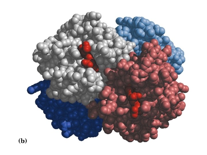
Haemoglobin-An example of quaternary structure i. e. complex formation by multiple subunits.
Protein domains Pairwise sequence comparison of proteins led to strange results • A domain is an independent folding unit • A domain is the next step up in complexity from a motif • There appear to be a limited number of folds (domains) that can be made from the 20 natural aa’s • Domain unit of evolution • Mixing and matching can create new function and regulation • Most proteins involved in cell signalling consist exclusively of small domains interspersed by linker regions. The linkers may be unstructured as described in the following section.
How proteins are made from domains. Some proteins consist only of domains that have no enzymatic activity. It is thought that they function as scaffolds for specific complex formation. SH 3 GRB 2 BRCT domains are a good example of divergent evolution. An ancient domain found in pro- and eukaryotes, it is characterised by a conserved fold despite significant sequence divergence. BRCTs are known to bind DNA and other proteins. Protein-protein interactions included self binding, binding BRCTs on other proteins, binding non. BRCT domains and binding to phoserine peptides.
Determining Domain Structure by Limited Proteolysis
Protein regulation by coordinated action of domains Having multiple domains in one protein can serve a variety of functions, one of which is illustrated here. The kinases, Src, Lck and Hck, all of which can cause aberrent growth signalling, are regulated by an internal Y phophorylation. When Y 527 is phosphorylated, SH 2 and SH 3 are “locked”, forcing lobes of kinase down and blocking access to the active site. Young et al. , 2001, Cell, v. 105, p. 115
Not all proteins are structured: Intrinsically Unstructured Proteins What are unstructured proteins? Proteins (segments of proteins) that are lacking wellstructured 3 -dimentional fold. They are referred as “natively denatured/unfolded”, “intrinsically unstructured/unfolded”. Why are they relatively obscure? Our view of protein universe was strongly determined by the tools we had: X-ray crystallography will not “see” such proteins, as they difficult to crystallize. How prevalent are unstructured proteins? About 35 -51% of the proteins have unstructured regions that are longer than 50 residues; 6 -17% of proteins in the Swiss-Prot are probably fully disordered. Determined by neural networks predictors (based on the protein sequence). This section of the lecture is not supported by any textbooks since it contains very new information.
What determines if the protein will be folded or unfolded? There is a sequence signature that describes unfolded regions. Signature: • low sequence complexity • bias toward polar and charged amino acids (Gln, Ser, Pro, Glu, Lys, and occasionally Ala and Pro) • bias away from bulky hydrophobic residues (Val, Leu, Met, Phe, Trp, Tyr) An array of programs are available now to predict disordered regions: PONDR (Dunker’s group) Fold. Index (Uversky’s group) Dis. EMBL (Gibson’s group) GLOBPLOT (Gibson’s group) DISOPRED (David Jones’s group) IUPred (Tompa’s group)
A continuum of protein structures Dyson and Wright, “Intrinsically unstructured proteins and their functions” (2005) Nature Review Molecular Cell Biology 6: 197 -208
Coupling of folding to target binding KID domain of CREB p. KID bound to KIX domain of CBP (CREB binding protein). Predicted a-helices in free peptide Experimentally determined a-helices in complex • Can provide tighter binding than similar sized, folded proteins. • Enthalpy-Entropy compensation. • Allows post-translational modification.
Unstructured proteins can adopt multiple structures upon target binding- they are “plastic” Hif 1 a peptide bound to the TAZ 1 domain of CBP. Here the peptide forms an a-helix. Hif 1 a peptide bound to asparagine hydroxylase. Here the peptide binds in an extended conformation.
Take-Home Lessons • Proteins are polymers of 20 naturally occurring, L-amino acids (aa). • The sequence of aa’s defines the structure and hence function, of a protein. • The aa’s can be divided into hydrophobic, polar and charged groups depending on the sidechain chemistry. This defines where in the 3 D protein structure a given aa is likely to be found. • Because of the sidechain, the rotation around the backbone bonds, defined by the dihedral angles f and y, is hindered with certain values being preferred. • Proteins fold into different levels of structure referred to as secondary through quaternary. You should know what each refers to. • Large proteins generally do not consist of one large structure but multiple, independently folding domains that not only provide specific functions, but interact to add a further level of regulation to protein function.
Take Home Lessons (cont) • Many proteins or portions of proteins within the cell are intentionally disordered.