
INTRODUCTION TO PHYSIOLOGY Kim Baily Ph D RN
INTRODUCTION TO PHYSIOLOGY Kim Baily Ph. D, RN.
Learning Objectives • By the end of the webinar, the learner will be able to: • Define the concepts of homeostasis, wellness and pathology. • List the main physiological systems of the body. • Describe the major components of the cardiovascular system.
Why Physiology? • The human body may be viewed as a system with constantly changing inputs and outputs. When existing systems fail to maintain body functions within normal limits, clinical signs and symptoms appear and the patient may become ill. • Healthcare professionals (HCPs) are trained to recognize, interpret and respond to these changes in a way that helps bring the system back into balance. • Simulation technologists manipulate the simulation environment to mimic clinical situations. In so doing, sim techs provide an opportunity for HCPs to practice and improve their clinical skills in a supportive and safe learning environment.
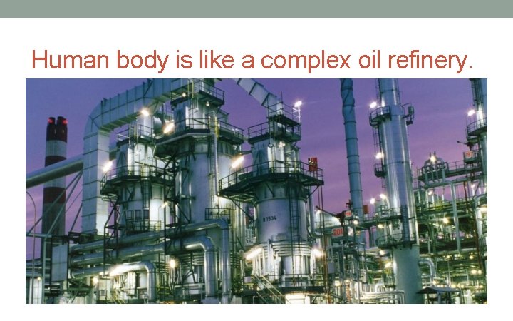
Human body is like a complex oil refinery.
The work of the body • A trillion cells each with their own work to do • Work includes: • Chemical work • Transport work • Mechanical • All work requires fuel (energy) • Waste products need to be eliminated
Homeostasis • Regulation of the body’s internal environment • Keeping internal environment within set limits • Interaction between • Outside world • Fluid surrounding cells (Extracellular fluid) • Fluid inside cells (Intracellular fluid) • Dynamic steady state • Not the same as equilibrium
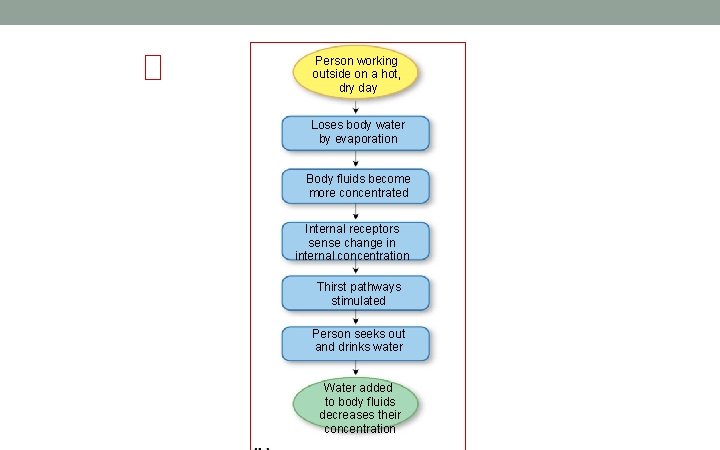
Person working outside on a hot, dry day Loses body water by evaporation Body fluids become more concentrated Internal receptors sense change in internal concentration Thirst pathways stimulated Person seeks out and drinks water Water added to body fluids decreases their concentration
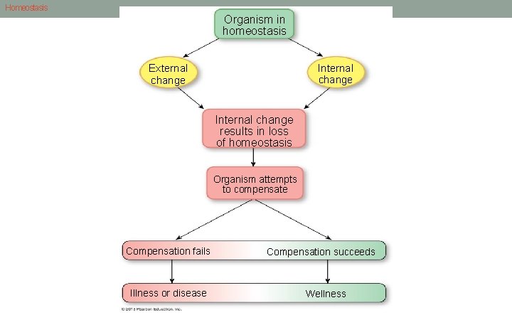
Homeostasis Organism in homeostasis Internal change External change Internal change results in loss of homeostasis Organism attempts to compensate Compensation fails Illness or disease Compensation succeeds Wellness
Control Systems and Homeostasis • Regulated variables are kept within normal range by control mechanisms • Keeps near set point, or optimum value • Control systems – local and reflex • Input signal • Integrating center • Output signal
Physiology • The study of the normal functioning of a living organism and its component parts, including all its chemical and physical processes. • Pathophysiology • The physiology of abnormal states • What happens if a particular process malfunctions • What signals does the body send out • Simulation is all about homeostasis, physiology, and pathophysiology
Levels of Organization • Organization of life • The cell is the smallest unit of structure capable of carrying out all life processes • Atoms, molecules, cells, tissues, organ systems, and organisms • Molecules • Lipids • Carbohydrates • Proteins • Nucleotides
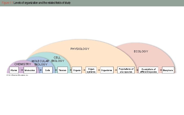
Figure 1. 1 Levels of organization and the related fields of study PHYSIOLOGY ECOLOGY CELL MOLECULAR BIOLOGY CHEMISTRY BIOLOGY Atoms Molecules Cells Tissues Organ systems Organisms Populations of one species Ecosystem of different species Biosphere
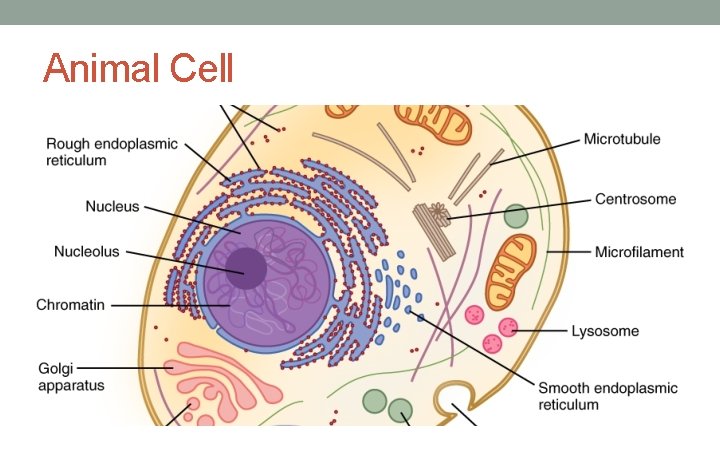
Animal Cell
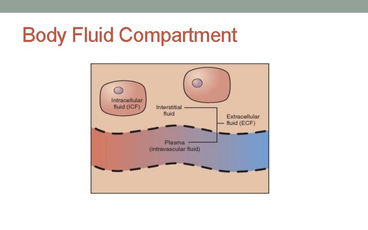
Body Fluid Compartment
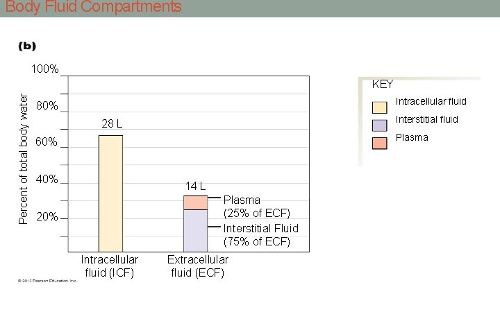
Body Fluid Compartments 100% Percent of total body water KEY Intracellular fluid 80% Interstitial fluid 28 L Plasma 60% 40% 14 L Plasma (25% of ECF) Interstitial Fluid (75% of ECF) 20% Intracellular fluid (ICF) Extracellular fluid (ECF)
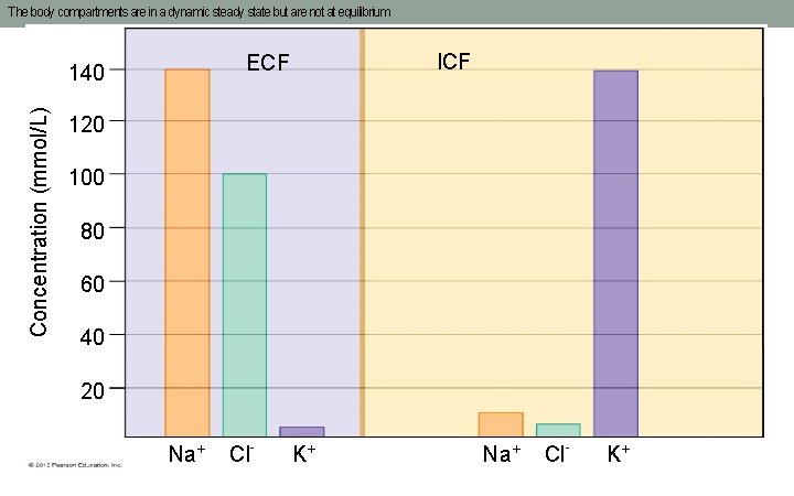
The body compartments are in a dynamic steady state but are not at equilibrium Concentration (mmol/L) ICF ECF 140 120 100 80 60 40 20 Na+ Cl- K+
Measuring Ions, Molecules and Concentrations • Can only measure extracellular concentrations • Lab results e. g. • Sodium, K, Ca, Cl, CO 2, concentration (osmolarity) BUN • Creatinine • Liver enzymes • Cardiac enzymes (proteins) released from damaged cardiac cells following a heart attack. • Thyroid hormones
Simulation • Simulation often requires HCPs to check lab results to indicate the fluid and electrolyte status of the patient • Specific imbalances cause distinctive changes in the patient which a student should be able to pick up from physical examination and lab data. • Simulation scenarios should always include lab results when appropriate.
Tissue types, organs and systems • Tissues types • Epithelial • Connective • Muscle • Neural/nerve • Tissue remodeling • Organs • Kidney, liver etc
Organ Systems – Think Homeostasis! • Musculoskeletal • Respiratory • Digestive • Urinary • Reproductive • Circulatory • Nervous • Endocrine • Immune © 2013 Pearson Education, Inc.
Cardiovascular System Function • Transport • Nutrients • Oxygen • Removal of waste products • Communication • Glucose + Oxygen = Energy + CO 2 • All cells need fuel • Without oxygen will die
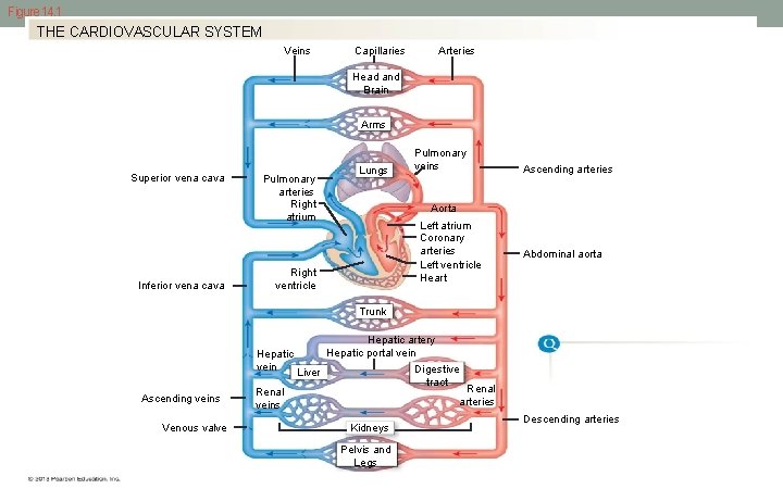
Figure 14. 1 THE CARDIOVASCULAR SYSTEM Veins Capillaries Arteries Head and Brain Arms Superior vena cava Inferior vena cava Pulmonary arteries Right atrium Lungs Pulmonary veins Ascending arteries Aorta Left atrium Coronary arteries Left ventricle Heart Right ventricle Abdominal aorta Trunk Hepatic vein Ascending veins Venous valve Renal veins Hepatic artery Hepatic portal vein Digestive Liver tract Renal arteries Kidneys Pelvis and Legs Descending arteries
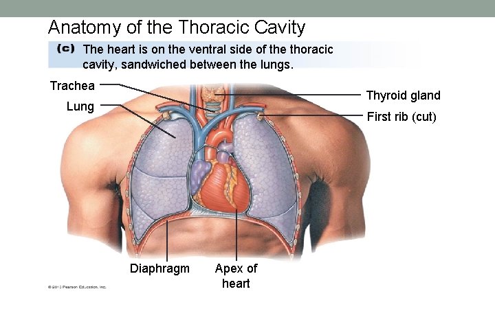
Anatomy of the Thoracic Cavity The heart is on the ventral side of the thoracic cavity, sandwiched between the lungs. Trachea Thyroid gland Lung First rib (cut) Diaphragm Apex of heart
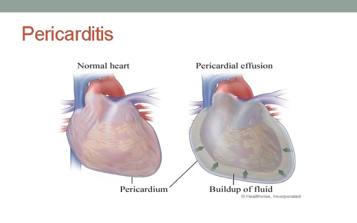
Pericarditis
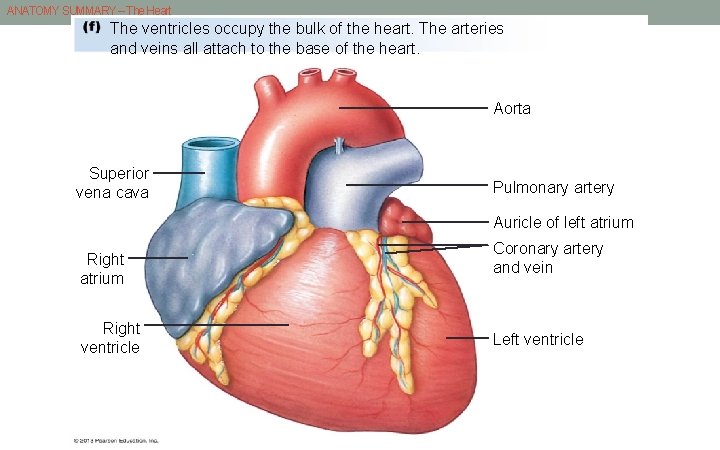
ANATOMY SUMMARY – The Heart The ventricles occupy the bulk of the heart. The arteries and veins all attach to the base of the heart. Aorta Superior vena cava Pulmonary artery Auricle of left atrium Right ventricle Coronary artery and vein Left ventricle
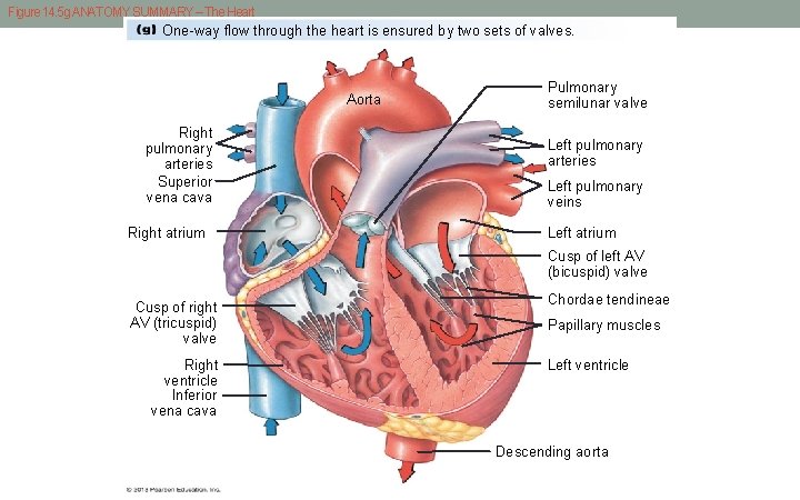
Figure 14. 5 g ANATOMY SUMMARY – The Heart One-way flow through the heart is ensured by two sets of valves. Aorta Right pulmonary arteries Superior vena cava Right atrium Pulmonary semilunar valve Left pulmonary arteries Left pulmonary veins Left atrium Cusp of left AV (bicuspid) valve Cusp of right AV (tricuspid) valve Right ventricle Inferior vena cava Chordae tendineae Papillary muscles Left ventricle Descending aorta
Blood flow in response to pressure gradient • Diastole – heart relaxed and filling with blood • Systole (heart contracted) • Atria contract first • Slight pause • Ventricles contract • Push blood into arteries which are elastic and expand • Artery has elastic recoil arteries contract. • Pressure wave created which moves around circulation • Pulse
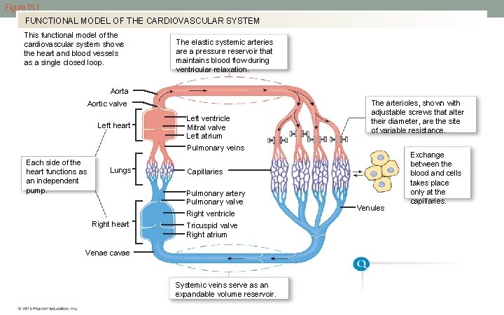
Figure 15. 1 FUNCTIONAL MODEL OF THE CARDIOVASCULAR SYSTEM This functional model of the cardiovascular system shows the heart and blood vessels as a single closed loop. The elastic systemic arteries are a pressure reservoir that maintains blood flow during ventricular relaxation. Aorta Aortic valve Left heart Left ventricle Mitral valve Left atrium The arterioles, shown with adjustable screws that alter their diameter, are the site of variable resistance. Pulmonary veins Each side of the heart functions as an independent pump. Lungs Capillaries Pulmonary artery Pulmonary valve Right ventricle Right heart Tricuspid valve Right atrium Venae cavae Systemic veins serve as an expandable volume reservoir. Venules Exchange between the blood and cells takes place only at the capillaries.
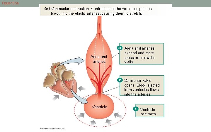
Figure 15. 5 a Ventricular contraction. Contraction of the ventricles pushes blood into the elastic arteries, causing them to stretch. Aorta and arteries expand store pressure in elastic walls. Semilunar valve opens. Blood ejected from ventricles flows into the arteries. Ventricle contracts.
Blood Pressure • Highest pressure in aorta during systole (120 mm. Hg) • Lower number because of elasticity of aorta and large blood vessel (80 mm. Hg) • Note this is not the pressure inside the ventricles which is typically very low.
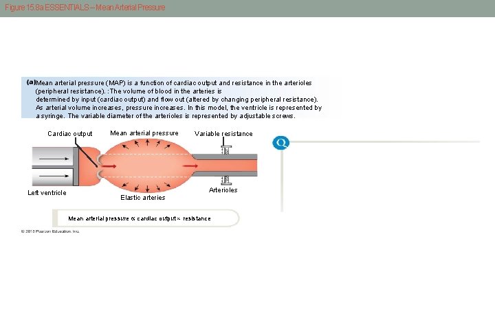
Figure 15. 8 a ESSENTIALS – Mean Arterial Pressure Mean arterial pressure (MAP) is a function of cardiac output and resistance in the arterioles (peripheral resistance). : The volume of blood in the arteries is determined by input (cardiac output) and flow out (altered by changing peripheral resistance). As arterial volume increases, pressure increases. In this model, the ventricle is represented by a syringe. The variable diameter of the arterioles is represented by adjustable screws. Cardiac output Left ventricle Mean arterial pressure Variable resistance Arterioles Elastic arteries Mean arterial pressure cardiac output resistance
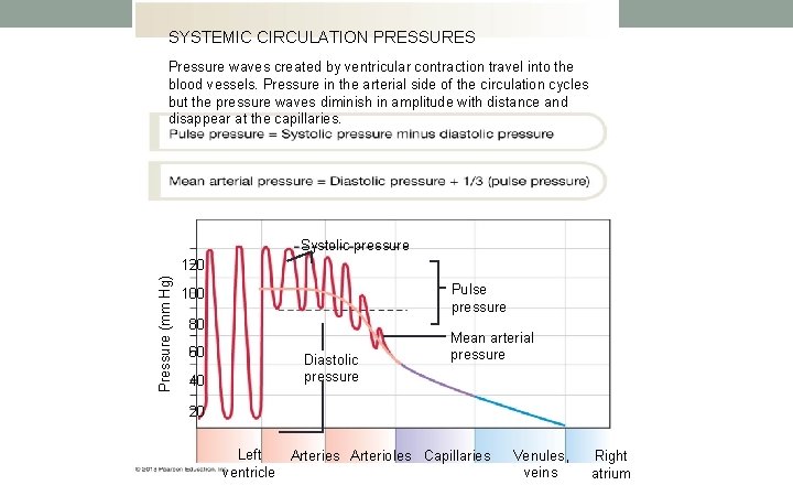
SYSTEMIC CIRCULATION PRESSURES Pressure waves created by ventricular contraction travel into the blood vessels. Pressure in the arterial side of the circulation cycles but the pressure waves diminish in amplitude with distance and disappear at the capillaries. Systolic pressure Pressure (mm Hg) 120 Pulse pressure 100 80 60 40 Diastolic pressure Mean arterial pressure 20 Left Arteries Arterioles Capillaries ventricle Venules, veins Right atrium
Blood return to the heart • Muscle pump • Respiratory Pump • Valves in veins ensure one-way flow in veins
Blood flow and resistance • Blood flow around the circulation is opposed by resistance • The Higher the resistance, the less the flow • Factors affecting blood flow • Length – usually a constant for an individual • One pound of fat add seven miles of blood vessels! • Viscosity - usually constant • Polycythemia • Radius of blood vessel • Narrower the blood vessel, higher resistance, less the blood flow (vasoconstriction) • Dilated as in allergic shock – blood pressure drops
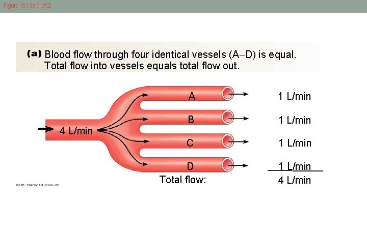
Figure 15. 13 a (1 of 2) Blood flow through four identical vessels (A D) is equal. Total flow into vessels equals total flow out. A 1 L/min B 1 L/min C 1 L/min 4 L/min D Total flow: 1 L/min 4 L/min
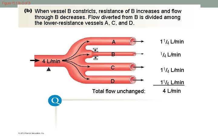
Figure 15. 13 b (2 of 2) When vessel B constricts, resistance of B increases and flow through B decreases. Flow diverted from B is divided among the lower-resistance vessels A, C, and D. A 1/ 1 L/min 4 B 1/ L/min 4 4 L/min C D Total flow unchanged: 1/ 1 L/min 4 4 L/min
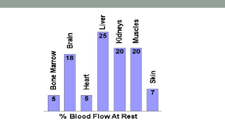
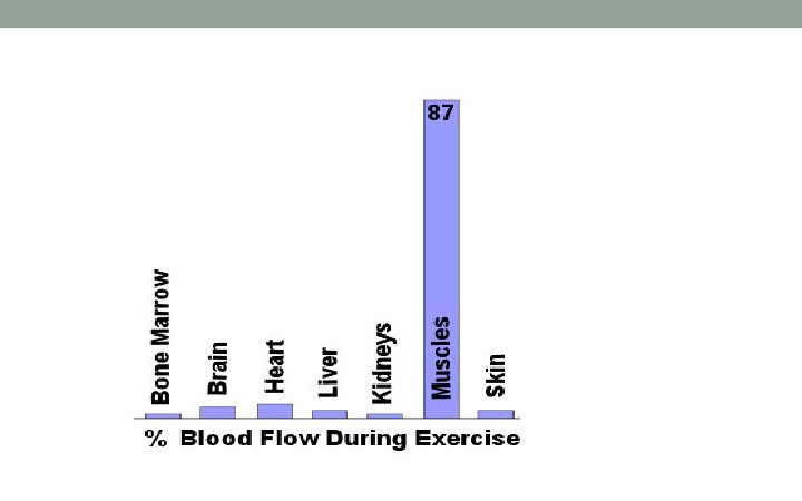
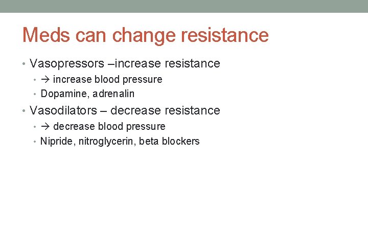
Meds can change resistance • Vasopressors –increase resistance • increase blood pressure • Dopamine, adrenalin • Vasodilators – decrease resistance • decrease blood pressure • Nipride, nitroglycerin, beta blockers
Simulation Technologists Rock! • ST create environmental and physiological fidelity which is critical for maximum HCP learning. • Without fidelity, learners can become confused and defensive. • Physiology is the bases of simulation – the more you know the more you can fully engage in scenario development, implementation and evaluation.
- Slides: 40