
Introduction to pathology lecture 1 Fatima Obeidat MD
Introduction to pathology lecture 1 Fatima Obeidat, MD
Mechanisms of cell Injury
Depletion of ATP • Consequences

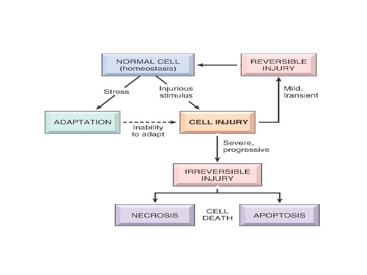
• Cells normally maintain a steady state called Homeostasis. • This means the intracellular environment is kept within a narrow range of physiologic state • Examples: temperature, p. H, chemical reactions, electrolyte concentration, water content… All are regulated and kept constant.
Cells and stress! • If cells are subjected to stresses ( like changes in electrolyte balance) then cells adapt and reach a new homeostatic state that will preserve cell function. • However if the stress is more severe and is beyond capability of adaptation then this will result in cell injury.
• Cell injury: at the beginning it is reversible within certain limits • Then it becomes irreversible. • Irreversible injury ends in cell death. • Two types of cell death: necrosis and apoptosis ( more details later)
Note • Not all injuries start as reversible then become irreversible • If the injury is severe enough it will be irreversible even in the early stages.
Adaptation in cells • Adaptation means: reversible changes in the a. number b. size c. phenotype, d. metabolic activity or e. function of cells in response to changes in their environment.
Adaptation • Adaptive changes are reversible. : if the insult is removed, things can go back to normal. • Can be physiologic or pathologic; depending on the cause

Cells and tissues adapt by four main mechanisms • • Hyertrophy Hyperplasia Atrophy Metaplasia
• Hypertrophy: Increased cell size. • Hyperplasia: increased number of cells occurs in labile cells, the cells that undergo. . Cell division. • Metaplasia: change from one adult cell type to another • Atrophy: decreased size.

Hypertrophy versus hyperplasia

Hypertrophy • Increased cell size. • Due to increased organelles and proteins. • Increased intracellular synthesis of the proteins and organelles. Caused by: increased demands, hormones or growth factors.

Examples of Physiologic hypertrophy a. Uterus during pregnancy… due to oestrogen effect on smooth muscles of the uterine wall. b. Skeletal muscle in body builders… due to increased demand. • Note: smooth muscles can divide. . So in the pregnant uterus there is hypertrophy and hyperplasia… whereas skeletal muscle is permanent tissue that can not divide, so there is pure hypertrophy without hyperplasia.

Physiologic hypertrophy Uterine wall
Pathologic hypertrophy • Cardiac enlargement due to hypertension = Hypertensive heart disease • Pathogenesis. . Two types of signals: mechanical: stretch and trophic: growth factors androgenic hormones cause the hypertrophy in the cardiac muscle • Note: the cardiac muscle can not undergo hyperplasia because it is a permanent cell.
• Cardiac hypertrophy during hypertension results in more contractile force that is needed to meet the demands of the increased pressure • This works to a certain extent. . But with time hypertrophy can not compensate for the increased burden and degenerate changes occur in the muscle.
• The myofibrllar contractile elements will be fragmented • Why hypertrophy can not compensate forever? ? ? • Maybe because there is limited blood supply and limited mitochondria to provide ATP. . So if there is more tissue than that can be supported by this blood supply and energy then degeneration occurs.

Pathologic hypertrophy

Pathologic hypertrophy
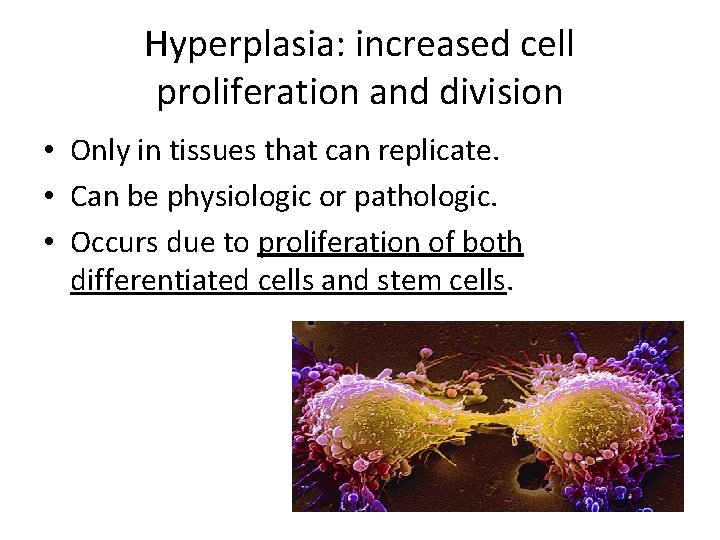
Hyperplasia: increased cell proliferation and division • Only in tissues that can replicate. • Can be physiologic or pathologic. • Occurs due to proliferation of both differentiated cells and stem cells.
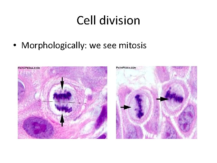
Cell division • Morphologically: we see mitosis

Physiologic hyperplasia • Hormonal: uterus, breast. • Compensatory: after removal or loss of part of tissue such as liver in transplantation. - In liver transplantation, after part of the liver is excised , the mitotic activity will start after 12 hours eventually restoring the normal size of the liver
Pathologic hyperplasia • Due to excess in hormones or growth factors. • E: g endometrial hyperplasia due to oestrogenprogesterone imbalance. • Hyperplasia is controlled. . it responds to normal growth stimuli and inhibitors. This differentiates it from cancer because cancer keeps growing regardless of normal growth mechanisms.
Note: However. . Because hyperplastic tissue divides there is more chance that it will acquire mutations that can lead to cancer. -This occurs more in pathologic hyperplasia, because the stimuli are long lived
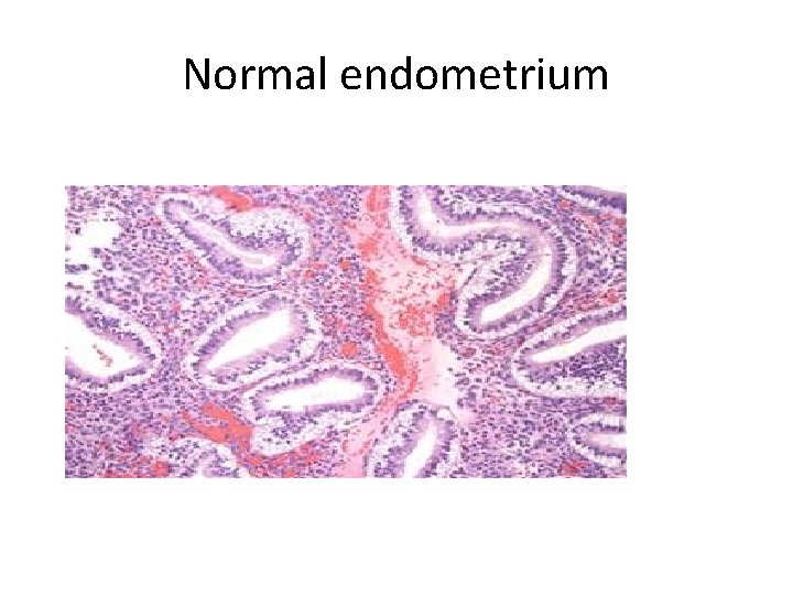
Normal endometrium
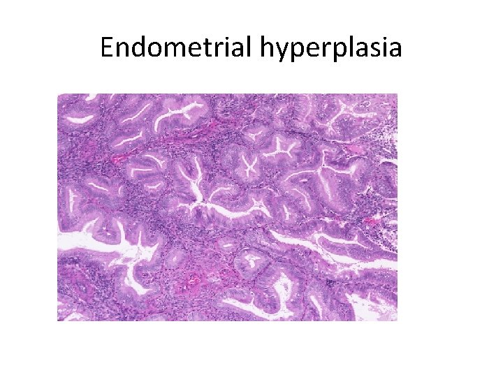
Endometrial hyperplasia

atrophy • Shrinkage in cell size due to loss of cell substance. Causes • Decreased work load. • Loss of innervation • Loss of endocrine stimulation. • Decrease blood supply • Aging
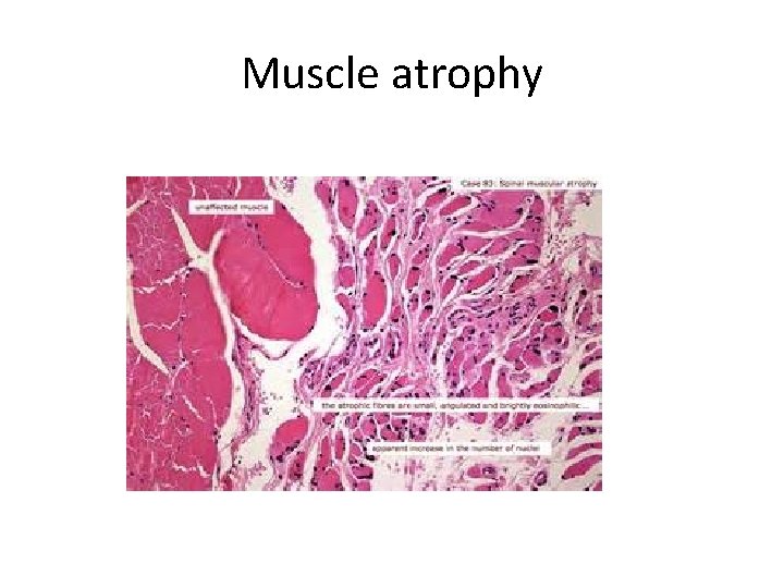
Muscle atrophy

Brain atrophy
Atrophy • Physiologic: endometrial atrophy during menopause • Pathologic: loss of innervation such as in poliomyelitis.
atrophy Mechanisms: • Decreased protein synthesis. • Degradation of cellular proteins, mainly by ubiquitin- proteasome pathway. . Proteins are attached to ubiquitin which targets them to be degraded by proteasome. • Autophagy…. Literally means self eating.
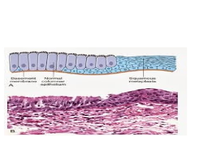
metaplasia • Adult cell type replaced by another adult cell type. • The cell type sensitive to a certain stress changes to another type which can better tolerate this particular stress. • Arise due to reprogramming of stem cells to differentiate along a new pathway.
Epithelial metaplasia/ example • Respiratory epithelium which is normally glandular, if exposed to smoking it changes to squamous epithelium, which can cope better with the smoke • However, this change means loss of the goblet cells that are present in the columnar glandular epithelium. . So no mucus secretion • Also there will be loss of the cilia. . So there is loss of important protective in metaplastic tissue.
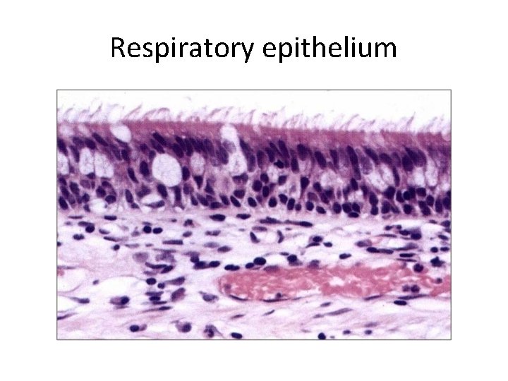
Respiratory epithelium
Barrett mucosa • This is another example of metaplasia • Normal esophageal mucosa is stratified squamous • If there is reflux of gastric content into the stomach, the esophageal epithelium changes to glandular. . So it can withstand the acidity.
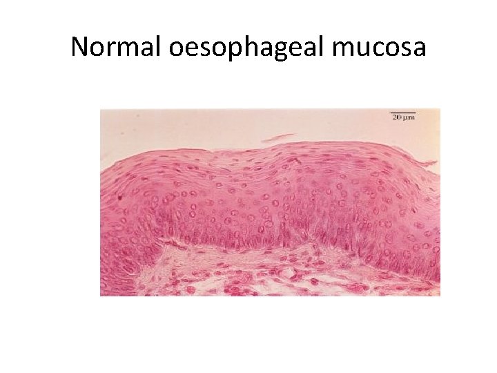
Normal oesophageal mucosa
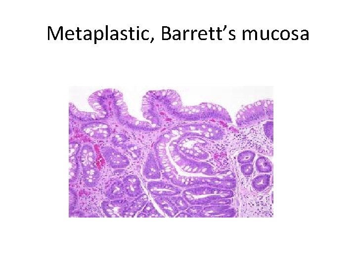
Metaplastic, Barrett’s mucosa
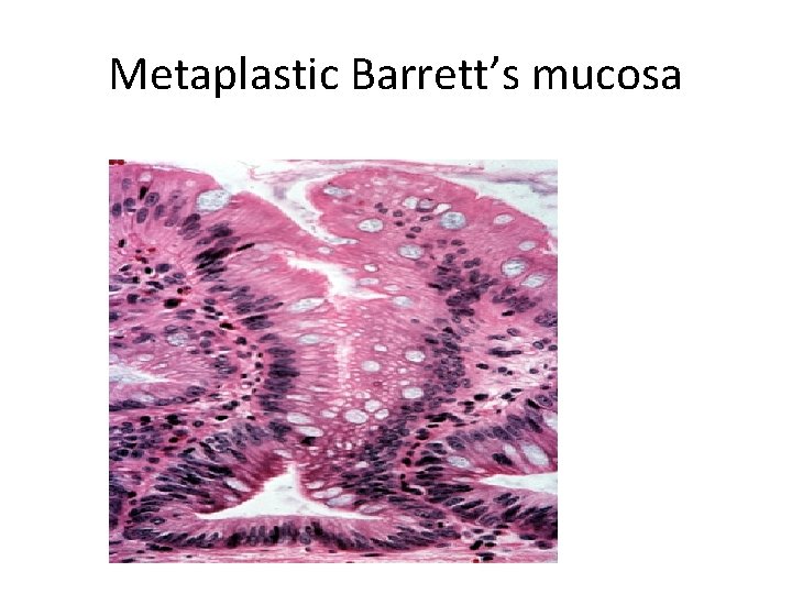
Metaplastic Barrett’s mucosa
Metaplasia in mesenchymal tissue • Usually pathologic • Ossification of soft tissue due to injury.
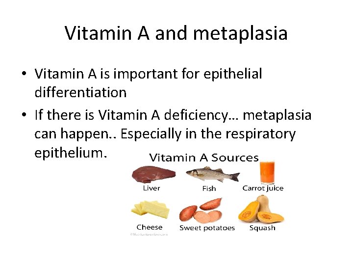
Vitamin A and metaplasia • Vitamin A is important for epithelial differentiation • If there is Vitamin A deficiency… metaplasia can happen. . Especially in the respiratory epithelium.
Metaplasia and cancer • The stimuli that cause metaplasia if persist for a long time can cause cancer • So metaplasia is considered premalignant.

Thank You
- Slides: 46