INTRODUCTION TO PATHOLOGY HISTOPATHOLOGY TECHNIQUES DR EKBAL ISKANDER

INTRODUCTION TO PATHOLOGY & HISTOPATHOLOGY TECHNIQUES DR. EKBAL ISKANDER

WHAT IS THE DISEASE? Disease may be defined as : an abnormal alteration of structure or function in any part of the body.
 ! –")
DEFINITION OF PATHOLOGY Pathology is the study of suffering (Latin word ) ! – Logos = study – Pathos = suffering It is the “Scientific study of disease" . "scientific study of the molecular, cellular, tissue, or organ system response to injurious agents. "

Pathology serves as a "bridge" or "link" between the preclinical sciences (anatomy, physiology, ……etc. ) and the courses in clinical medicine.

MEDICAL KNOWLEDGE: § § § § Epidemiology – Where & When. . Etiology – What is the cause? Pathogenesis - Evolution of dis. Morphology - Structural Changes Clinical Significance – relate. . Management Prognosis Prevention

PATHOLOGY FOCUSES ON 4 ASPECTS OF DISEASE: l ETIOLOGY: Cause of disease. l PATHOGENESIS: Mechanisms of development of disease. l MORPHOLOGY: The structural alterations induced in cell and tissues. l FUNCTIONAL CONSEQUENCES: Functional results of the morphologic changes, as observed clinically.

LEARNING PATHOLOGY: * General Pathology Common changes in all tissues. E. g. . Inflammation, cancer, ageing. * Systemic Pathology Specific changes in organs. E. g. . Goiter, pneumonia, breast cancer.

Diagnostic Microscopy There are two methods of obtaining samples for diagnostic microscopy: * Histopathology is the study of the cellular changes within tissues that are caused by disease. *Cytopathology is a diagnostic technique that examines sampled cells from various body sites to determine the cause or the nature of disease. pap smear. . fine-needle aspiration. fluids,

Method of Biopsy Taking: 1. Incisional biopsy, 2. Excisional biopsy 3. Punch biopsy 4. Core needle biopsy 5. Curettage biopsy

1 - Incisional biopsy: * It is performed when removal of entire lesion is impossible. * Often performed prior to major surgical procedure. * Is strictly a diagnostic nature. 2 - Excisional Biopsy: * In this technique, the entire lesion is removed, usually with a rim of normal tissue. * It is performed when the lesion is smaller in size. * The procedure serves the diagnostic and therapeutic function.

3 - Punch biopsy: * It is done by biopsy forceps. * It is performed in the lesion of uterine cervix, oral cavity, esophagus, stomach, intestine and bronchus. 4 -Core needle biopsy: * It is done with special type of wide bore biopsy needle. It permits a percutaneous approach to internal structures 5 -Curettage biopsy: Curetting are usually done for diagnosis of endometrial disease,

HANDLING OF SPECIMEN * Tissue specimens received in the surgical pathology laboratory have a request form that containing patient’s name, age, sex, relevant clinical data, surgical findings, nature of operation and name of tissue submitted.

• Specimen should be transported in glass, plastic containers The container should have an opening larger enough so that the tissue can be removed easily after it has hardened by fixation • The specimens are accessioned by giving them a number that will identify each specimen for each patient.

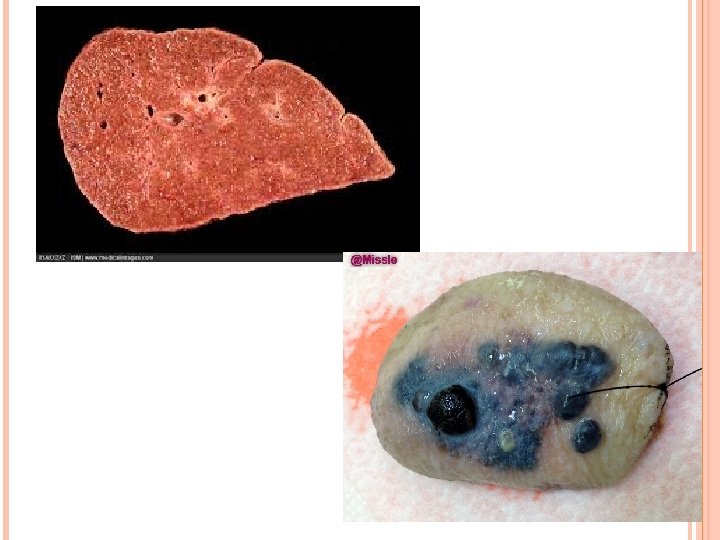
Handling of Specimen Gross appearance: Types of specimen Structure included Dimensions, Size Shape Weight Color Consistency Surface Edge, section

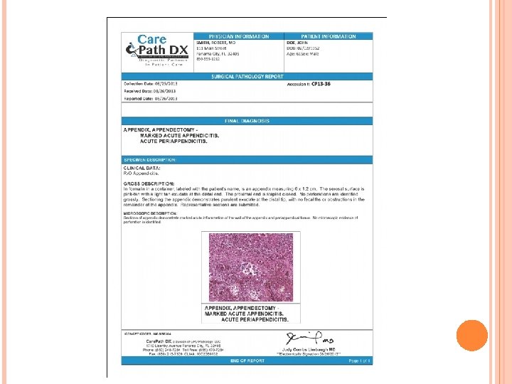
Handling of Specimen Fixation Cut-up Embedding Blocking Microtomy Staining Mounting Microscopy Report writing Reports will follow a format of: Clinical history, macroscopic appearance, microscopic appearance, conclusion

1 -Fixation. * Fixation ensures that the tissue is preserved in its natural state until processing. Prevents autolysis and bacterial decomposition. * This should be approximately 10 -20 times the volume of the specimen. Duration about 24 hours A. Tissue fixatives a. Buffered formalin 10%formalin. , c. Zenker’s formal saline d. Bowen’s fluid B. Cytological fixatives a. Ethanol 95% b. Methanol c. Ether

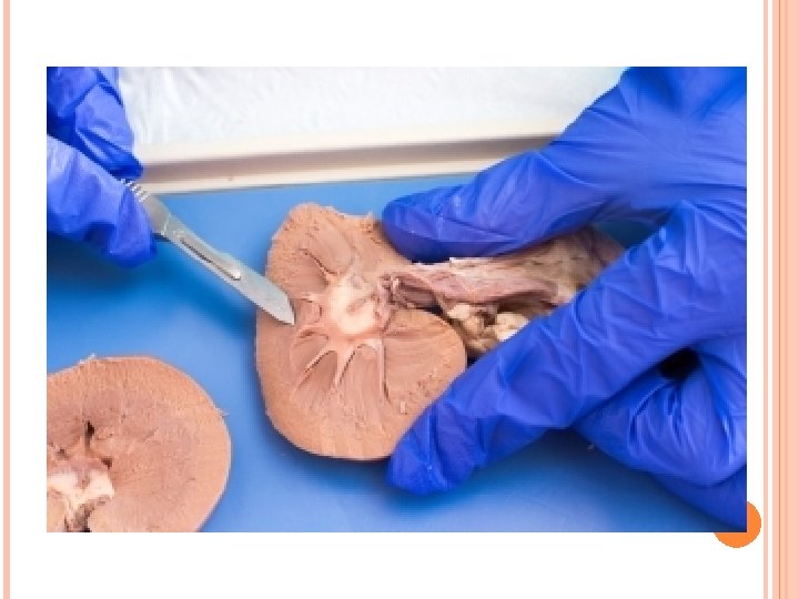
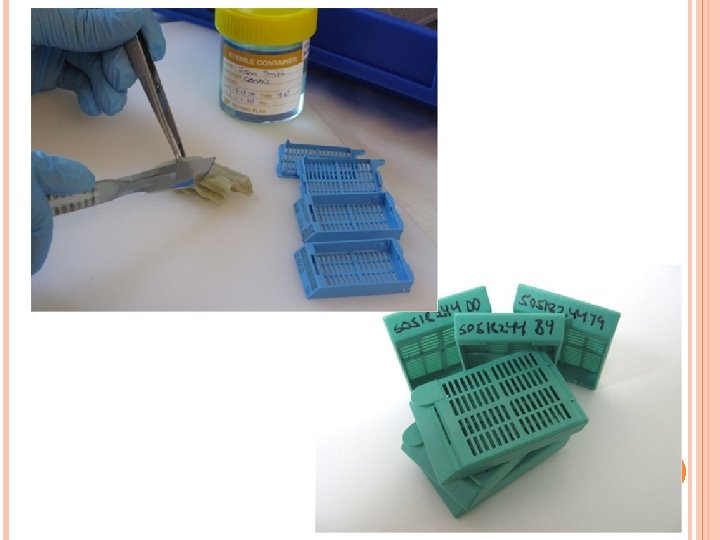
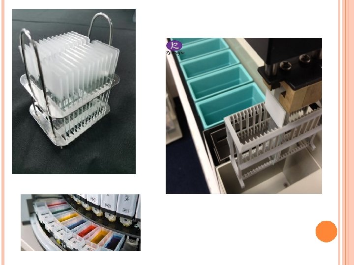
2 -Cut-up The tissues are cut up and placed into a cassette and then placed in racks of formalin

Tissue Processing: *It is a long procedure and required 24 hours. *It is done in stages. It can be subdivided into; Dehydration: Using alcohols, say 70% to 95% to 100%. Clearing, alcohol is replaced with xylene (which can mix with wax). Impregnating and embedding. The xylene is finally replaced with molten paraffin wax, which will permeate the cells to fix them.

Tissue processor

Blocking The embedded tissues are removed from their cassettes and put into metal blocks, which are then filled with more molten paraffin wax. The wax is allowed to cool and the metal tray is moved. The tissues are now embedded in blocks of wax and are hard enough to be cut

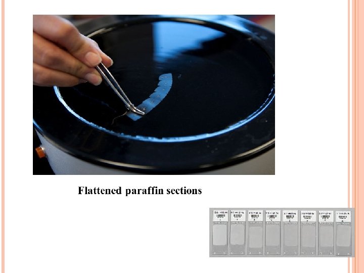
Microtomy The blocks are cut into very thin sections using a machine called a microtome. The sections are 4 -7 microns thick, which is thin enough to be seen through with a light microscope.

Staining is a process by which we give color to a section. Haematoxylin and Eosin staining is the most common used routine stain in histopathology laboratory Haematoxylin stains nuclei purple and eosin stains the cytoplasm and connective tissues pink.

Microscopy
 stain: This stain demonstrates glycogen 2. Stains")
SPECIAL STAINS: 1. PAS (Periodic Acid Schiff) stain: This stain demonstrates glycogen 2. Stains for micro-organism: a. Gram-stain: b. Ziehl_Neelsen stain: This stain detect acid fast bacilli. c. PAS stain: It is used for fungi, amoeba and Tricomonas. d. Modified Giemsa (2% Giemsa in water): For Helicobacter pylori. 3. Congo-red: It is used for identification of amyloid. 4. Sudan-Black: It is used for fat staining. 5. Masson’s Trichrome: It is used for differentiation of connective tissue

Modified Giemsa. For Helicobacter pylori.

Immunohistochemistry Ag-Ab specific reaction Estrogen Receptor, , breast
")
Leiomyosarcoma Actin (+)
 Filtering membrane")
ULTRASTRUCTURAL OBSERVATION TEM (transmitting electron microscope) Filtering membrane

Frozen sections: In the case of an urgently required diagnosis to giving a result it takes roughly 10 minutes. Cryostat will be used to rapidly freeze a section and skip the embedding process Example: To detect whether a breast nodule is benign or malignant to decide lumpectomy or mastectomy.

Thank you
- Slides: 36