Introduction to Pathology for 3 rd yr Medical


 1.")
 B. The meaning and general knowledge concern with study the")
 Pathology is the study of")
of organ system of body (=cell; tissue; organ). or")
 Clinical")


")


 Biopsy Any time During operation in OR.")

- Slides: 38
Introduction to Pathology for 3 rd yr. Medical student, Medical Faculty Naresuan University By S. Pongsabutra MD. 13 June 06
LEARNING OBJECTIVE OF INTRODUCTION TO PATHOLOGY Student must understand can explain the followings : 1. Course Syllabus of Pathology I(405311) (ประมวลการเรยนการสอนรายวชาพยาธวทยา 1 ชแจงรายละเอยดในชวโมงแนะนำพยาธวทยาภาคป ฏบต ) 2. The meaning and general knowledge concern with study the pathology
LEARNING CONTENT OF INTRODUCTION TO PATHOLOGY A. Course Syllabus Pathology I (ประมวลหวขอรายวชา ) 1. Content And Learning objective 2. Topic of learning 3. Organization of teaching & learning 4. Teaching media 5. Learning place 6. Assessment 7. Time table study 8. References
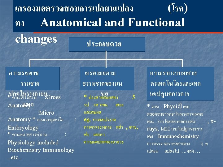
LEARNING CONTENT (CONT. ue) B. The meaning and general knowledge concern with study the pathology : 1. The meaning of pathology and scope of pathology. 2. How to study the pathology? * Strategy of learning * Tactics of learning 3. The general main causes of diseases. 4. Applied pathology to solve clinical problems.
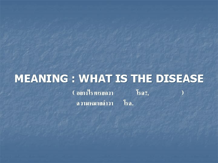
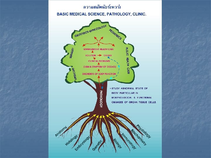
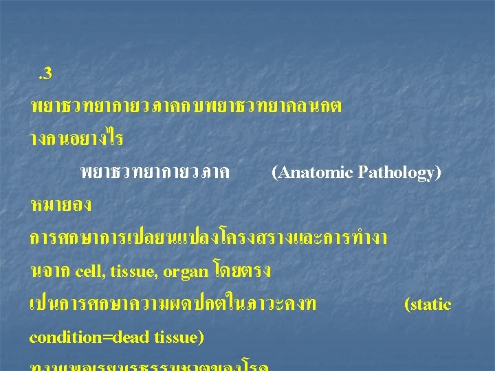
MEANING : WHAT IS THE PATHOLOGY (พยาธวทยา ? ) Pathology is the study of abnormal state of body particulary in morphological and functional changes of organ, tissue, cell. (Pathology; Gr. =Pathos+Logos) Pathology is the medical science specialty practice concerned with all aspects of Diseases
Disease is the disorderly function(symptom, sign)of organ system of body (=cell; tissue; organ). or Disease= dis+ease =(suffering, torture of that body which show off as abnormal sign and symptom)
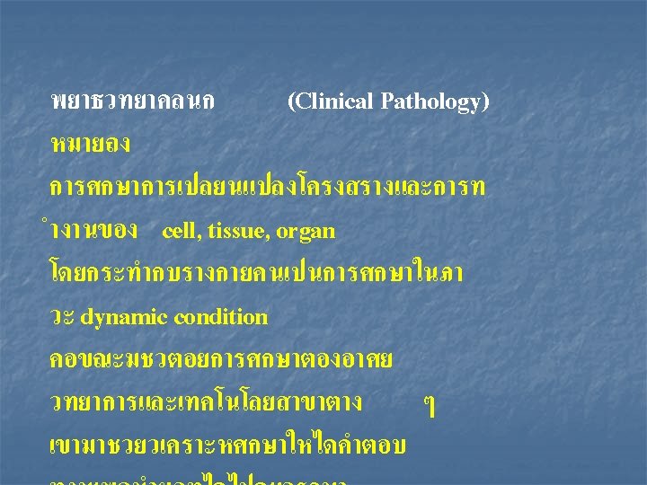
Scope of Pathology General Pathology Special Pathology AP + CP Anatomic Pathology (AP) Clinical Pathology (CP) Basic Pathology Systemic Pathology * Autopsy Pathology * Forensic Pathology * Surgical Pathology * Cyto Pathology * Molecular Pathology * Hemato Pathology ……. ฯลฯ. . APPLY TO SOLVE PROBLEMS OF CLINICAL DISEASES
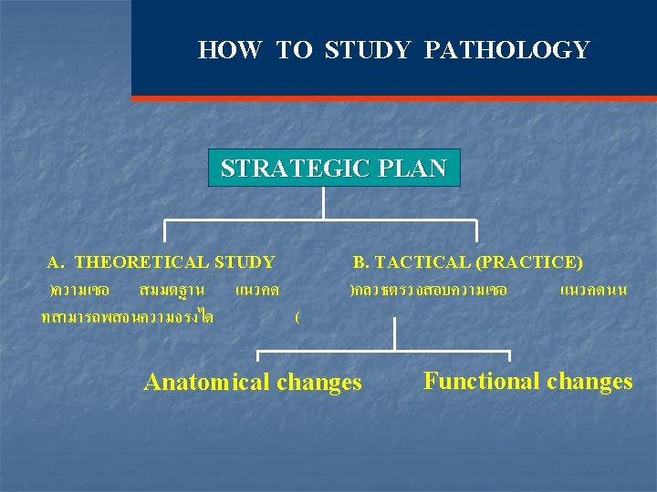
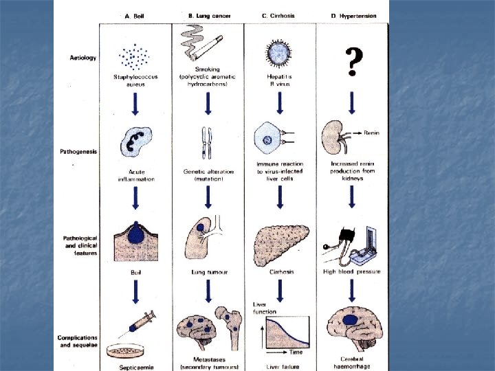
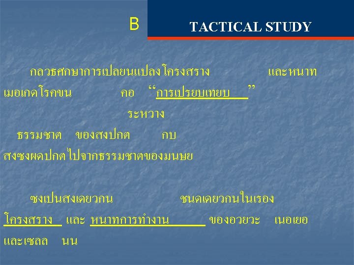
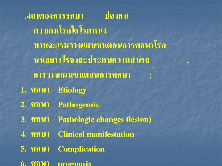
A THEORETICAL STUDY Main object is to know the nature of DISEASE by study as the following : 1. Etiology( causes of diseases ) 2. Pathogenesis (mechanism of diseases) 3. Pathological changes (structural change = Lesion) 4. Clinical feature (functional & structural changes presented as symptom and sign of patient) 5. Complication – ภาวะแทรกซอน – ผลกระทบกบอวยวะระบบอน ๆ
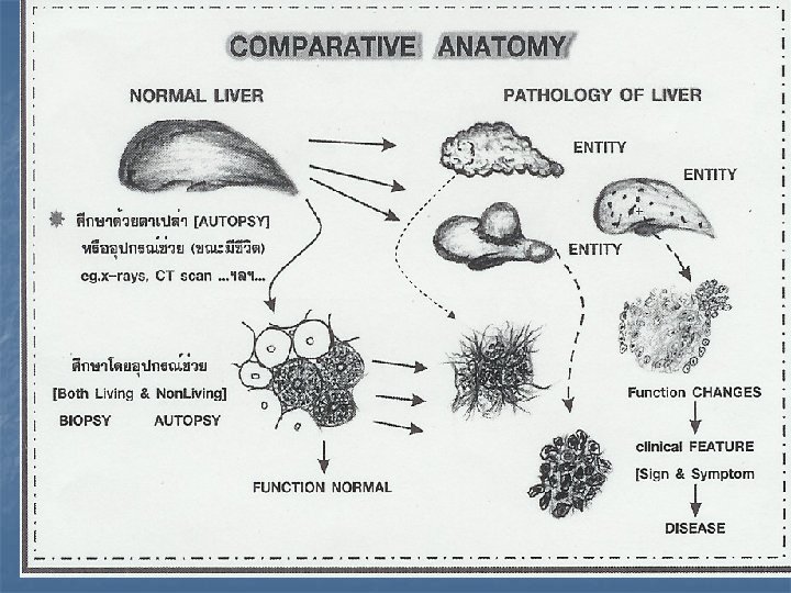
ตวอยางการเปรยบเทยบ Normal squamous cell vs abnormal squamous cell? Structure Normal mucosal small bowel vs abnormal mucosal small bowel? Normal liver vs abnormal liver Function Normal blood sugar vs abnormal blood sugar of …. . etc…. . organ, tissue, cell
กลม ทมาของ พยาธสภาพ ชนดตางฯ The general main causes of disease กลม 1 Disorders Congenital Hereditary Disease Congenital anomaly * Familial diseases * Abnormal Growth & Development กลม 2 Disorders กลม 3 of Formation Tumor of * Non – Neoplastic Self defense * Neoplastic mechanism * inflammation * Immune disorder (Biological or. Non Biological causes) กลม 4 Unclassified: *Degeneration. * Metabolic Disorders eg. DM. …. ฯลฯ. . . .
Basic Medical Science with Clinico-pathological Correletion Prognosis Surgery Medical Rx. Prophylaxis or Prevention Management Other Clinical Dx. Aortic Valve Stenosis c Lt. Sided Failure Heart, lungs changes Solution of problem s Clinical Problem Pathology (disease) หอบ เหนอยงาย เสยงหวใจผดปกต เสยงหายใจผดปกต หวใจเตนเร (Murmu (Crepitat - Heart sound Structural Changes Heart V. (Sclerosis Stenosis) Functional Changes - Murmur Circulation Disturb Cardiac out put Hypertrophy Strength of Heart rate heart Muscle heart pump Compensate to be normal Complication เลอดคงในปอด Fail to pump Over Compensate นำไหลเขาถงลม Irregular heart rate หายใจไมสะดวก หอบ มเสยงก
Macroscopic : พยาธสภาพลน Aortic Valve Acute bacterial vegetative endocarditis
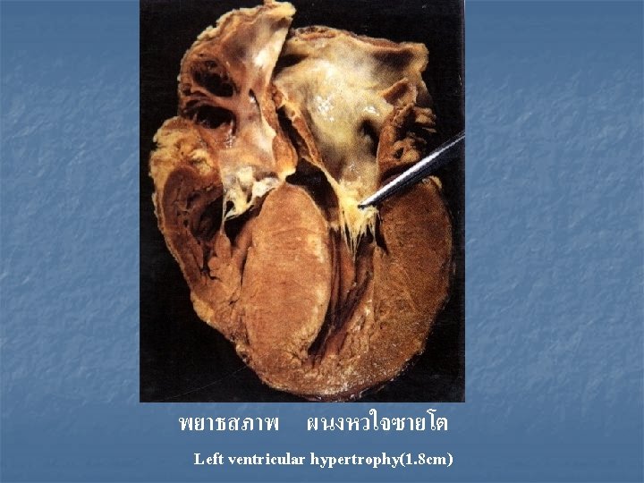
พยาธสภาพ hypertrophy heart
Macroscopic : Abnormal cut surface characterized by multiple , varying in size , up to 1 cm. , grey – white , masses
Microscopic : Abnormal both in size , shape of liver Cell
Macroscopic : Focals loss Liver parenchyma ; Multiple lesions
Microscopic : Foreign body in Hepatic bile duct ( Fluke – พยาธในทอนำดตบ )
Macroscopic : Severe , Diffuse dilatation of sinusoid with reddish – brown color Passive congestion of liver
Microscopic : Changes characterized by loss of Liver cord around central vein – meaning ? Central necrosis of Liver
Liver Macroscopic : Abnormal surface – diffuse nodular lesions , size 1 -2 mm. and Shrinkage
Liver Macroscopic : Abnormal cut surface show diffuse grey-white spots size vary to ~ 1 -3 mm. through out cut surface
Necropsy Chemical Fixation for light microscopy (formalin) Biopsy Any time During operation in OR. Specimen Freezing Dehydration (ethanol) Clearing (xylene) Infiltration (xylene/paraffin) Embedding (paraffin) Sectioning (microtome) Mounting (glass slide) Removal of paraffin Rehydration Staining (H & E) and / or histochemical reaction Light Microscopy Sectioning (cryostat)
REFERENCES 1. Anderson’ s Pathology, 10 th edition Edited by Ivan Damjanov, James Linder, Vol. I, “The history of pathology” pp 1 – 11. 2. Concise Pathology, 3 rd edition, Edited by P. Chandrasoma ; C. R. Taylor, Introduction : The discipline of Pathology, pp xiii – xiv 3. Robbins and Cotran Pathologic Basis of Disease, 7 th edition, Edited by Kumar; Abbas; Fausto; “Introduction to Pathology” pp 4