Introduction to Microbiology Microbiology is the biology of

Introduction to Microbiology

Microbiology is the biology of microorganisms. It is a bioscience for the study of the evolution, Classification , morphology, physiology, genetics, ecology of microbes under certain definite conditions, The law of their life activities, and their interaction with human being, animals or plants as well as with natural environment.

MICROBIOLOGY The study of little things ØVIROLOGY - Viruses ØBACTERIOLOGY - Bacteria ØMYCOLOGY - Fungi and yeast ØIMMUNOLOGY

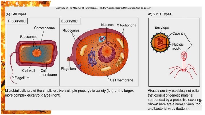
What is Microorganisms • Microorganisms are creatures that are not directly visible to the unaided eye, with dramatically biologic diversity. • Viruses , bacteria, fungi, protozoa and some algae are all in this category • All with the exception of plants and animals

Distribution of microorganisms • • • Air Soil Water Animals Human body

Microorganisms and Human Beings • Beneficial activities: Most microbes are of benefit to human beings, some are necessary (nitrogen, carbon cycles, etc. ) • Pathogenic : A portion of microbes cause diseases and are poisonous to human, and these are really that concern us in the study of medical microbiology, etc.

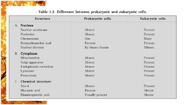
The Bacterial Cell EUKARYOTES PROKARYOTES BACTERIA ARCHAEA

NOMENCLATURE Binomial nomenclature Genus and species; e. g. Staphylococcus aureus Classification Heirarchy Order Family Genus Species

BASICS OF CLASSIFICATION üMorphological Characteristics üStaining Properties üBiochemical Characteristics üSerological Properties üGenetic Characteristics üProteins cell structure

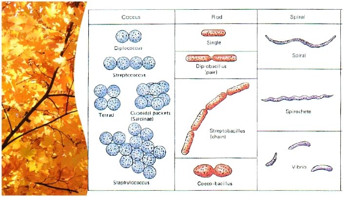
Bacterial Morphology and Structure Bacterial Morphology Size Arrangement Shape

SIZE OF BACTERIA • Unit for measurement : Micron or micrometer, μm: 1μm=10 -3 mm • Size: Varies with kinds of bacteria, and also related to their age and external environment.

Cocci: sphere, 1μm l Bacilli: rods , 0. 5 -1 μm in width -3 μm in length l Spiral bacteria: 1~3 μm in length and 0. 3 -0. 6 μm in width l

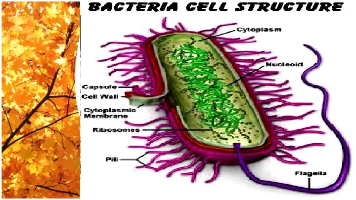
Bacterial Ultrastructure

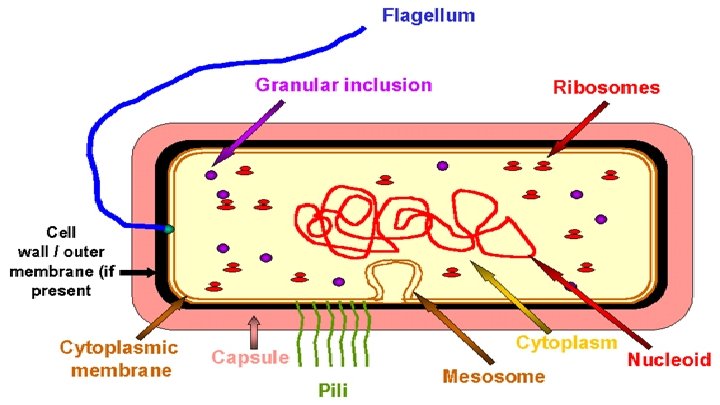
Essential components basic structure present in all bacteria, e. g. ♣ Cell wall. ♣ Cytoplasmic membrane. ♣ Cytoplasm. ♣ Nuclear material. Non essential components present in some bacteria species, e. g. µ Capsule. µ Fimbria. µ Flagella. µ Spores

Cell Wall The cell wall is the outermost component of bacterial cell. Functions: 1 - Maintain shape. 2 - Support cytoplasmic membrane. 3 - Role in cell division. 4 - Protection against antibiotics & antibodies. 5 - Antigenicity. 6 - Staining reaction to Gram.
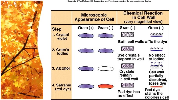

4 groups based on cell wall composition • • Gram positive cells Gram negative cells Bacteria without cell walls Bacteria with chemically unique cell walls

Composition of cell wall I- Gram positive cell wall Two layers 1 - Peptidoglycan: constitute 80% of cell wall thickness. It is a polymer of N-acetyl muramic acid and N-acetyl glucose amine, joined together by a tetra peptides side chain. 2 - Teichoic acid: it is a polymer of glycerol or ribitol. It is located in the outer layer of the Gram positive cell wall. It is antigenic.

I- Gram positive cell wall

II- Gram negative cell wall 1 - Thin layer of peptidoglycan: represents 5 -20% of Gram negative cell wall. 2 - Periplasmic space: filled with gel-like substances. This space separates the peptidoglycan layer from outer lipopolysaccharides layer. 3 - Outer layer: composed of lipopolysaccharides (LPS). It is subdivided into 3 layers: Inner highly toxic lipid A. Middle polysaccharide Core. Outer polysaccharides side chain.
 is usually called endotoxin because it is firmly bounded to the cell surface")
(LPS) is usually called endotoxin because it is firmly bounded to the cell surface and is released only when the cell lysed. Toxicity is mainly associated with lipid A. The outer polysaccharide side chain is known as somatic or (O) antigen.

Wall Deficient Bacteria 1 - Spheroplasts 2 - protoplasts - G-ve +penicillin * G-ve + lysozyme - Inhibit peptidoglycan* Removal of cell wall - Damaged cell wall due to damaged - May multiply peptidoglycan * Can’t multiply Lysozyme can't act on G-ve cell wall due to outer membrane unless treated with EDTA to disrupt it

3 - L-forms May develop from cells that normally possess cell walls when they are exposed to hydrolysis by lysozyme or by blocking peptidoglycan biosynthesis with antibiotics, e. g. penicillin. They may revert to bacterial form producing relapses of infection. 4 - Mycoplasma - The only group of bacteria that exist naturally without cell wall. - It has no defined shape due to lacking the rigid cell wall. - It is resistant to antibiotics which destroy bacterial cell wall, e. g. penicillin

Cytoplasmic Membrane • It lies just inside the peptidoglycan layer. • It is a phospholipids bilayer that contains protein. • It is similar to eukaryotic cell membrane but does not contain sterol except in mycoplasma. • Mesosomes are invagination of the cytoplasmic membrane inside the cytoplasm. Two types of mesosomes are known as lateral and septal. Septal mesosomes are involved in cell division where bacterial DNA is attached.

Function of Cell Membrane: • Maintaining the cell's characteristic shape- the rigid wall compensates for the flexibility of the phospholipid membrane and keeps the cell from assuming a spherical shape. • Countering the effects of osmotic pressure. • Providing attachment sites for bacteriophages.

• Providing a rigid platform for surface appendages- flagella, fimbriae, and pili all emanate from the wall and extend beyond it. • Play an essential role in cell division. • Be the sites of major antigenic determinants of the cell surface. • Resistance of Antibiotics.

Cytoplasm • The cytoplasm of the bacterial cell is a viscous watery solution or soft gel that contains a variety of organic and inorganic solutes. • A- Mesosomes: • These are invagination of cytoplasmic membrane. • It is the site of attachment of DNA chromosome during cell division. • Site of respiratory activity of the cell. • Increase surface area of the membrane, thus increase efficiency of active transport.

B- Ribosomes: • • They are complex structure composed of 60% RNA and 40% protein. They are the site of protein synthesis in the cell. They have sedimentation constant of 70 S being composed of 30 S and 50 S subunits. The two subunits separate except in protein synthesis, and aggregate to form polyribosomes.

C- Inclusion granules: • Round granules observed in cytoplasm of many bacteria. These are not permanent or essential structure. • They appear to be either stored energy or nutrient reserve concerned with cell metabolism, e. g. volutin granules (Metachromatic granules of Corynebacterium diphtheria)

D- Plasmids: • Extra chromosomal double stranded circular DNA that carry certain genetic information, e. g. antibiotic resistant toxin production, virulence, … etc. • Dispensable: not necessary for life of the cell. • Autonomous: multiply independent of the host. • Transmissible: can transfer to other bacteria by conjugation, transformation, or transduction.

E- The Nuclear Body: - The genetic information of the bacterial cell is contained in single circular DS-DNA molecules, which constitutes the bacterial chromosome. - The nuclear body does not have nuclear membrane, mitotic apparatus, or histones.

Structures outside the Cell Wall Capsule • It is present only in some bacteria outside the cell wall. It is gelatinous in nature. The capsule may be polysaccharide as in pneumococci, meningococci or polypeptide as in Bacillus anthrax or hyalouronic acid as in streptococci. • Capsules usually formed in vivo only and not stained by ordinary stain. The capsule is antigenic. • The capsule has antiphagocytic function so it determine the virulence of many bacteria. It also plays a role in attachment of the organism to mucous membrane.

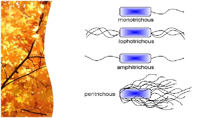
Flagella • Flagella are the organs of motility. They are thread like appendages, protein in nature, formed of flagellin protein which is antigenic (H Ag). • They can not be stained by gram stain. They have special stain. • According to their number, they may be monotrichate, amphitrichate, lophotrichate, peritrichate.
 • They are a hair like filaments, formed of protein called pillin")
Fimbriae (pili) • They are a hair like filaments, formed of protein called pillin which is antigenic in nature. Fimbriae are responsible for the attachment of bacteria to specific receptors of human cell (Adherence). • There are special types of pili called sex pili involved in the process of conjugation (transfer of DNA between bacteria). • Fimbriated bacteria can cause haemagglutination of RBCs. • Fimbriae are shorter and thinner than flagella.
 • Some bacterial genera are capable of forming highly resistant resting")
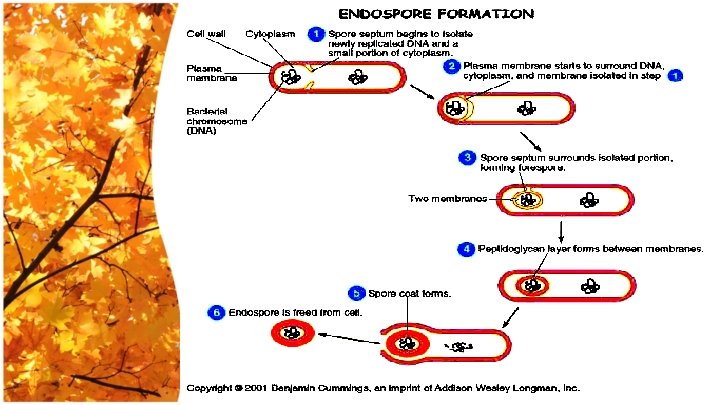
Bacterial Spores (Endospores) • Some bacterial genera are capable of forming highly resistant resting phase or endospores, e. g. Bacillus group and Clostridium group. • Endospores do not reproduce and exhibit absolute dormancy. Spore formation occurs in response to unfavorable conditions, e. g. depletion of nutrition, accumulation of metabolites or unsuitable temperature or moisture. • When the unsuitable conditions changed, the spore germinates to the vegetative form which can multiply.

Stages of Sporulation: • The plasma membrane invaginate enclosing section of cytoplasm that contain bacterial chromosome, some ribosomes and other cytoplasmic materials • It forms a thick hard protective coat. • The spores formed outside the body, and can not be stained by ordinary stain. • The spore is highly resistant to dryness, heat disinfectant. • Spore may be oval or round; terminal, central, or subterminal.

Endospores • • Resting, dormant cells Produced by some G+ genera: Clostridium, Bacillus & Sporosarcina Have a 2 -phase life cycle – vegetative cell & an endospore Sporulation -formation of endospores Germination- return to vegetative growth Hardiest of all life forms Withstand extremes in heat, drying, freezing, radiation & chemicals not a means of reproduction

• • • Resistance linked to high levels of calcium & dipicolinic acid Dehydrated, metabolically inactive Thick coat Longevity verges on immortality 25, 250 million years. Pressurized steam at 120 o. C for 20 -30 minutes will destroy.
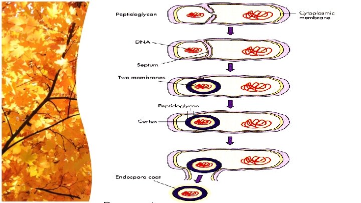

Spore structure Core: one copy of DNA and cytoplasmic contents Inner membrane and Spore wall Cortex: peptidoglycan layer Coat: Keratine-like protein which protect the spore. Exosporium

Microbial Metabolism

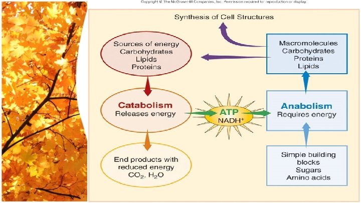
Metabolism The sum total of all chemical reactions & physical workings occurring in a cell

2 types of metabolism • Anabolism - biosynthesis – Building complex molecules from simple ones – Requires energy (ATP) • Catabolism - degradation – Breaking down complex molecules into simple ones – Generates energy (ATP)

Binary Fission
 Energy Nutrients Optimal temperature")
Bacterial requirements for growth • • • Oxygen (or absence) Energy Nutrients Optimal temperature Optimal p. H

Aerobes • Grow in presence of oxygen • No fermentation • Oxidative phosphorylation • EX: Mycobacterium tuberculosis Anaerobes • • No oxidative phosphorylation Fermentation Killed by oxygen Example: genus Clostridium

Aerotolerant anaerobes • • • Respire naerobically Not killed by oxygen Ex: Clostridium perfringens Facultative anaerobes • • Fermentation Aerobic respiration Survive in oxygen EX: E. coli

Microaerophilic bacteria • Grow – low oxygen • killed – high oxygen – Example: Campylobacter

Optimal growth temperature • Mesophiles: – human body temperature * pathogens * opportunists • Pyschrophile – close to freezing • Thermophile – close to boiling

Psychrophiles – optimum temperature below 15 o. C, capable of growth at 0 o. C. Mesophiles – optimum temperature 20 o. C-40 o. C, most human pathogens. Thermophiles – optimum temperature greater than 45 o. C.

PH • Many grow best at neutral p. H • Some can survive/grow - Acidophilic ex: helicobacter pylori - Alkaliphilic ex: vibrio cholera
")
Nutrient Requirements • • • Carbon Nitrogen Phosphorus Sulfur Metal ions (e. g. iron)

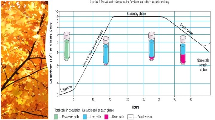
Growth Curve Stationary COLONY FORMING UNITS Death Log Lag TIME

۩ ۩ Lag phase – “flat” period of adjustment, enlargement; little growth. Exponential growth phase – a period of maximum growth will continue as long as cells have adequate nutrients & a favorable. ۩ Stationary phase – rate of cell growth equals rate of cell death cause by depleted nutrients & O 2, excretion of organic acids & pollutants. ۩ Death phase – as limiting factors intensify, cells die exponentially in their own wastes,
- Slides: 63