INTRODUCTION TO MEDICAL ULTRASONOGRAPHY Basic Introduction to Ultrasound

INTRODUCTION TO MEDICAL ULTRASONOGRAPHY Basic Introduction to Ultrasound

Definition: Ultrasound imaging, called ultrasound scanning or ultrasonography involves exposing part of the body to highfrequency sound waves to produce pictures of the inside of the body.

Sound waves n Sound is the transmission of mechanical vibrations in a medium Sound waves don’t exist in a vacuum and propagation in gases is poor. The closer the molecules, the faster the sound wave through the medium, so bone and metals conduct sound exceedingly well Frequency of soind is the Number of oscillations per second, measured in Hertz (HZ)

Audible sound waves lie between 20 and 20, 000 Hz. Ultrasound is sound with a frequency in excess of 20 k. Hz, which is the upper limit of human hearing

ULTRASOUND TISSUE INTERACTION Can be described in terms of -Attenuation. -Absorption. -Reflection. -Scattering -Refraction. -Diffraction

ATTENUATION Sound energy is attenuated or weakened as it passes through tissue because parts of it are reflected, scattered, absorbed, refracted or diffracted
 A reflection of beam is called an echo. The production and")
REFLECTION (ECHO PRODUCTION) A reflection of beam is called an echo. The production and detection of echoes form the basis of ultrasound.

ACOUSTIC IMPEDENCE Reflection occurs at the boundary between two materials provided that a certain property of the materials is different. This property is known as the acoustic impedance

ULTRASOUND IMAGING The creation of an image from sound is done in three steps - producing a sound wave, receiving echoes, and interpreting those echoes. The probe plays the major role in constructing image.

Contd. The probe contains a large number of transmitters set in a line along its length. Typically up to five of these firing simultaneously generate a short pulse of ultrasound that travels in a narrow column away from the probe. The transmitters then act as receivers and record. the intensity of the reflected sound.

Contd. The process is repeated sequentially along the length of the probe The time taken for an echo to return is used determine the distance from the probe and is calculated assuming that sound has a constant speed (1540 m/s). The strength of the echoes returning from any point is represented by the brightness of that point on the screen

DISPALYING MODES OF ULTRASOUND A-mode. - Simplest type of ultrasound. - A single transducer scans a line through the body with the echoes plotted on screen as a function of depth. - Used to measure distances within the body and the size of internal organs. - Therapeutic ultrasound aimed at a specific tumor or calculus is also A-mode, to allow for pinpoint accurate focus of the destructive wave energy.

B-Mode - In B-mode ultrasound, a linear array of transducers simultaneously scans a plane through the body that can be viewed as a twodimensional image on screen. - Ultrasound probes containing more than 100 transducers in sequence form the basis for these most commonly used scanners.

M-Mode. - The M stands for motion. - A rapid sequence of B-mode scans whose images follow each other in sequence on screen enables doctors to see and measure range of motion, as the organ boundaries that produce reflections move relative to the probe. - M mode ultrasound has been put to particular use in studying heart motion.

Doppler Mode. - Includes the capability of accurately measuring velocities of moving material, such as blood in arteries and veins. The principle is the same as that used in radar guns that measure the speed of a car on the highway. - Doppler capability is most often combined with Bmode scanning to produce images of blood vessels from which blood flow can be directly measured. - This technique is used extensively to investigate valve defects, arteriosclerosis and hypertension, particularly in the heart, but also in the abdominal aorta and the portal vein of the liver.

B MODE COLOURS BLACK ANECHOIC GREY HYPOECHOIC WHITE ECHOGENIC
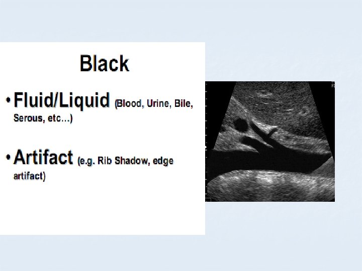
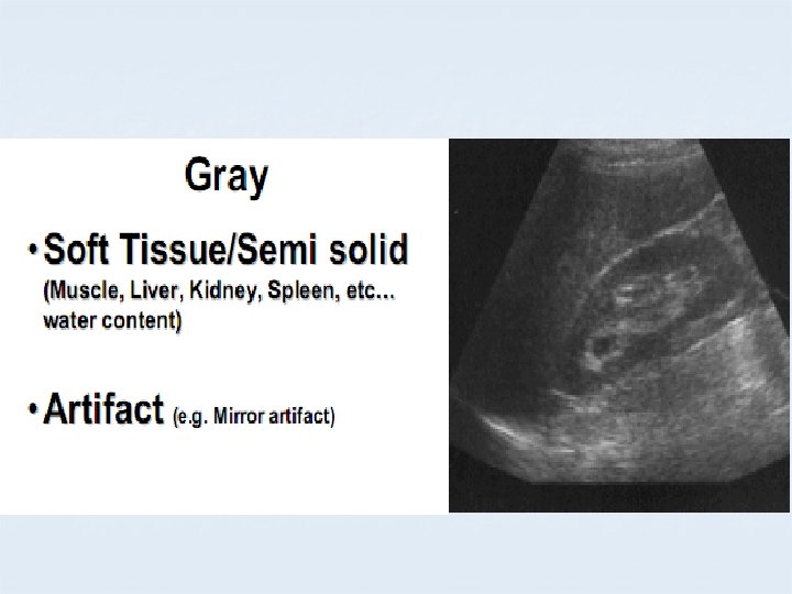
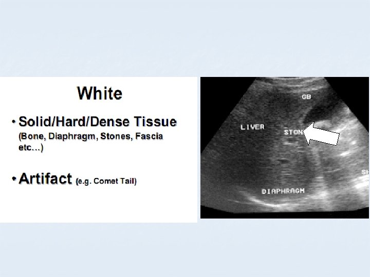

APPEARANCE OF DIFFERENT TISSUES STRUCTURE APPEARANCE Viscera ; Liver Spleen Hypoechoic Muscles Hypoechoic with echogenic lines Blood, Urine, Bile, ascites, water Anechoic Bone, Stone Hyperechoic

ULTRASOUND APPARATUS
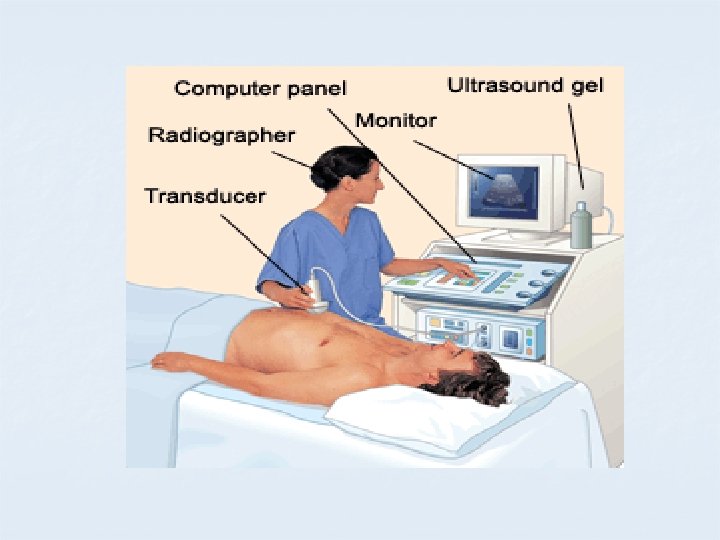

The Ultrasound Equipment Components: 1. The probe. 2. The control panel. 3. The freeze frame. 4. Measuring facilities. 5. A means of storing images.

CONTROL PANEL Adjusting the controls: - Freeze botton so that measurments & Print can be taken - Depth Increasing depth allows deeper structures to be viewed. - Gain The overall brightness can be adjusted.

CONTROL PANEL Adjusting the controls: - Focus so that a particular area can be examined in more detail - Zoom This takes a portion of the screen & magnifies it. - Measurment Cursers are used & are calibrated so that reasonably accurate measurements can be made.

CONTROL PANEL Adjusting the controls: - Taking pictures & labelling Images are of value as aid memoirs and for demonstration and discussion. Attension please !!! Any conclusions should be drawn while actually scanning the patient.
 Five main components n The transducer crystal , converts the electrical")
Probe (Transducers ) Five main components n The transducer crystal , converts the electrical voltage into acoustic energy upon transmission, and acoustic energy to electrical energy upon reception. n The matching layers Provide an acoustic connection between the transducer element and the skin. n Damping material such as rubber is attached to the back of the transducer. n The transducer case n The electronic cable used to excite the trancducer elements and receive the returned electrical impulses

Various Ultrasound Probes

Probe Movements Abdominal Probe n Sliding n Rotating n Angling n Dipping

Examination Technique 1. External Probe Sonography 2. Vaginal probe Sonography 3. Rectal probe Sonography.

Value &Benefits n n n Noninvasive No ionizing radiation Clear picture of soft tissues Monitoring of pregnant women Guiding minimally invasive procedures

Ultrasound is energy and is absorbed by tissue, causing heating 2 D ultrasound has been used to image the foetus for about 50 years. It is thought to be completely safe and does not cause significant heating 4 D ultrasound is new, requires more energy and therefore generates more heating. We think it is safe.

Mini Atlas Of Normal and Abnormal Sonographic images

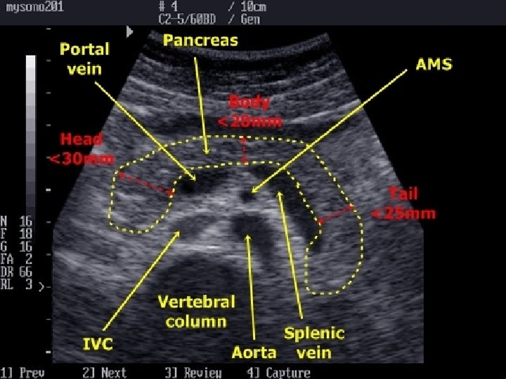
Sonographic anatomy Of Liver

Normal Gall bladder

Gall calculus with cholecystitis

Gall calculus

Gall bladder calculi and sludge

Gall calculi

Normal Renal sonographic anatomy

Normal Renal sonogram

Kidney LS image

Renal calculus

Nephrocalcinosis

U B study

Urin Bladder - Calculus

Vesicoureteric jn calculus
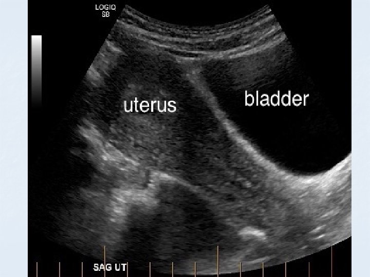

Pelvis sonogram TS UB /Uterus/ Ovary
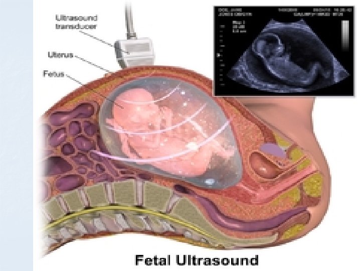

Fetus in Sagittal

Twin gestation

Multiple gestation

Sonogram of Appendix

Appendicolith

Colour doppler image

Colour doppler –Duplex

- Slides: 59