Introduction to Light and Electron Microscopy Neu 259

 • Convolution with")


 Effects Resolution slide courtesy of Edgar Garduno")
 Object Image")
 (PSF) where J 1=Bessel function")


 by applying")

 and the Fourier transform")


- Slides: 26
Introduction to Light and Electron Microscopy Neu 259 Spring 2006 Introduction to Deconvolution Image Processing James Bouwer UCSD
Outline Convolution: 2 D: • Airy Disk (Light Point Spread Function) • Convolution with an Image 3 D: • 3 D Point Spread Function • Convolution with a Volume Deconvolution: • Frequency space decomposition • Fourier Transforms • Application of the Deconvolution Theorem Some Examples:
The Microscope Optical Train is Complex • Every lens element alters the image in some way • We call this the image transfer function: h(x, y, z) • The image transfer function of the system is a convolution of all of the image transfer functions of all the lens elements and apertures in the optical train
The Definition of Convolution: Con-vo-lut-ed: adj. 1. Having numerous overlapping coils or folds: a convoluted seashell. 2. Intricate; complicated: convoluted legal language; convoluted reasoning. The American Heritage® Dictionary of the English Language, Fourth Edition
Formation of an Airy Disk Pattern A point in the object space A point convolved with the transfer Microscope function Impulse Response Function [ Point Spread Function (psf)]
Impulse Response Function Applied to a line A great advantage is afforded by the ability to express the response of the optical system to an arbitrary input in terms of the response to certain “elementary” functions into which the input has been decomposed Line Transfer Function
How the psf (Impulse Response) Effects Resolution slide courtesy of Edgar Garduno
Image Transfer Function h(x, y) Object Image
Theoretical Model of the Impulse Transfer Function, h(x, y) (PSF) where J 1=Bessel function of the first kind
The Definition of Convolution: Con-vo-lut-ed: adj. 1. Having numerous overlapping coils or folds: a convoluted seashell. 2. Intricate; complicated: convoluted legal language; convoluted reasoning. The American Heritage® Dictionary of the English Language, Fourth Edition Convoluted Mess!
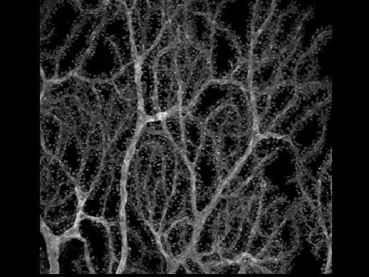
Spatial Frequency Decomposition Fourier Transform 0. 25µm myelin • Any image can be decomposed into a series of sines and cosines added together to give the image Amplitudes Phase Fourier Transform
Fourier Transform of the Myelin Image Low frequency High frequency
Mathematical Formulation of the Fourier Transform 2 -D Fourier Transform: Real Space to. . Frequency Space Inverse 2 -D Fourier Transform: Frequency Space to. . Real Space
Inverse Fourier Transform of the Fourier Transform Returns the original Image Fourier transform of myelin -1 F = Very Powerful Tool !
The Wonderful, Great and Amazing Convolution Theorem If and Then: Remembering that the image intensity is a convolution of the impulse function h(x, y) and the object Iobject(x, y) The object intensity can be easily de-convolved from the Smear of the impulse function (PSF)
Deconvolution using the Convolution Theorem The image is a convolution of the impulse function h(x, y) (PSF) and the object (the sample): Therefore, the Fourier transform of the image is just the Fourier transform of object times the Fourier transform of the impulse function (PSF) in frequency space So, to obtain the object, we simply divide by the Fourier transform of the impulse function (PSF)
Transform back to real space Finally, we obtain the deconvolved object (sample) by applying an inverse Fourier transform A simple division in frequency space yields the Object intensity !
Let’s Run though it… Step 1: Acquire the image and it’s Fourier transform Convoluted Image Fourier Transform
Steps to Deconvolution… Step 2: Obtain the impulse function (PSF) and the Fourier transform of the impulse function (PSF) Point spread function Fourier transform
Steps in the Deconvolution… Step 3: Divide and inverse Fourier transform -1 F
3 D Out-of-focus Point Spread Function 3 D Impulse Response z x y 2 -D PSFs vs. z-height
A Real 3 -D PSF From the RTS-2000 100 nm Diameter Fluorescent Latex Bead Imaged with 50 nm Steps in z
… Plenty more to this story … Many commercial software packages are available in at the NCMIR