Introduction to immunity and innate immunity Dr Eman


 that is")


 can also develop later in life e. g. antigens")


")


")
; T cells (60%), B")


. – They are")
 PRR are 3 types 1. Toll like receptors")
 3. opsonin receptors • Act indirectly by binding")


 • have rounded or kidney-shaped nuclei")


 of NK cells detect alteration in host")


")


 give rise to two kinds of multipotent progenitor")


 • function")


- Slides: 78
Introduction to immunity and innate immunity Dr. Eman Albataineh, Associate Prof. Immunology College of Medicine, Mu’tah university Immunology, 2 nd year students
History • Immunity: protection from infectious microbes or foreign macromolecules; proteins and polysaccharides • Immune system constitutes of cells, tissues and small molecules • The first application in immunology is done by edward jenner’s vaccination against smallpox when he injected parts of cowpox microbe into small boy who is later became resistant to smallpox disease in 1798 (vaccine) • This was crowned in 1980 when the WHO announce the smallpox have been eradicated worldwide.
• The first line of defense is provided by – barriers , Mechanical; skin, Chemical; acidic stomach, Biological; commensal microbes – Enzymes and anti-microbial peptides as defensins and surfactants – Complements and cytokines, Phagocytes and natural killers (Innate immune system)
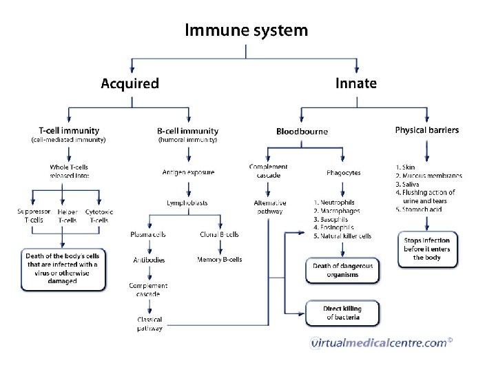
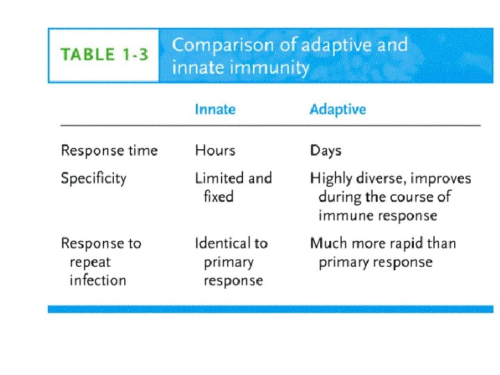
Immunity in practice – Defense system evolved to prevent or limit invasion of infectious agents, non infectious particles as protein and lipid macromolecules and cancerous cells – The innate immune system ( the first to act; complements and cells), • • • present and act the same in all people against general antigens. it is monotonic; the same magnitude and speed of response each time, not specific, act against common microbial antigens. . – The second is adaptive immune system (T and B cells), • • it is specific act against certain antigen, increasing in magnitude and speed of response in re-exposure to the same antigen (memory) – Innate immune response is better than adaptive in recognizing self from non-self
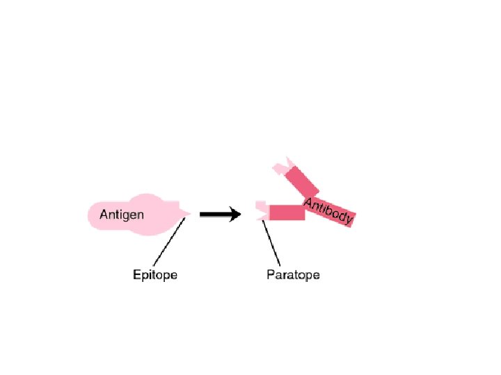
Antigen and immunogen • Any particle (organism, molecule or part of molecule) that is recognized by the immune system called antigen but not always stimulate the immune response (hapten, tolerogen) • Each antigen contain one or more antigenic determinant that are specific for binding to immune componenets; called epitopes • Epitopes are the smallest part of an antigen that is seen by antigen receptors on immune cells, or antibodies.
immunogen • Whereas any antigen capable of inducing immune response called immunogen
Haptens • Very low molecular weight antigen need to conjugate to a carrier protein (or immunogen) to induce the immune response, then the immune response will direct against both the carrier and the hapten • Examples 1. Drugs as penicillin 2. Food additives, lipids, nucleic acids, small peptides and carbohydrates
tolerogen • Tolerance (antigen- insensitive) can also develop later in life e. g. antigens that administered orally or exposed to in early life. Tolerogens induce negative or diminished immune response.
Factors determine the antigenicity 1. More Foreignness more response 2. Chemical complexity Proteins are most potent, polysaccarides are both antigenic or non antigenic whereas nucleic acids and lipids are non antigenic but can be antigenic when bind protein carriers 3. High molecular weight antigen, whereas hapten have very low molecular weight 4. Biodegradable 5. Mode of contact more response in Intra venous than subcutaneous or Intramuscular injections. 6. Host genetics like HLA type
Types of antigens • Auto-antigen • Iso-antigen; found in genetically identical twins, like HLA antigens • Allo-antigens; found in members of the same species; blood groups in human • Xeno-antigen; found in different species like animals and human
Phases of immune response – Antigen enter tissue cause inflammation which activate the local innate cells ( neutrophils, macrophages) – Movement of the innate cells then activation and movement of adaptive immune cells – Movement from all body toward the site of infection called chemotaxis, and mediated by molecules secreted from local cells. – Functional activities of the immune cells • Recognition of the foreign antigen through cell receptors • Response – Effector or activated cells – Memory ( only in adaptive response)
Inflammation • Inflammation is the process by which circulating leukocytes and plasma proteins are brought into sites of infection in the tissues and are activated to destroy and eliminate the offending agents. Inflammation is also the major reaction to damaged or dead cells and to accumulations of abnormal substances in cells and tissues. • It is the major way by which the innate immune system deals with infections and tissue injury
Changes in inflammation A. Increased blood supply to the area B. expression of endothelial adhesion molecules on blood vessel lining C. Increase capillary permeability D. Activation of local innate cells to secret chemotactic factors that recruit leukocytes,
inflammation • Signs of inflammation 1. Swelling 2. Pain 3. Redness 4. Loss of function 5. Heat • Cells of inflammation – Local macrophages and Mast cells that secret mediators help in chemotaxis and vascular permeability
– All of these changes are induced by cytokines (IL-1, IL 6, and TNF) and small-molecule mediators initially derived from resident cells in the tissue, such as mast cells, macrophages,
Innate immune cells A. phagocytic cells • Neutrophil polymorphs, main • Mononuclear phagocytes, phagocytic, and help in acquired immune response. They have many names; kupffer cells in liver, histiocytes in connective tissues, macrophage in bone marrow, spleen and lymph nodes, langerhans’ cells in skin, osteoclast in bone, mesangial cell in kidney, microglial cells in brain and monocytes in blood. B. Non phagocytic cells; Extracellular killing • Eosinophils and basophils • Natural killer cells,
DCs • Their main function is to phagocytose antigen material and present it on the surface to lymphocytes, thus functioning as antigen-presenting cells. • Dendritic cells are present in small quantities in tissues that are in contact with the external environment, mainly the skin (where there is a specialized dendritic cell type called Langerhans cells) and the inner lining of the nose, lungs, stomach and intestines. They can also be found in an immature state in the blood. • Once activated, they migrate to the lymphoid tissues where they interact with T cells and B cells to initiate and shape the adaptive immune response. they grow branched projections for that they are called DC,
DC or dentritic cells • 4 types – Myeloid DC, macrophage origin, common, diffuse localization, phagocytose antigen and activate T cells – Lymphoid DC, lymphocyte origin, recruit cells to site of infection – Follicular DC, mesenchymal origin, present in peripheral lymph nodes, do B cell activation. – plasmacytoid dendritic cells, are early cellular responders to viral infection. They have potent antiviral activities.
Adaptive immune cells • T cells • B cells
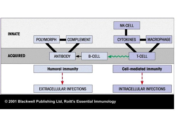
Innate immunity • Innate immunity is the initial response to microbes that prevents, controls, or eliminates infection, eliminate damaged cells and initiate the process of tissue repair. • Innate immunity stimulates adaptive immune responses • the major types of responses of the innate immune system that protect against microbes are inflammation, innate cells, complements and cytokines.
Innate and Adaptive • Innate immune response is better than adaptive in recognizing self from non-self. • Innate immune responses to a microbe are immediate and do not require prior exposure to the microbe • effective adaptive immune responses to a newly introduced microbe develop over several days as clones of T and B lymphocytes that activated by some activated innate cells undergo expansion and differentiate into functional effector Th and Tc cells and antibody producing B cells.
• The receptors of the innate immune system are encoded by inherited (germline) genes, whereas the genes encoding receptors of adaptive immunity are generated by somatic recombination of gene segments in the precursors of mature lymphocytes • In Innate cells the receptors are present in nature, and are less in number (103) and variety than adaptive cells receptors
Cellular Components of all immune system in percents 1. Lymphocytes(30%); T cells (60%), B cells (30%) (high N: C ratio) and large granular lymphocytes called natural killer cells(10%) low N: C ratio and granular) 2. Mononuclear phagocytes; macrophages (5. 3%) 3. Granulocytes; neutrophils (62%), eosinophils(2. 3%) and basophils (0. 4%)
Cell chemotaxis v Recruitment of phagocytes to site of infection – Follow chemotactic factors gradient(C 3 a, C 5 a, AND IL-8) produced by resident macrophages and phospholipids and peptides of bacteria, they migrate by • Capture and rolling; enhance adhesion molecules on both endothelial and innate immune cells • enhance the strength of binding of interacting molecules on both endothelium and leucocytes that make the leukocyte flatten • Extravasation to site of infection. First neutrophils then macrophages. activated T cells migrate in the same way
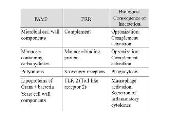
Innate cells Recognition of microbe v Recognition phase -Innate immune cells recognize structures found only on microbes called Pathogen-associated molecular pattern (PAMPs) by pattern recognition receptors (PRR). The PAMPs may be; 1. double stranded RNA in viruses 2. DNA in bacteria 3. Lipo-polysaccharides or endotoxins in G- bacteria 4. Teichoic acid in g+ bacteria 5. Mannose rich oligo-saccharides -The targets of the innate immune response is essential for the survival of the microbes -The innate immune system also recognizes endogenous molecules that are produced by or released from damaged and dying cells. These substances are called damage-associated molecular patterns (DAMPs)
Receptors of innate immune cells • Via Pattern recognition receptors (PRR). – They are proteins expressed by cells of the innate immune system to identify pathogen-associated molecular patterns (PAMPs), which are associated with microbial pathogens or cellular stress, as well as damageassociated molecular patterns (DAMPs), which are associated with cell components released during cell damage – PAMP is general conserved microbial molecules as sugar, protein, lipid and nucleic acids. – the receptors are similar in all humans, – And bind to the target : 1. directly through cell surface receptors binding to the antigen 2. indirectly by binding to soluble molecules that engage the microbe( opsonins as complements)
Receptors of innate immune cells (PRR) PRR are 3 types 1. Toll like receptors (TLR), binding results; signaling; phagocyte activation and secretion of immune mediators called cytokines 2. Scavenger receptors they help in internalization of bacteria in the phagocytic cells
Receptors of innate immune cells (PRR) 3. opsonin receptors • Act indirectly by binding to soluble molecules that engage the microbe( opsonins as complements or antibodies). the receptors are called depending on opsonin that bind as; antibody receptor or complement receptors. The result is microbe internalization to the cell. Opsonins: Opsonization of microbe (coating to make it obvious) 1. Opsonization of microbe (coating to make it obvious) using IGG or IGM. 2 types 1. 2. Direct opsonization by IGG Indirect opsonization by IGM + complement 2. Opsonization using C 4 b, C 3 b and C 3 bi complements 3. Opsonization by fibronectin and C-reactive proteins 4. Or by Cytokine
Opsonins
The job of innate cells; 1 -Ingestion in macrophages and neutrophils • Intracellular killing, mechanisms of lysosomal killing – O 2 dependent; the process called respiratory burst. O 2 metabolites are; hydrogen peroxide, singlet oxygen, hydroxyl radica, hypohalite ( Ocl or OI) and nitric oxide – O 2 independent; using granules contents as proteases, hydrolases and nucleases
2 -Secretion of cytokines • Help in attraction of cells to site of infections • Induce signs of inflammation as high temperature
3 - activation of the adaptive immune sys. • Mediated by mainly Macrophages • By either – Indirect way; secretion of molecules that attract adaptive cells to site of infection – Direct way; present antigen to T cells.
• The link between innate and adaptive immunity 1. The innate stimulate the adaptive ( macrophage secret IL-12 and/ or IL-4 that activate T cells. C 3 d complement activate B cell. Antigen presentation to T cell by macrophages. 2. The adaptive immune response use some innate cells to eliminate the antigen (T cells secret IFN gamma that activate macrophages)
Innate immune cells 1 -Mononuclear phagocyte system (macrophages) • have rounded or kidney-shaped nuclei with finely granular cytoplasm • Mononuclear phagocyte’s primary function is phagocytosis – Originate in BM, and first to leave. When monocyte becomes settled in tissue they are called macrophages. Some mononuclear cells may differentiate to dentritic cells. some joint to form multi-nucleated giant cells
Activation and function of macrophages • Functions in Natural and adaptive Immunity 1. Phagocytosis of foreign particles the same as neutrophils – Scavenger receptors – opsonin dependant phagocytosis; engulf antigen antibody complex as in viruses via receptor for opsonizing Ig. G and complement C 3 b, No receptors for IGM 2. Secretion of enzymes and oxidative metabolites if antigen is big, cause tissue damage (respiratory burst- oxygen radicals, NO, prostaglandins) 3. Cytokine production which recruit other inflammatory cells, 4. Antigen-Presentation to T cells,
2 -Neutrophils • Granulocytes contain nucleus segmented into 3 -5 connected lobes, hence the name polymorphonuclear leukocyte and cytoplasmic granules. Neutrophils (95% of granulocytes) respond w/I 24 hours of stimulus (the earliest). have 20 times as many receptors as macrophages. They have Fc receptor to IGG and IGA as well as complement receptors. • Intracellular killing by azurophil lysosomal granules and specific granules. • Cytokine production which recruit other inflammatory cells,
phagocytosis
NK cells • are a type of lymphocyte critical to the innate immune system. are defined as large granular lymphocytes (LGL) and constitute third kind of cells differentiated from the common lymphoid progenitor-generating B and T lymphocytes. • 10 % of mononuclear cells in blood and spleen and rare in lymphoid organs • Act very early against viruses and intracellular microbes and tumor cells or altered expression of surface MHC 1 molecule until T cells become activated. • There activity increase by IFN alpha and beta (secreted by virally infected cells). • activated cells secrete IFN gamma
NK receptors 1. Killer activation receptors (KAR) of NK cells detect alteration in host cells as cancers. recognize stress related molecules as MICA, MICB 2. Killer inhibition receptors (KIR) of NK cells, used to detect the presence of MHC 1 protein on host cells any binding means inhibition killing is the net result from the activating receptors (KAR) and inhibitory receptors (KIR) The action depend on the 2 nd receptor 3. , Opsonin receptors for antibodies; low affinity IGG receptors ( IGG 1 and IGG 3)( Called low affinity FCγR 111 or CD 16) , and kill these coated cells, this is called antibody dependent cell mediated cyto-toxicity (ADCC) 4. Expression fas ligands that bind fas on target cells and activation of caspases, this is a way in killing activated T cell (activation induced cell death)
NK cells • Effecter functions of NK cells – Direct extracellular killing by secretion • Perforins; making pores then osmotic lysis • Granzymes, enzymes enter through perforin pores and activate caspases leading to cell death – Indirect killing. increase macrophage killing of phagocytic microbe by secreting IFN gamma
When a KAR binds to MICA and MICB molecules on the surface of an infected cell (or a tumor cell), a KIR examines the levels of MHC class I of this target cell. If the MHC class I levels are enough, killing of the cell doesn't proceed (left), but if they aren't, the killing signal proceeds and the cell is eliminated
Direct killing by NK cells
NK killing
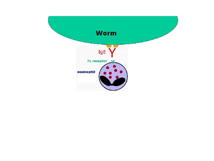
EOSINOPHILS • These cells are eosinophilic or "acid-loving" as shown by their affinity to coal tar dyes: Normally transparent, it is this affinity that causes them to appear brick-red after staining with eosin, a red dye, • Eosinophils kill extracellularly • 2 functions – When eosinophils bind to Ig. E on the surface of a worm, the cell is triggered to degranulate. The contents of the granules cause damage to the worm. – Other function is in allergy. • There are many hydrolytic enzymes present in the granules responsible for the anti-helminthic activity. One component which is unique to the eosinophils - and highly toxic to worms - is a substance known as Major Basic Protein (MBP).
Basophils and mast cells • Granulocytes, have acidic proteoglycan, Lobed nucleus--more variable, large coarse granules stain blue with basic dye methylene blue. • Mast cells is the cessile form whereas basophils is the circulating form • 2 types of mast cells – Connective tissue – Mucosal mast cells, act in allergy and is T cell dependent to degranulate. • Mast cells degranulation and release of the mediators the acidic granules, which help in Inflammatory cell response, allergy. • 2 receptors on mast cells that mediate degranulation – High affinity IGE receptor. IGE dependent; – Receptors for anaphylatoxins. C 3 a and C 5 a. IGE in-dependent;
EOSINOPHIL Cells of the blood RED BLOOD CELLS NEUTROPHILS MONOCYTE BASOPHIL LYMPHOCYTES
receptors of acquired immune- cells • expressed on B and T (BCR and TCR) cells of the adaptive immune system, each cell do random DNA rearrangement to develop – unique receptor able to recognize single structure. – Each human has its won receptors depending on what antigens invade his body – Formed continuously through out life – The total pool of receptors are capable of recognizing more than 1010 different structures. – Some cells may develop receptors recognize self as a result T and B cells undergo a process of education to remove those expressing receptors against self.
Organs of the immune response • Primary lymphoid organs A. Bone marrow; where the immune cells originate B. Thymus; where T cells differentiation to mature • Secondary lymphoid organs – – – maintain mature naive lymphocytes and initiate an adaptive immune response. the sites of lymphocyte activation by antigen. It is exemplified by the lymph nodes, and the lymphoid follicles in tonsils, Peyer's patches, spleen, adenoids, skin, etc. that are associated with the mucosa-associated lymphoid tissue (MALT).
Tissues of the immune system Dr. Eman Albataineh, Associate Prof. Immunology College of Medicine, Mu’tah university Immunology, 2 nd year students
Primary immune organs; Bone marrow and thymus • Bone marrow functions – Leukocytes production, B cells maturation. hematopoiesis start in childhood (YOLK SAC AND mesenchyme, then liver and spleen and finally the bone marrow in puberty) and get maximum in adult age, most common site of BM is sternum, vertebrae, iliac bones and ribs. – In cases of excess demand liver and spleen help the BM (the extramedullary hematopoiesis). • Thymus – T cell maturation and formation of T cell antigen receptors
Bone marrow components • The two components of bone marrow are – "red marrow" which consists mainly of hematopoietic tissue, Red blood cells, platelets, and most white blood cells arise in red marrow Red marrow is found mainly in the flat bones, such as the pelvis, sternum, cranium, ribs, vertebrae and scapulae, and at the epiphyseal ends of long bones such as the femur and humerus – and "yellow marrow", which is mainly made up of fat cells. At birth, all bone marrow is red. With age, more and more of it is converted to the yellow type; only around half of adult bone marrow is red. . Yellow marrow is found in the hollow interior of the middle portion of long bones. In cases of severe blood loss, the body can convert yellow marrow back to red marrow to increase blood cell production – Stroma; any tissue not associated to blood production as fatty marrow, fibroblast, osteoclast and osteoblast.
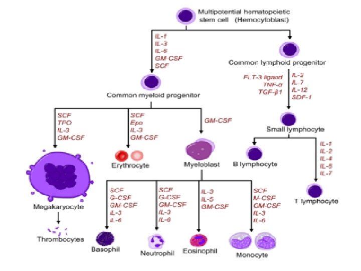
• Hematopoietic stem cells (HSCs) give rise to two kinds of multipotent progenitor cells, one that generates lymphoid another that produces myeloid cells, • The common lymphoid progenitor gives rise to committed precursors of T cell, B cell • The common myeloid-megakaryocyte- erythroid progenitors give rise to committed precursors of the erythroid, megakaryocytic, granulocytic, and monocytic lineages, • Stem cells express 2 main proteins, CD 34 and stem cell antigen-1 • Hematopoietic Cytokines called Colony stimulating factors are the influencing factors for stem cell differentiation and maturation e. g; G-CSF, M-CSF and GM-CSF
BM
Blood cells precursors
Thymus • The thymus gland is found in the thorax in the anterior mediastinum. It gradually enlarges during childhood but after puberty it undergoes a process of involution resulting in a reduction in the functioning mass of the gland. It continues to function throughout life, however. • The thymus has a rich vascular supply and efferent lymphatic vessels that drain into mediastinal lymph nodes. The thymus is derived from invaginations of the ectoderm in the developing neck and chest of the embryo, forming structures called branchial clefts.
Thymus • A. Anatomy; The thymus is composed of two identical lobes and is located anatomically in the anterior superior mediastinum, in front of the heart and behind the sternum • B. Histology: The thymus gland is surrounded by a fibrous capsule, and arranged into an outer, more cellular, cortex and an inner, less cellular, medulla. Cells involved – The most immature T cells in the cortex. As thymocytes or T cells mature, they migrate toward the medulla, then to circulation – Epithelial cells – Macrophages and lymphoid dentritic cells • Digeorge syndrome ( genetic defect in development of 3 rd pharyngeal pouch in embryo); T cell deficient as a result of impaired thymus development, plus parathyroid gland defect
Thymus
Lymph nodes and lymphatic system (peripheral or 2 nd lymphatic sys. ) • function to concentrate antigens that are introduced through the common portals of entry (skin and gastrointestinal and respiratory tracts). • They Are places where the innate cells carry the antigen and present it to the adaptive immune system • Site of lymphocyte activation by antigen
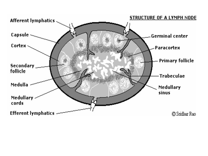
• Secondary lymphoid tissues consist of the – lymph nodes, which are clustered at sites such as the groin, armpits and neck and along the small intestine, and collect antigen from the tissues; – the spleen, which collects antigen from the bloodstream; – and the mucosa-associated lymphoid tissues (MALT), which collect antigen from the respiratory, gastrointestinal and urogenital tracts and are particularly well organized in the small intestine, in structures known as Peyer’s patches • The node is made up of three components: – lymphatic sinuses the Lymph flows from afferent vessels cortical sinuses, into the medullary sinuses and into efferent lymphatic vessels – , blood vessels – parenchyma (cortex, paracortex, medulla)
Structure of the lymph node Cortex • Cortex consists of primary follicles and secondary follicles ( with germinal center). • Germinal center formed from stimulated B cells and follicular dendritic cells. whereas primary follicles have only mature but not activated B cells • Stimulated mature B cells change into plasma cells or memory B cells which reside in medulla and antibody that move to the circulation.
• Paracortex • The paracortex contains T lymphocytes and macrophages • T cells: The various types of T cell enter the node from the blood via the HEVs. When activated they form lymphoblasts which divide to produce a clone of T cells responding to a specific antigen. Activated T cells then pass into the circulation to reach peripheral sites. • Medulla • The medulla comprises: – large blood vessels – medullary cords and sinuses – plasma cells
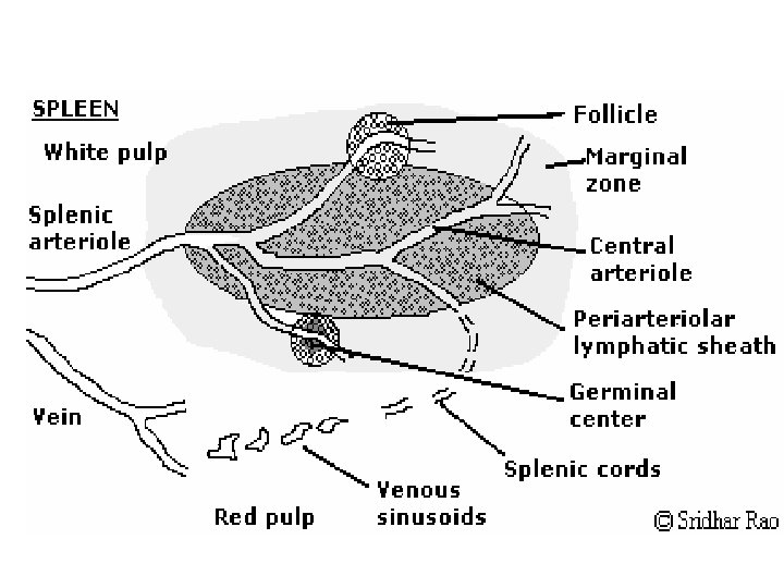
spleen • Weigh 150 g, in left upper quadrant • Immune response against blood borne antigens • Consist of white pulp(inner) – peri-arteriolar lymphoid sheath; PALS ( T cell Zone) – follicles (B cells zone). – Marginal zone in between red and white pulp, have both B and T cells and macrophages. • Red pulp; outer, splenic artery, vascular sinusoid, splenic vein. consist of old erythrocytes and macrophages, It is the place where aged RBC is destroyed by macrophages • The splenic artery enters the red pulp through a web of small blood vessels, and blood-borne microorganisms are trapped in this loose collection of cells until they are gradually washed out through the splenic vein • No afferent lymphatic vessel in spleen.
Spleen functions • Functions – It is the major site for killing antibody coated microbesand destroying the damaged RBC – Storage of RBCs and lymphocytes – Individuals lacking a spleen are extremely susceptible to infections with encapsulated bacteria such as pneumococci and meningococci because such organisms are normally cleared by opsonization and phagocytosis, and this function is defective in the absence of the spleen
Immunity against certain microbe • Immunity can be active or passive – Active; induced by previous exposure to an antigen and host immune cells respond and form antibodies and memory cells – Passive, transfer serum or lymphocytes from specifically immunized individual to not-exposed person (naïve). Maternal Ab to fetus
Important definitions • The immune system Cells in our bone marrow, thymus, and the lymphatic system of ducts and nodes, spleen, and blood that function to protect us. • Antigen Anything causing an immune response, usually foreign material but may be our own tissues. • Pathogen Any disease causing micro-organism. • Tolerance Non-reactivity of the immune system, usually refers to "self" but may include foreign tissue in organ transplants. • Autoimmunity A failure of tolerance, the immune system reacts to self. • Chemokines Molecules released by pathogens and infected tissues to attract cells of the immune system. • Cytokines Signaling molecules released by one cell to cause a response in another. Signaling is extremely important in our immune response. • Innate immunity Protection that is always present. Includes phagocytic (cells that eat other cells) macrophages and dendritic cells. • Adaptive immunity Protection that arises by an immune response, including humoral immunity producing antibodies and cellular immunity.