INTRODUCTION TO ICU INTENSIVE CARE UNIT It can


 There should")
, torniquets and arm board Airways, endotracheal tubes and")


 Admitted to ICU criteria in general � � � � ♯compromized airway ♯GCS")
 Essential management in ICU: (FAST HUG) � F : feedings A : analgesia")


 Hypoxia classification < pao 2 < 60 mmhg 1 - Hypoxic Hypoxia in")

 Blood pressure : is the pressure of circulating blood on")

 Arterial cannulation is used in patients in ICU to")
 The CVP cannula is inserted in to the internal or")


")

")
 and the level of oxygen")
 Partial pressure")


 shivering – not under voluntary control vasoconstriction")
 reduced level of consciousness loss of fine")
 casualty falls to the ground, curls up")


- Slides: 68
INTRODUCTION TO ICU
INTENSIVE CARE UNIT It can be defined as a “service for patients with potentially recoverable diseases who can be benefit from more detailed observation and treatment than is generally available in standard wards and departments”] Or The intensive care unit is a designated area of a hospital facility that is dedicated to the care of patients who are seriously ill.
ICU: intensive care unit � � � ●Highest level of continuous patient (maintenance, care, treatment) ●nurse : patient 1: 1 ●nurse in change 24 hr ●resident doctor 24 hr ●consultant frequent (visit 2 -4 hr) ●facilities to support organ system failure with full monitor care
� � � � HDU: high dependency unit Specialist advice Supportive care ●nurse : patient 1: 2 ●nurse in change 24 hr ●resident doctor availbe immediately ●appropriate level of monitor ●consultant frequent: per call or shift or round
� � � Ward: ●nurse : patient : NURSE/shift ●nurse in change : NURSE/room ●resident doctor availbe ●consultant frequent: per call or round ●facilities to support organ system failure: not necissery
DIFFERENT UNITS There are verity of names depends on specific purpose and the degree of dependency of the patient Many different hospitals have many different terms. Frequently seen are MICU = Medical ICU SICU = Surgical ICU TICU = Trauma ICU or Transplant ICU NICU = Neuro ICU or Neonatal ICU PICU = Pediatric ICU CVICU = Cardiovascular ICU CCU = Coronary Care. Unit CICU = Cardiac ICU BICU = Burn ICU RCU = Renal care unit
The main functions of any ICU is to � Provide optimum life support � � & Provide adequate monitoring of vital � functions.
ICU PATIENTS critical patients (multiple diagnoses, multi-organ failure, immunocompromised and major trauma and post surgery) Move less Malnourished More obtunded / deaden (Glasgow coma scale) Heart, kidney, liver failure etc…
PREPRATION OF THE UNIT The unit should be kept ready all the time which should include the following 1. special bed having the following facilities Head board should be detachable to facilitate intubation (in case of cardio pulmonary arrest) Bed should be firm and non yielding to facilitate cardiac massage Should have a tilting mechanism (to keep position of patient)
Should have side rails to prevent falling (psychiatric and anxious patient) There should be a bed side locker an over bed table and a foot stool kept adjacent to the bed 2. Cardiac monitor system with alarm that may be connected to the central console 3. Oxygen and suction apparatus (preferably pipe line model) 4. Resuscitation unit containing the following Syringes, needles, IV cath, intravenous administration sets, blood sets, scalp vein sets and intra venous fluid
Spirit, swabs, adhesive plaster (micropore/transpore), torniquets and arm board Airways, endotracheal tubes and laryngoscopes of different sizes Ambu bag and suction catheters Oxygen cylenders special trays such as tracheostomy tray, and catheterization tray Drugs such as (antiarrhythmics, antianginals, antihypertensive, diure tics, anticoagulents, antibiotics, anticonvulsants etc… Infusion pump
Following equipments should be easily available Defibrillator in working mode with electrodes and jell Cardiac pacemaker with pacing catheters in the sterile tray Mechanical ventilators (to ventilate the lungs in case of resp: arrest) Facility for invasive and non invasive procedure like (CVP line, intra arterial pressure monitor ) Portable X-Ray machine ECG machine Oxygen therapy
Indication s f o r admission Pre and post-operative patients and who underwent major surgeries. Craniotomy patients. Thoracotomy patients. Ultra major surgeries. Unstable multiple trauma patients. Patients with head or spine trauma requiring mechanical ventilation. Any surgical patient who requires continuous monitoring or continuous life support
(A) Admitted to ICU criteria in general � � � � ♯compromized airway ♯GCS ˂= 12 ♯Unstable vital sign ♯p. H ˂=7. 2. . or 7. 5 ♯paco 2 ˃45 -55 ♯pao 2˂60 HYPOXIA ♯Fio 2˃60%needed
(B) Essential management in ICU: (FAST HUG) � F : feedings A : analgesia S : sedation T : thrombo-embolism prophylaxis � H : head of bed elevation (30 - 45 ) U : ulcer prophylaxis ( stress ulcer , bed sore ) G : glucose control
Intubation indications ☻apnea central decrease GCS ≤ 8 absent gag reflex induced general anesthesia poisoning ☻ Resp. failure drugs arrest disease ARDS , COPD, ASTHMA , sepsis ☻ Resp. airway protection risk of aspiration burn patient ☻ Resp. obstruction trauma , tumor , laryngeal oedema , epiglottitis ☻hemodynamic instability /seve shock , cardiac arrest , sever hypothermia
� � ♥hypoxemia S&S : restlessness , tachycardia , tachypnea, cyanosis Cardiac irritability Obtundation Bradycardia Hypotension and Ischemia cardiac arrest. ♥ hypovolemia S&S : Dehydration+ Decrease B. P , Orthostatic hypotension ± decrease Hb level or blood volume
(D) Hypoxia classification < pao 2 < 60 mmhg 1 - Hypoxic Hypoxia in inadequate ventilation and oxygenation. altitude , asthma , Resp. disease 2 - Anemic Hypoxia in inadequate hemoglobin content OR function. anemia , carboxy Hb , methemo Hb 3 - Circulatory Hypoxia in inadequate perfusion CHF 4 - Histotoxic Hypoxia in inability of the cells to utilize oxygen. Cyanide , salicylic acid poisoning 5 - DEMAND HYPOXIA increase O 2 requirement fever , seizure.
Monitorin g system Topics : 1 - Non-Invasive BP measurement 2 - cental line ( central venous catheter ) 3 - Invasive arterial line 4 - Capnography 5 - pulse oximeter 6 - Arterial Blood gas (ABG) 7 - Temperature 8 - Oxygen analyzer 9 - Urine catheter
Noninvasive Blood Pressure (NIBP) Blood pressure : is the pressure of circulating blood on the walls of blood vessels. In ICU we monitor blood pressure every (1 -4) hours. Why do we monitor BP ? ! 1. It Provides data for therapeutic decisions. 2. Important for determining organ perfusion. 3. One of the vital signs.
Non-invasive methods 1. Palpation 2. Auscultatory 3. Oscillometry
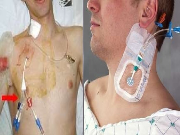
INVASIVE BLOOD PRESSURE MONITORING (IBP) Arterial cannulation is used in patients in ICU to access arterial blood sample , for checking ABG and for arterial pressure monitoring Arterial cannula is not used for intra vascular drug administration A saline-filled tube is used to connect the cannula to the transducer, to the display. It measures IBP on beat to beat basis.
CENTRAL VENOUS PRESSURE (CVP) The CVP cannula is inserted in to the internal or external jugular vein or subclavian vein The tip is situated approximately 2 cm above the right atrium in the superior vena cava They provide access for intra venous drugs particularly which produce irritation to peripheral veins eg-strong potassiumchloride
Complications of Central Line : � Pneumothorax - 30% of all mechanical adverse events of Central Line insertion - (for central lines placed in the chest); the incidence is thought to be higher with subclavian vein catheterization. In catheterization of the internal jugular vein, the risk of pneumothorax is minimized by the use of ultrasound � Haemothorax � Arterial puncture � Air embolism
Arterial Line
Ø Definition: An arterial line or art-line is a thin catheter inserted into an artery Ø Most commonly used in I. C. U and anesthesia to monitor the BP and it’s more accurate than measurement of BP by noninvasive means, and to obtain continuous samples for ABGS Ø Arterial lines can be placed in multiple arteries, including: the Radial, Ulnar, Brachial, Axillary, Posterior tibial, Femoral, and Dorsalis pedis Ø � � Mostly inserted in Radial artery because its larger and more superficial, Low complication The second Most common is Femoral artery: larger and strong pulsation
Allen’s test q q Allen’s test is recommended before the insertion of radial arterial line. It is used to determine collateral circulation between the Ulnar and Radial arteries to the hand. � q q Collateral flow should retain within 5 -10 sec More than 10 sec >> insufficient collateral blood flow Disadvantage : Need Cooperation with the patient Alternative : Doppler probe, Pulse oximeter , pletysmography
Allen test Technique v The hand is elevated and the patient is asked to clench their fist for about 30 seconds Pressure is applied over the ulnar and the radial arteries so as to occlude both of them
v Still elevated, the hand is then opened, It should appear blanched
v Ulnar pressure is released while radial pressure is maintained the color should return
Indications for Arterial Catheterization v Patient Factors: � The need for continuous BP measurement in patients who are morbidly obese and severe burn � Patient with severe sepsis or shock. � Cardiac diseases. � Respiratory Failure. � Vasopressor requirement. � Hemodynamic instability. �
Surgical considerations: � Cardiac Surgery. � Major surgery on aorta or carotid artery. � Neurosurgery as craniotomy or aneurysm clipping. v Anesthetic considerations: � Controlled hypotensive technique. � Inability to measure blood pressure noninvasively. v
� Arterial line Contraindications v Absolute Contraindications: Absent Pulse. Thromboangitis obliterans. Full thickness burns over the Raynaud syndrome. v Relative Contraindications: Hemorrhage Angiopathy Coagulopathy Atherosclerosis cannulation site.
� Complication of Arterial line insertion � Common complications of arterial line placement are as: - Temporary radial artery occlusion (19. 7%) - Hematoma /Bleeding (14. 4%) � Less common and rare complications include the following: - Localized catheter site infection Hemorrhage Sepsis Permanent ischemic damage Thrombosis Air embolism Carpel tunnel syndrome Areteriovenous fistula
Oxygen saturatio n Sp. O 2 The pulse oximeter measures the oxygen saturation It is noninvasive and risk free when used properly, the pulse oximeter should be used in all clinical settings in which there is a potential risk of arterial hypoxemia It provide an early and immediate warning of hypoxaemia If Sp. O 2 is below 95% means the O 2 delivering system is inadequate to meet the needs of the tissue or poor cardiac output Start O 2 if SPO 2 less than 95%
pulse oximeter
CAPNOGRAPHY *Capnography is a valuable monitor of the pulmonary, cardiovascular, and anesthetic : breathing systems using 1 -A numerical value of the Et. CO 2 measured by capnometer AND 2 -A waveform of the concentration of CO 2 present in the airway presented by capnogram *Capnographs in common use rely on the absorption of infrared light by CO 2. *As with oximetry, absorption of infrared light by CO 2 is governed by the Beer– Lambert law. �
ETCO 2 *is the partial pressure or maximal concentration of carbon dioxide (CO 2) at the end of an exhaled breath, which is expressed as a percentage of CO 2 or mm. Hg. *The normal values are 5% to 6% CO 2, which is equivalent to 35 -45 mm. Hg. *ETCO 2 Less Than 35 mm. Hg -“Hypocapnia” - Respiratory Alkalosis *ETCO 2 Greater Than 45 mm. Hg -“Hypercapnia” - Respiratory Acidosis
Indications for Use End-Tidal CO 2 Monitoring 1 -Validation of proper endotracheal tube placement 2 -Detection and Monitoring of Respiratory depression 3 -Hypoventilation 4 -Obstructive sleep apnea 5 -Procedural sedation 6 -Adjustment of parameter settings in mechanically ventilated patients
ARTERIAL BLOOD GAS (ABG)
Arterial blood gases ABG test measure the acidity (PH) and the level of oxygen and carbon dioxide I in the blood from an artery. � This test is used to check how well your lungs are able to move oxygen in to the blood and remove carbon dioxide from the blood. � Blood drown from an artery , where the oxygen and carbon dioxide levels can be measured before thay enter body tissues. �
ABG measurs: � � � Partial pressure of oxygen (Pa. O 2) Partial pressure of CO 2 (Pa. CO 2) PH Bicarbonate (HCO 3) Oxygen saturation value (O 2 Sat)
Normal ABG values � PO 2 80 – 100 mm. Hg � PCO 2 35 – 45 mm. Hg � p. H 7. 35 – 7. 45 � HCO 3 22 – 26 mmol/L � Sa. O 2 >95%
contraindication � � � Bleeding diathesis AV fistula Severe peripheral vascular disease, absence of an arterial pulse
complication � The most common complication from anarterial puncture is : � Hematoma at the site. � Less common but important complications are thrombus in the artery and infection at the site
What artery to choose? The radial artery � is superficial, has collaterals and is easily compressed. It should almost always be the first choice. � Other arteries (femoral, dorsalis pedis, brachial) can be used in emergencies. �
Patien t Temperature regulation is important to the survival of the patient Although uncommon, hypothermia below 32° C is ominous Ventricular irritability increases, and if the temperature decreases to 28° C cardiac arrest is likely shivering can increase oxygen demand 135% to 468%, when respiratory and cardiovascular systems may be unable to respond normally to the increased demand
Sites f o r monitoring body temperature 1. Oral. 2. Tympanic membrane 3. Esophageal 4. Nasopharyngeal 5. Pulmonary arterial blood 6. Rectal 7. Bladder 8. Axillary 9. Forehead
Hypothermia � Hypothermia is a potentially dangerous drop in body temperature, usually caused by prolonged exposure to cold temperatures.
What Causes Hypothermia? � Cold exposure. � Certain medical conditions such as and thyroid diabetes conditions, some medications, severe trauma, or using drugs or alcohol all increase the risk of hypothermia.
Temperatures for mild, moderate, and severe hypothermia generally range from:
� � � Mild Hypothermia (35 -32°C) shivering – not under voluntary control vasoconstriction to periphery (fingers, toes etc)
� � � Moderate hypothermia (32 -28) reduced level of consciousness loss of fine motor coordination, particularly in hands due to restricted peripheral blood flow slurred speech violent shivering
� � � Severe hypothermia (< 28°C) casualty falls to the ground, curls up into a fetal position to conserve heat muscle rigidity develops because peripheral blood flow is reduced and due to lactic acid and carbon dioxide buildup in the muscles the skin becomes pale pupils dilate pulse rate decreases
What Is the managem ? for Hypothermia � Hypothermia is a potentially life-threatening condition that needs emergency medical attention. � Remove any wet clothes, hats, gloves, shoes, and socks. Protect the person against wind, drafts, and further heat loss with warm, dry clothes and blankets. Move gently to a warm, dry shelter as soon as possible. Begin rewarming the person with extra clothing. Use warm blankets. � � �
Take the person's temperature if a thermometer is available. � Offer warm liquids, but avoid alcohol and caffeine, which speed up heat loss. Don't try to give fluids to an unconscious person. � � � CPR should be continued, in the absence of signs of breathing or a pulse, rewarm the core temperature. Hypothermia treatment may include warmed IV fluids, heated and humidified oxygen, peritoneal lavage (internal "washing" of the abdominal cavity), and other measures.
Monitoring the Urine Output
� Urine output is the best indicator of the state of the patient’s kidneys. If the kidneys are producing an adequate amount of urine it means that they are well perfused and oxygenated. Otherwise, it is a sign that the patient is suffering from some complication
Physical examination of urine Examination of physical characteristics � � � • Volume • Color • Odor • p. H • Specific gravity
Volume: � � �Normal- 1 -2. 5 L/day �Oliguria- Urine Output < 400 ml/day Seen in – Dehydration – Shock – Acute glomerulonephritis – Renal Failure �Polyuria- Urine Output > 2. 5 L/day Seen in – Increased water ingestion – Diabetes mellitus and insipidus. �Anuria- Urine output < 100 ml/day Seen in renal shut down Volume
colour �Normal- pale yellow in color due to pigments urochrome, urobilin and uroerythrin. � � Cloudiness may be caused by excessive cellular material or protein, crystallization or precipitation of non pathological salts upon standing at room temperature or in the refrigerator. � � Colour of urine depending upon it’s constituents. � Abnormal colors: • Colorless – diabetes, diuretics. • Deep Yellow – concentrated urine, excess bile pigments, jaundice Color �
Odor � � Normal - aromatic due to the volatile fatty acids, On long standing – ammonical (decomposition of urea forming ammonia which gives a strong ammonical smell) • Foul, offensive - pus or inflammation • Sweet - Diabetes • Fruity - Ketonuria
PH � � Reflects ability of kidney to maintain normal hydrogen ion concentration in plasma & ECF Normally it is slightly acidic lying between 6 – 6. 5. • Acidic Urine – Ketosis (diabetes), UTI- E. coli Specific gravity • the ability of the kidney to concentrate the urine relative to the plasma from which it is filtered. • Normal : - 1. 001 - 1. 040. �Increase in Specific Gravity - Low water intake, Diabetes mellitus, Albuminuruia, Acute nephritis. �Decrease in Specific Gravity - Absence of ADH, Renal Tubular damage.
� � � To measure urine output in critical care units, a Foley catheter is introduced through the patient’s urethra until it reaches his/her bladder. The other end of the catheter is connected to a graduated container that collects the urine. Periodically the nursing staff manually records the reading of the container of every patient, and operates a valve which releases the urine into a larger container. To avoid urine reflux and minimize the risk of infection, the chamber should remain at a level below the bladder. Keep the urinary catheter bag lower than the patient!!!
� often only burn patients—for whom urine output monitoring is of paramount importance—have this parameter recorded every hour, while the remaining critical patients have it recorded every 2 or 3 hours.
Complications of catheterization, urethral trauma & UTI. Rapid decompression of a distended bladder can cause hypotension. An additional advantage of placing Foley catheter is the ability to include a thermistor in the catheter tip so that bladder temp can be monitored. As long as urinary output is high, bladder temp. accurately reflects core temp.
Thank You