Introduction to human embryology gametogenesis fertilization cleavage Dr

Introduction to human embryology gametogenesis, fertilization, cleavage Dr. Zita Puskár
 Vitelline membrane Size: ~125 µm Akoury et al. , 2014,")
Female gamete: Oocyte (egg) Vitelline membrane Size: ~125 µm Akoury et al. , 2014, Hum Reprod
 ZP: specialized extracellular matrix (ECM) which is produced by the oocyte")
Zona Pellucida (ZP) ZP: specialized extracellular matrix (ECM) which is produced by the oocyte and follicular granulosa cells Main glycoproteins: ZP 1, ZP 2, ZP 3, ZP 4 Function: protection, transport, sperm binding, prevention of polyspermy, support of blastocyst development, prevention of early implantation ZP 3 -ZP 4 matrix formation ZP 3: primary (species specific) sperm binding receptor ZP 2: secondary sperm binding (recognition) receptor (It is cleaved by ovastacin released from cortical granules after fertilization and other sperm can not bind to the oocyte) It plays an important role in defence against polyspermy. embryology. med. unsw. edu. au

Without species specific ZPs…
 farok")
Male gamete: Sperm (spermatozoon) farok

Gametogenesis: conversion of germ cells into male and female gametes 3 rd week 4 th-5 th week Prenatal events: Primordial germ cells appear in the wall of the yolk sac at the end of the 3 rd week of development. Germ cells migrate from the yolk sac toward the developing gonads (primitive sex glands). They arrive at the end of the 4 th or the beginning of the 5 th week.
 meiosis (gamets) 46 46 92 92 46 23 46 46 46")
mitosis (somatic cells) meiosis (gamets) 46 46 92 92 46 23 46 46 46 23 23 23
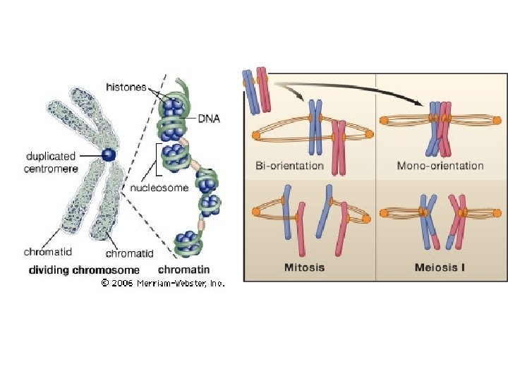

! Double chromatids
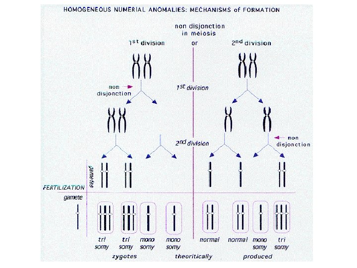

Amniocentesis

21 - trisomy: Down syndrome 21/03/2014

Spermatogenesis: is the sequence of events by which spermatogonia are transformed into mature gametes: sperm (spermatozoa). 1. Spermatocytogenesis : asymmetric division of spermatogonia. Some of them remain „stem cell”, others start the differentiation process. 2. Meiosis: meiotic division of spermatocytes (I and II) 3. Spermio (histo)genesis: spermatids are transformed to sperm that can fertilize eggs Classification depends on books!

Spermatocytogenesis and meiosis Postnatal events - at puberty Spermatogonia → mitosis, growth → primer spermatocytes → first meiotic division → secunder spermatocytes → second meiotic division → spermatids
 Formation of acrosome cap (lysosome-hydrolytic enzymes for")
Spermiohistogenesis: transformation of spermatids into spermatozoa (a) Formation of acrosome cap (lysosome-hydrolytic enzymes for acrosomal reaction of fertilization) (b) Condensation and elongation of the nucleus (c) Development of flagellum (formation of neck, middle piece and tail) (d) Shedding of most of the cytoplasm

Sperm under the microscope

Oogenesis 700 000 -2 million 400 000 500 <

Follicular growth

Menstrual cycle proliferative secretory Contraceptive pills - combination of estrogen and progesteron or progesteron only (mini pills) - prevent (i) the release of FSH and LH from the pituitary gland → inhibition of ovulation (but permit menstruation) (ii) preparation of the lining of the uterus for implantation and (iii) changing the uterine mucus to avoid sperm penetration into the egg.

Ovulation

Fertilization: process by which male and female gamets fuse

Before fertilization 200 -300 million spermatozoa arrive in the female genital tract → 300500 reach the site of ferilization → only 1 fertilize Oocyte and spermatozoa remain viable for approximately 24 hours and 4 - 5 days respectively Spermatozoa are not capable of fertilizing oocyte!!! They must undergo (a) Capacitation (b) Acrosomal reaction

Capacitation Heparin FPP from prostate: fertilization promoting peptide Increased Ca 2+-influx → increased motility
,")
Capacitation and acrosomal reaction Capacitation: conditioning in the female reproductive tract (approx 7 hours), glycoprotein coat and seminal plasma proteins are removed that overlies the acrosomal region → hyperactivation of motility Acrosomal reaction: afer binding to zona pellucida, release of enzymes (acrosin, trypsin like substances) induced by zona pellucida proteins

Cannabinoids and male fertility Chronic administration of THC is correlated with male infertility (Wang et al 2006). Sperm express CB 1 receptor and its activation inhibits capacitation and acrosome reaction (Rossato et al 2005).

The 3 phases of fertilization Phase 1: Penetration of the corona radiata Phase 2: Penetration of the zona pellucida Phase 3: Fusion of the oocyte and sperm cell membranes

Phase 1: Penetration of the corona radiata Capacitated sperm penetrates the barrier corona radiata by „the corona radiata penetrating enzyme”, hyaluronidase, phosphatase and acrosine. The enzymes are released from the plasma membrane of the sperm, that break apart the cell junctions and ECM.

Phase 2: Penetration of zona pellucida The outer membrane of the sperm binds to ZP 3 receptor of the ZP → (acrosomal reaction begins) formation of composite membrane → release of enzymes (neuramidase and acrosine) → breaking down ZP → sperm reaches the vitelline membrane of the oocyte
 connects to integrin")
Cortical reaction When fertilin (on the head of the fastest sperm) connects to integrin of the oocyte vitellin membrane, Ca 2+-influx (depolarization) increases and then exocytosis of cortical granules (CG) occurs. Enzymes released from the CG cleave ZP molecules and inhibit the docking of other sperm. Other peptides from CG facilitate the increase of osmotic pressure → the periveitelline space takes up water and expands. The structural composition of the membrane changes. These events prevent polyspermy.

Phase 3: fusion of the oocyte and sperm cell membrane Membrane of the oocyte and the inner acrosomal membrane of the sperm fuse → head and tail of the sperm enter the cytoplasm of oocyte but the plasmamembrane is left behind. „Only maternal mitochonria will be in the zygote. ” The oocyte finishes its second meiotic division (definitive oocyte and 2 nd polar body). Female pronucleus and sperm pronucleus are in close contact (DNA replicated) and loose their nuclear envelope → metabolic activation, initiation of cleavage

Results of the fertilization 1. Restoration of the diploid number of chromosomes (new combination of chromosomes that differs from both parents) 2. Determination of sex (XX- female embryo, XY- male embryo) 3. Initiation of cleavage

Cleavage Zygote in 2 -cell stage → series of mitotic division → increase in cell number – cells become smaller and arranged loosely (blastomeres) → following the 3 rd cleavage blastomers form a compact ball (tight junctions are among the cells) → segregation of inner and outer cells (communication with gap junctions) → morula, mulberry (16 -cell stage)
 4")
Microscopic view fusion 8 -cell stage compactation pronuclei 2 -cell stage (30 hour) 4 -cell stage (40 hour) 16 -cell stage (3 days) morula, mulberry blastocyst

Blastocyst formation Morula enters the uterine cavity, fluids began to penetrate through the zona pellucida into the intercellular spaces → single cavity the blastocele is formed → blastocyst stage : inner cell mass-embryoblast, outer cell mass-trophoblast (epithelial wall). Zona pellucida disappears → implantation begin

After fertilization

Events in the female genital tract

Scale of prenatal development

In vitro fertilization
- Slides: 39