Introduction To Hematology READ Chapter 12 AP Chapter


")


 is found in early developing")


- Slides: 52
Introduction To Hematology READ: Chapter 12 A&P Chapter 2 Lab Pro
Learning Objectives • Formation and maturation of blood • Composition of blood • Functions of blood • Hemoglobin structure • Staining procedures
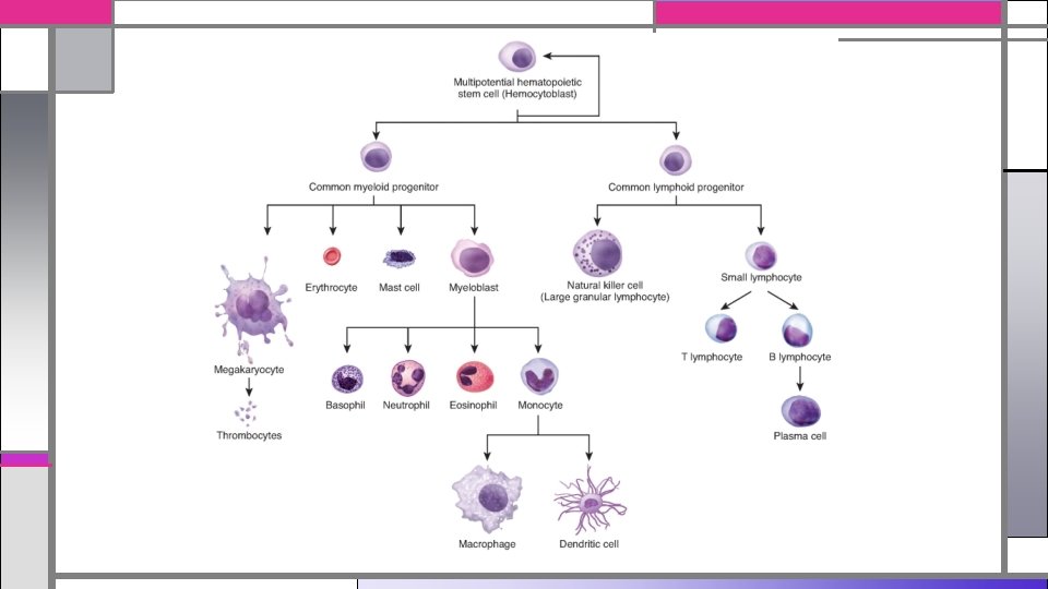
Where does blood come from? * Hematopoiesis is the production of ALL blood cells that occur as a continuous process throughout an animal's life. – Where does that happen? • Fetus: Liver and spleen • Newborn animal: Red bone marrow • Adult: Red bone marrow (skull, ribs, sternum, vertebra, pelvis, proximal femurs
Where do ALL blood cells come from? • Pluripotential or multipotential stem cells are primitive stem cells that have the potential to replicate and differentiate into many different types of unipotential stem cells. • These stem cells then commit to one of three maturation processes. – erythropoiesis – leukopoiesis – thrombopoiesis
Erythropoiesis
Erythropoiesis • This is the process by which RBCs are created. – Unipotential stem cells are stimulated to differentiate into proerythroblasts. – The rate at which this happens is controlled by hormones, mainly Erythropoietin; EPO and the availability of the materials needed to make RBCs: iron, folic acid, vitamin B 12 and protein – Where is EPO produced? kidneys – To consider: What does hypoxia have to do with this process? ? – Regulated by blood O 2 levels in the kidney
Maturation cycle of a RBC
Morphological Changes During Maturation • Rubriblast – Very large with royal blue cytoplasm – Contains a large purple nucleus in a loose chromatin pattern • Prorubricyte – Slightly smaller, with a basophilic cytoplasm – Chromatin composing the nucleus is slightly smaller and denser and stains purple
Morphological Changes During Maturation, continued… • Rubricyte – Smaller still with either a basophilic or slightly purple cytoplasm as it begins to acquire the hemoglobin molecules • Metarubricyte – Cytoplasm is similar in color to mature RBC – Nucleus is now considered pyknotic (more dense) and stains dark blue – Polychromasia may be seen here
Morphological Changes During Maturation, continued… • Reticulocyte – Nucleus is ‘pushed out’ of cell, leaving behind reticulum composed of RNA – Cytoplasm stains slightly blue, and Howell-Jolly bodies may be seen if stained with Wright’s stain. – Mature RBC is seen after ALL reticulum is lost from reticulocyte.
Structure of a mature RBC • Lack a nucleus, mitochondria and ribosomes • Contain: – Water – Hemoglobin – Other structural elements • When viewed microscopically, mature RBCs appear as nonnucleated, biconcave discs with a central zone that is thinner and appears lighter ( central pallor ) on a stained blood smear. • Since they have no mitochondria, RBCs utilize glucose from plasma for energy • Hemoglobin dissolved in RBC cytoplasm makes them stain red
Structure of a mature RBC, continued… • An RBCs membrane is ‘deformable’, it can change shape to move through vessels, but it is not elastic • The bioconcave shape provides more surface area to allow for gas exchange to take place • Disk shape allows for shorter diffusion distance in and out of the cell, compared to a spherical shape – Animals with less bioconcave shape usually have higher number of RBCs on average.
RBC differences among species • Dogs have the largest red blood cells with a prominent central zone of pallor • Cats, horses, cow, sheep, and goats have smaller RBCs • Cats and horses do not have a prominent central zone of pallor • Camelids (llamas, camels, and their relatives) have elliptical or oval RBCs • Deer have sickle-shaped RBCs • Birds, fish, amphibians, and reptiles have elliptic RBCs that are nucleated even when mature
Thrombopoiesis
Thrombopoiesis • This is the process where platelets are created. • Production starts when there is a specific stimulant acting upon a unipotential stem cell in the red bone marrow. The first phase of growth is called a megakaryocyte. • The megakaryocyte is a large multinucleated cell that never leaves the bone marrow. • Pieces of cytoplasm from the megakaryocyte are released into the peripheral blood as platelets. • Thrombopoiesis can take up to 7 days to reach completion
Leukopoiesis
Leukopoiesis • This is the general term for the formation of WBCs. They all start out with the same pluripotential stem cells, but then each WBC has its own stimulus for production. Granulopoiesis Lymphopoiesis Monopoiesis • Neutrophils • Eosinophils • Basophils • Lymphocytes • Monocytes
Blood 101: Quick facts • Blood is a fluid connective tissue that flows throughout the entire body • Whole blood is contained within the cardiovascular system • Peripheral blood is whole blood circulating in blood vessels carrying oxygen, nutrients, and waste materials. – Blood samples are taken from peripheral blood sites
What is in that blood? • Liquid portion: Plasma – What is it made of? – Primarily water and solutes – Cellular portion contains: – Erythrocytes, leukocytes and platelets
Composition of Blood Chart Whole blood _____93__% fluids and tissues _____7____ % blood Percentages based on body weight Plasma ___55___% Percentages based On volume Lets talk first about plasma Formed elements __45____%
Formed elements, what’s are they? 60 -77% 12 -30% 3 -10% 2 -10% rare
What happens when you take whole blood and centrifuge it down? plasma Buffy coat erythrocytes
Plasma Colors
Close up of a lipemic sample
Functions of the Blood
Blood Functions • Blood has three main functions • Transportation • Regulation • Defense
Transportation • Erythrocytes contain _______hemoglobin_____ which carry oxygen to every cell in the body. They do this by using hemoglobin. • Hemoglobin is made up of _____4______ heme units associated with one globin chain. Each heme unit is the pigmented portion of hemoglobin. • Each heme unit contains an iron atom to which _____1_______ molecule of oxygen molecule can attach. • Therefore, one hemoglobin molecule can carry ____4______ molecules of oxygen. – What is oxyhemoglobin? Hemoglobin with O 2 bound – What is deoxyhemoglobin? When hemoglobin gives up its oxygen
Transportation, continued… • Nutrients and other essential elements are dissolved in the blood plasma and transported to tissues via arteries and capillaries • Blood carries waste from cellular metabolism via veins to the lungs and kidneys • Blood transports hormones from the endocrine glands to target organs as well as WBCs to various sites • Blood transports platelets to sites of damage in blood vessel walls to form a plug to control bleeding, this is a mechanism called hemostasis. They are also involved in the activation of blood-clotting cascade
Regulation • Blood aids in regulation of body temperature. Where are your body temperature regulators? _____brain_______ • Blood plays a part in tissue fluid content. – What is hemoconcentration? Less plasma in the bloodstream, cells are more concentrated (vomiting, diarrhea, profuse sweating, other pathologic conditions – What is hemodilution? Extra plasma in the bloodstream; excessive SQ fluids
Regulation, continued… • Blood aids in the regulation of blood p. H (acid-base balance) • What is the normal p. H range? 7. 35 – 7. 45 • Which part of the blood is slightly more alkaline because most of the acidic waste products are carried there? Arterial blood Death 6 Acidosis 7 Normal p. H 7. 35 Death Alkalosis 7. 45 7. 8 9
Defense • Blood carries WBC to tissues exposed to foreign invaders. • Blood carries platelets to sites of vessel damage to aid in hemostasis so that the animal does not bleed excessively.
Hemoglobin
Attaches to erythrocytes to carry _____oxygen_______ Hemoglobin and how it assists the RBCs ___heme______ = pigment portionheme Produced in mitochondria globin = protein portion Produced by ribosomes
Hemoglobin Types • Each type of hemoglobin is found during different stages of development of the animal – Embryonic hemoglobin (Hb. E) – Fetal hemoglobin (Hb. F) – Adult hemoglobin (Hb)
Embryonic and Fetal Hemoglobin • Embryonic hemoglobin (Hb. E) is found in early developing fetuses • Fetal hemoglobin (Hb. F) is present in fetal blood during mid to late gestation – It is also present up to a couple of months after birth – Highest concentration of Hb. F is at birth • Adult hemoglobin (Hb) is found in the RBCs of all animals beginning a couple of weeks to months after birth – They gradually replace Hb. F as primary type being produced by the body – Inside normal RBCs, Hb is conjugated
Adult Hemoglobin Function • Transporter of ______oxygen_____ to the tissues • Exists in two normal physiological states: • Oxyhemoglobin: carrying O 2 • One oxygen molecule is associated with each iron molecule • p. H, temperature, and O 2 and CO 2 levels influence ability of Hb to carry O 2 – Deoxyhemoglobin: having given up O 2 • Also known as empty hemoglobin • CO 2 is transported directly and indirectly in the RBC and is dissolved in the plasma
Senescence
Erythropoietic Cycle: Senescence • What is senescence? The process of aging • What is the average lifespan of a RBC in a dog? _____120_________ – What is the average lifespan of a RBC in a cat? ______68_________ – What happens as RBCs wear out, age and die? They are replaced by young RBCs from the red bone marrow
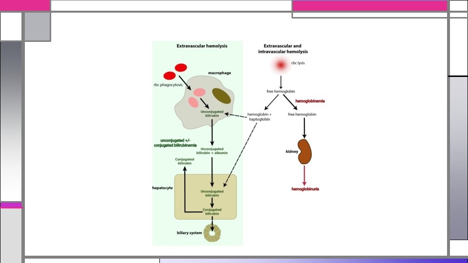
Senescence, continued… • When an RBC membrane ruptures in the blood vessel, hemoglobin is released directly into the blood. • Unconjugated (unbound) Hb is picked up by a transport protein called haptaglobin • Haptaglobin carries the unconjugated Hb to macrophages in the liver • Once in the liver, extravascular hemolysis continues
Senescence, continued… Extravascular Hemolysis About 90% of the destruction of senescent RBCs occurs by extravascular hemolysis. • Once inside a macrophage, the membrane of the RBC is destroyed. • Contents are recovered from the macrophages and iron is transported back to the red bone marrow • Heme is eliminated from the body, but is converted to bilirubin and carried to the liver by albumin (plasma protein), it is called unconjugated or ‘free’ bilirubin – Bilirubin: The yellow breakdown product of normal heme catabolism, caused by the body clearing out aged RBCs which contain Hb – Serum albumin: Globular protein that helps important molecules attach to the cell.
Senescence, continued… Extravascular Hemolysis • Once in the liver, bilirubin is conjugated to flucuronic acid, making the combination water soluble • Conjugated bilirubin is excreted as bile pigment into the intestines. – Some conjugated bilirubin is converted into urobilinogen by bacteria and eliminated in the urine as urobilin (yellow) – Other conjugated bilirubin is converted into stercobilinogen and excreted in the stool as stercobilin (brown)
Senescence, continued… Intravascular Hemolysis About 10% of normal RBC destruction takes place by intravascular hemolysis. Meaning that the destruction takes place within blood vessels. • When a RBC membrane ruptures within a vessel, hemoglobin is released directly into the bloodstream. • The hemoglobin released is unconjugated, and is quickly picked up by haptaglobin, which is a transport protein in plasma, to form haptoglobinhemoglobin complex • This complex travels to the macrophages in the liver for further breakdown, similar to what happens in extravascular hemolysis.
Senescence, continued… Intravascular Hemolysis • When haptoglobin is filled with unconjugated hemoglobin, as in cases of severe hemolysis, excess unconjugated hemoglobin appears in the plasma, making it pink, red or brown in color. This is referred to as hemoglobinemia • The excess of unconjugated hemoglobin has no way to get to the liver, so it is carried to the kidney, where it is eliminated in the urine, making it red in color. This is referred to as hemoglobinuria
What will you see in your patient when excess RBCs blood cells breakdown? Patient will exhibit jaundice or icterus. This can be associated with liver diseases. If the bile ducts are obstructed the conjugated bilirubin can’t be passed with bile into the intestines, conjugated bilirubin will eventually back up into the blood stream and then the tissues.
Staining of Blood
Staining of Blood Different structures within the cell stain specific colors There are many different hematology stains • Different names but have similar staining characteristics • Many are Romanovsky-type stains (Wright’s stain, Giemsa, Leishman’s, Wright. Giemsa, and May-Grunwald) • All are polychromic stains • Combination of basic blue and acidic red dyes dissolved in ethyl alcohol • Allows acidic structures to stain _______red____ • Allows alkaline structures to stain _______blue_____ • Supervital stain is a type of stain used when components of a cell need to be visualized, but cannot be picked up by the Romanovsky-type stains
Staining of Blood • Stain depends on what test you are doing • Each test we do here will have specific staining instructions • Wright’s stain is the most widely used hematology stain – Alkaline part is methylene blue – Acidic part is eosin • Modified Wright’s stains offer faster staining times, but don’t stain some cellular structures as effectively – Diff-Quick is a Modified Wright’s stain
Staining of Blood • Reticulocytes are immature erythrocytes that contain organelles that are lost as the cell matures • Most species have just an aggregate form, except the cat, which has a unique punctate form of reticulocyte • The punctate form contains just 2 -8 small basophilic granules.
Staining of Blood • Performing a reticulocyte count: – Purpose: Performed on the blood of all anemic domestic animals (except horses). This count is useful in assessing the bone marrow’s response to anemia – Procedure: Prepare two blood films and count 500 RBCs on each slide, keeping track of how many out of the 500 cells are reticulocytes (if you have a cat, you do not count the punctate reticulocytes as a reticulocyte) – Results: Reticulocyte count is an expression of the percentage of RBCs that are reticulocytes.