
Introduction to fracture Ass Prof Dr Zaidw AlShahwanii

- Slides: 30
Introduction to fracture Ass. Prof. Dr. Zaidw. Al-Shahwanii Consultant Orth. Surgeon MB. CHB. FICMS (orth. )
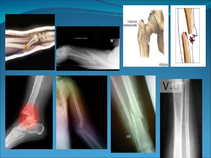
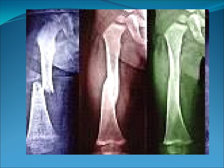
Definition A fracture is a complete or incomplete break in a bone resulting from the application of excessive force. ( i. e a fracture usually results from traumatic injury to bones causing the continuity of bone tissues or bony cartilage to be disrupted or broken ). Up to the age of 50, more men suffer from fractures than women because of occupational hazards. However, after the age of 50, more women suffer fractures than men because of osteoperosis
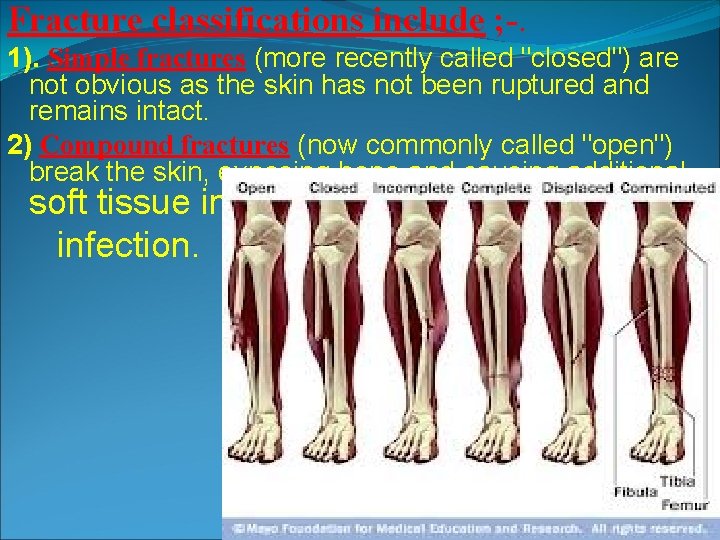
Fracture classifications include ; -. 1). Simple fractures (more recently called "closed") are not obvious as the skin has not been ruptured and remains intact. 2) Compound fractures (now commonly called "open") break the skin, exposing bone and causing additional soft tissue injury and possible infection.
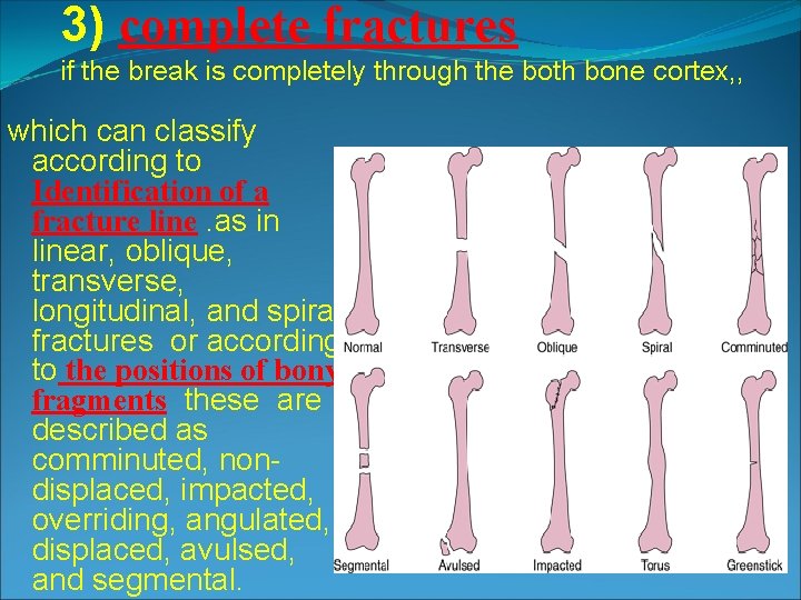
3) complete fractures if the break is completely through the both bone cortex, , which can classify according to Identification of a fracture line. as in linear, oblique, transverse, longitudinal, and spiral fractures or according to the positions of bony fragments these are described as comminuted, nondisplaced, impacted, overriding, angulated, displaced, avulsed, and segmental.
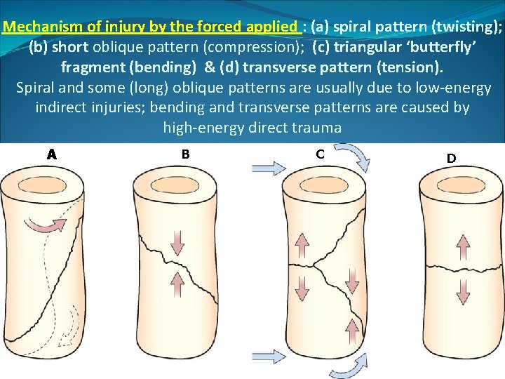
Mechanism of injury by the forced applied : (a) spiral pattern (twisting); (b) short oblique pattern (compression); (c) triangular ‘butterfly’ fragment (bending) & (d) transverse pattern (tension). Spiral and some (long) oblique patterns are usually due to low-energy indirect injuries; bending and transverse patterns are caused by high-energy direct trauma A B C D
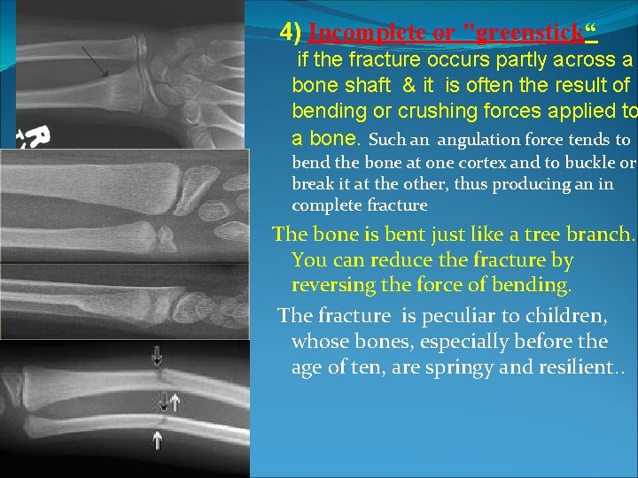
4) Incomplete or "greenstick“ if the fracture occurs partly across a bone shaft & it is often the result of bending or crushing forces applied to a bone. Such an angulation force tends to bend the bone at one cortex and to buckle or break it at the other, thus producing an in complete fracture The bone is bent just like a tree branch. You can reduce the fracture by reversing the force of bending. The fracture is peculiar to children, whose bones, especially before the age of ten, are springy and resilient. .
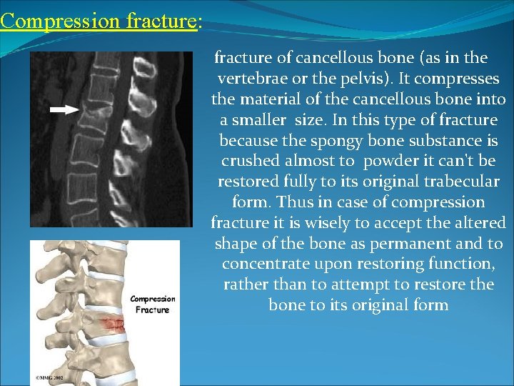
Compression fracture: fracture of cancellous bone (as in the vertebrae or the pelvis). It compresses the material of the cancellous bone into a smaller size. In this type of fracture because the spongy bone substance is crushed almost to powder it can't be restored fully to its original trabecular form. Thus in case of compression fracture it is wisely to accept the altered shape of the bone as permanent and to concentrate upon restoring function, rather than to attempt to restore the bone to its original form
5) A single fracture means that one fracture only has occurred and multiple fractures refer to more than one fracture occurring in the same bone. 6). Fractures are also classify according to the specific part of the bone involved and the nature of the break. eg: # neck femur 7) Lastly, an injury may be classified as a fracture-dislocation when a fracture involves the bony structures of any joint with associated dislocation of the same joint FRACTURE DISPLACEMENT ; - The fracture may be undisplaced or usually 1. 2. 3. 4. 5. displaced according to the type of injury and the direction of fracture line, also depends on localized muscle action and gravity; displacement described according to the direction of distal segment, this can be in form of: Shift (the fragments are shifted to one side as anterior, medial or lateral. . . and the axis of the two pieces is just the same). Overlap (some part of the bone overlapping on the segment above) Impaction (pieces getting jumped into each other) Tilt (when there is an angulation) Twist/ rotation: the limb may be aligned well but if you look at the foot or knee there is either internal or external rotation.
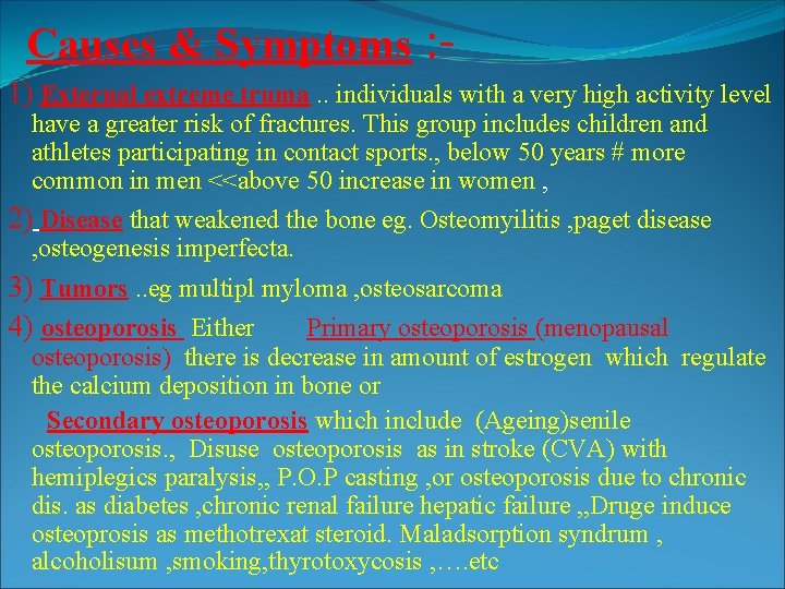
Causes & Symptoms : 1) External extreme truma. . individuals with a very high activity level have a greater risk of fractures. This group includes children and athletes participating in contact sports. , below 50 years # more common in men <<above 50 increase in women , 2) Disease that weakened the bone eg. Osteomyilitis , paget disease , osteogenesis imperfecta. 3) Tumors. . eg multipl myloma , osteosarcoma 4) osteoporosis Either Primary osteoporosis (menopausal osteoporosis) there is decrease in amount of estrogen which regulate the calcium deposition in bone or Secondary osteoporosis which include (Ageing)senile osteoporosis. , Disuse osteoporosis as in stroke (CVA) with hemiplegics paralysis, , P. O. P casting , or osteoporosis due to chronic dis. as diabetes , chronic renal failure hepatic failure , , Druge induce osteoprosis as methotrexat steroid. Maladsorption syndrum , alcoholisum , smoking, thyrotoxycosis , …. etc
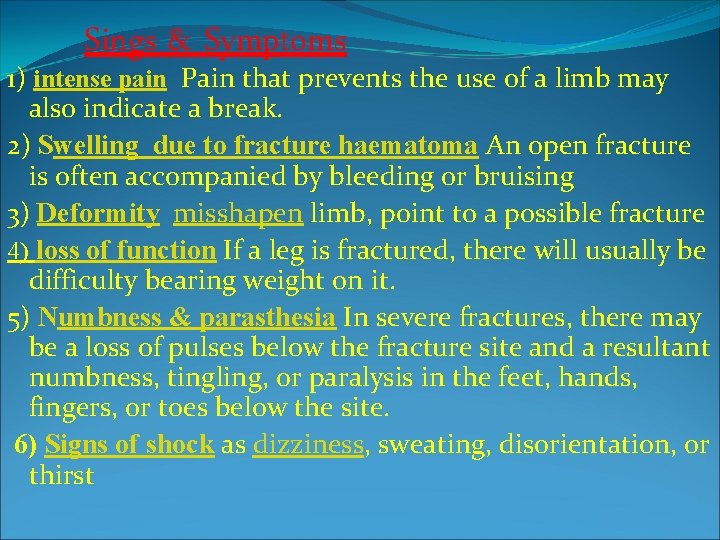
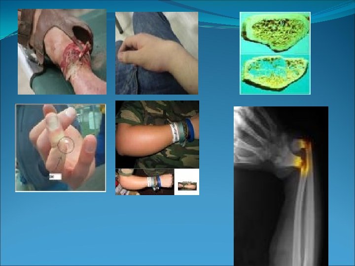
Sings & Symptoms 1) intense pain Pain that prevents the use of a limb may also indicate a break. 2) Swelling due to fracture haematoma An open fracture is often accompanied by bleeding or bruising 3) Deformity misshapen limb, point to a possible fracture 4) loss of function If a leg is fractured, there will usually be difficulty bearing weight on it. 5) Numbness & parasthesia In severe fractures, there may be a loss of pulses below the fracture site and a resultant numbness, tingling, or paralysis in the feet, hands, fingers, or toes below the site. 6) Signs of shock as dizziness, sweating, disorientation, or thirst
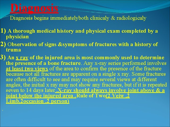
Diagnosis begins immediatelyboth clinicaly & radiologicaly 1) A thorough medical history and physical exam completed by a physician 2) Observation of signs &symptoms of fractures with a history of truma 3) An x ray of the injured area is most commonly used to determine the presence of a bone fracture. Any x-ray series performed involves at least two views of the area to confirm the presence of the fracture because not all fractures are apparent on a single x ray. Some fractures are often difficult to see and may require several views at different angles, the initial x ray may not show any fractures, but if it is repeated seven to 14 days later. X-ray should always involve joint above & a joint below the injured area. Rule of Two(2 Veiw , 2 Limb, 2 occasion , 2 person).
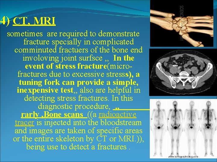
4) CT, MRI sometimes are required to demonstrate fracture specially in complicated comminuted fractuers of the bone end involoving joint surfsce , , In the event of stress fracture(microfractures due to excessive stresss), a tuning fork can provide a simple, inexpensive test, , also are helpful in detecting stress fractures. In this diagnostic procedure, , , rarly , Bone scans ((a radioactive tracer is injected into the bloodstream and images are taken of specific areas or the entire skeleton by CT or MRI. )), being use to detect a fractures.
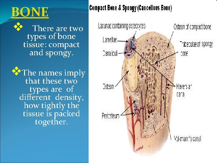
BONE v There are two types of bone tissue: compact and spongy. v. The names imply that these two types are of different density, how tightly the tissue is packed together.
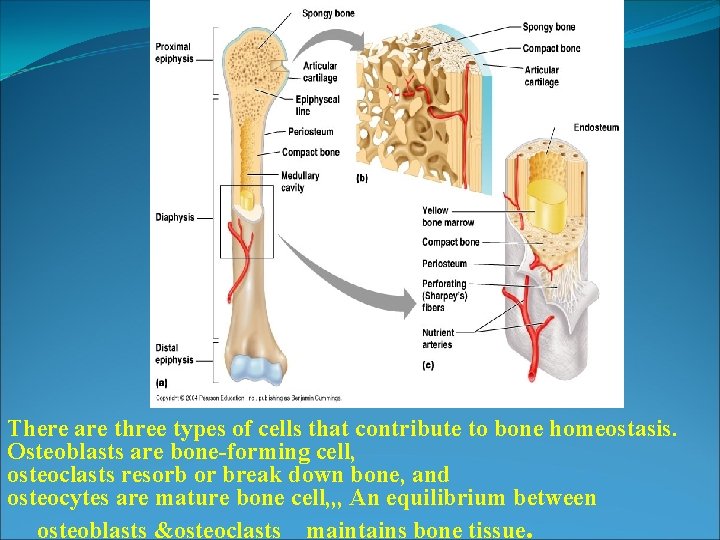
There are three types of cells that contribute to bone homeostasis. Osteoblasts are bone-forming cell, osteoclasts resorb or break down bone, and osteocytes are mature bone cell, , , An equilibrium between osteoblasts &osteoclasts maintains bone tissue.
Bone healing, or fracture healing, is a proliferative physiological process in which the body facilitates the repair of a bone fracture. FRACTURE HEALING OR UNION Healing process depends upon: Type of bone fr. , site of fr. . age of the patient, vascularity of the Fr. Bone & Degree of movement at fracture site
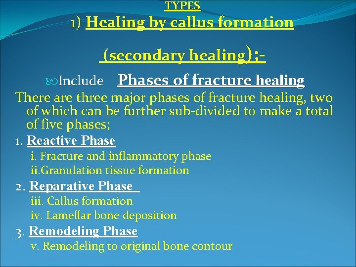
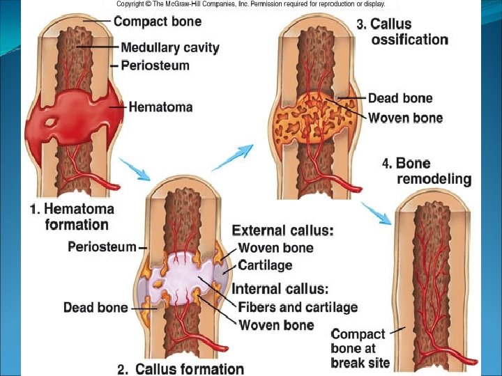
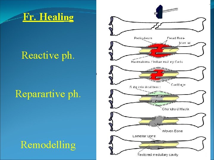
TYPES 1) Healing by callus formation (secondary healing); Include Phases of fracture healing There are three major phases of fracture healing, two of which can be further sub-divided to make a total of five phases; 1. Reactive Phase i. Fracture and inflammatory phase ii. Granulation tissue formation 2. Reparative Phase iii. Callus formation iv. Lamellar bone deposition 3. Remodeling Phase v. Remodeling to original bone contour
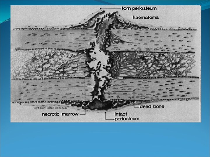
Reactive phase 1. After fracture, the first change seen by light and electron microscopy is the presence of blood cells within the tissues which are adjacent to the injury site. 2. Soon after fracture, the blood vessels constrict, stopping any further bleeding. . Within a few hours after fracture, these extravascular blood cells, known as a "hematoma", form a blood clot. 3. All of the cells within the blood clot degenerate and die. . , only the fibroblasts survive and replicate. They form a loose aggregate of tissue cells, interspersed with small blood vessels, known as granulation tissue
Reparative phase 1. 2. 3. 4. 5. ii- Callus formation In (3 -7 )Days after fracture, the cells of the periosteum replicate and transform. The periosteal cells proximal to the fracture gap develop into chondroblasts and form hyaline cartilage. The periosteal cells distal to the fracture gap develop into osteoblasts and form woven bone( internal & external callus ). The fibroblasts within the granulation tissue also develop into chondroblasts and form hyaline cartilage. . These two new tissues grow in size until they unite with their counterparts from other pieces of the fracture. This process forms fracture callus, By the end the fracture gap is bridged by the hyaline cartilage and woven bone, restoring some of its
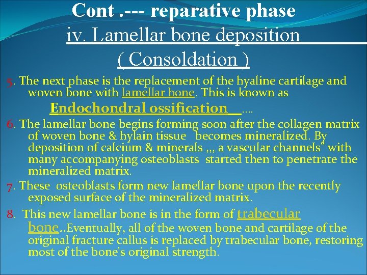
Cont. --- reparative phase iv. Lamellar bone deposition ( Consoldation ) 5. The next phase is the replacement of the hyaline cartilage and woven bone with lamellar bone. This is known as Endochondral ossification …. 6. The lamellar bone begins forming soon after the collagen matrix of woven bone & hylain tissue becomes mineralized. By deposition of calcium & minerals , , , a vascular channels" with many accompanying osteoblasts started then to penetrate the mineralized matrix. 7. These osteoblasts form new lamellar bone upon the recently exposed surface of the mineralized matrix. 8. This new lamellar bone is in the form of trabecular bone. . Eventually, all of the woven bone and cartilage of the original fracture callus is replaced by trabecular bone, restoring most of the bone's original strength.
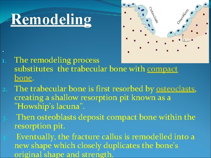
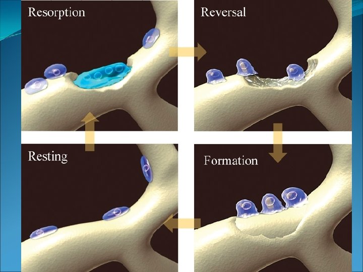
Remodeling. 1. The remodeling process substitutes the trabecular bone with compact bone. 2. The trabecular bone is first resorbed by osteoclasts, creating a shallow resorption pit known as a "Howship's lacuna". 3. Then osteoblasts deposit compact bone within the resorption pit. 4. Eventually, the fracture callus is remodelled into a new shape which closely duplicates the bone's original shape and strength.
Fr. Healing Reactive ph. Reparartive ph. Remodelling
Time factor Repair of fractures is a continuous process and no specific even signifies the moment of union. Rate of repair depends on: i. Age: healing in children is as twice faster as in adult. ii. Type of bone: fracture of scaphoid or neck of the femur is iii. iv. v. vii. delayed Type of fracture: transverse fracture takes more time to heal than spiral fracture because in spiral fracture there greater fr. Surface area reacting than in transverse fracture so there are more osteoblastic and osteoclastic activity. State of blood supply & Patient general condition Proper contact and apposition of fractured fragments Adequate immobility or fixation of the fracture Absence of infection viii. Early and good management
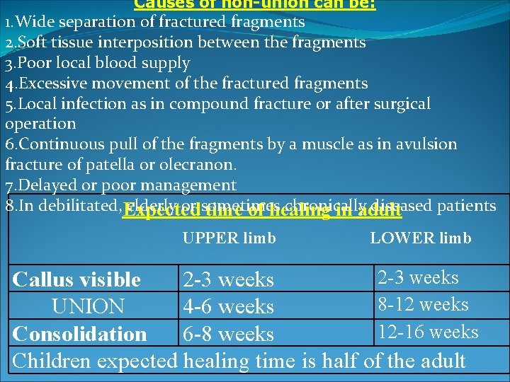
Causes of non-union can be: 1. Wide separation of fractured fragments 2. Soft tissue interposition between the fragments 3. Poor local blood supply 4. Excessive movement of the fractured fragments 5. Local infection as in compound fracture or after surgical operation 6. Continuous pull of the fragments by a muscle as in avulsion fracture of patella or olecranon. 7. Delayed or poor management 8. In debilitated, elderly or sometimes chronically diseased patients Expected time of healing in adult UPPER limb LOWER limb 2 -3 weeks Callus visible 2 -3 weeks 8 -12 weeks UNION 4 -6 weeks 12 -16 weeks Consolidation 6 -8 weeks Children expected healing time is half of the adult
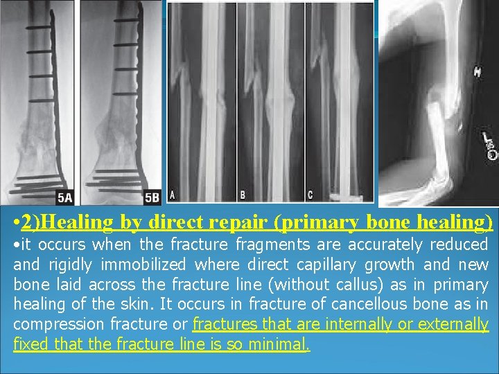
• 2)Healing by direct repair (primary bone healing) • it occurs when the fracture fragments are accurately reduced and rigidly immobilized where direct capillary growth and new bone laid across the fracture line (without callus) as in primary healing of the skin. It occurs in fracture of cancellous bone as in compression fracture or fractures that are internally or externally fixed that the fracture line is so minimal.