Introduction to EEG Rachel Garvin MD Neurocritical Care


 • Amplitude: size of")


- Slides: 33
Introduction to EEG Rachel Garvin, MD Neurocritical Care UTHSCSA
Neuronal Arrangement in Cortex
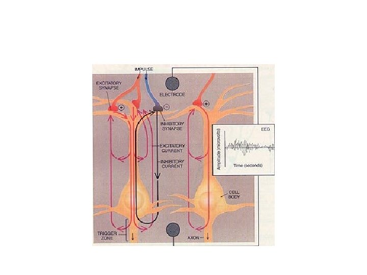
How does EEG work? • EEG sets up a circuit: lead is an electrode and plasma membrane acts a capacitor • Negative charges line up on inside of cell membrane and positive on outside completed circuit
How does EEG work? • Neurons in a column in the brain behave as a group • EPSP correlate with surface negative EEG waves • IPSP correlate with surface positive EEG waves • Electrical activity of the brain ranges from 10150 microvolts
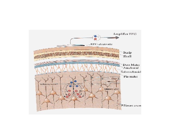
What areas of the brain does EEG detect • Cortex best – more superficial areas better • Does not detect deep structures such as BG, thalamus, brainstem • Also does not detect well in sulci, sylvian fissure, interhemispheric fissure or skull base area
Electrode Application • Electrodes must make good contact with skin • Electroconductive gel is used for low impedence of current • Max impedence of 5 Kohms – checked before EEG recording • Increased impedence = increased noise
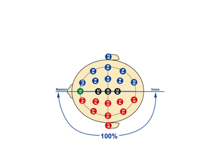
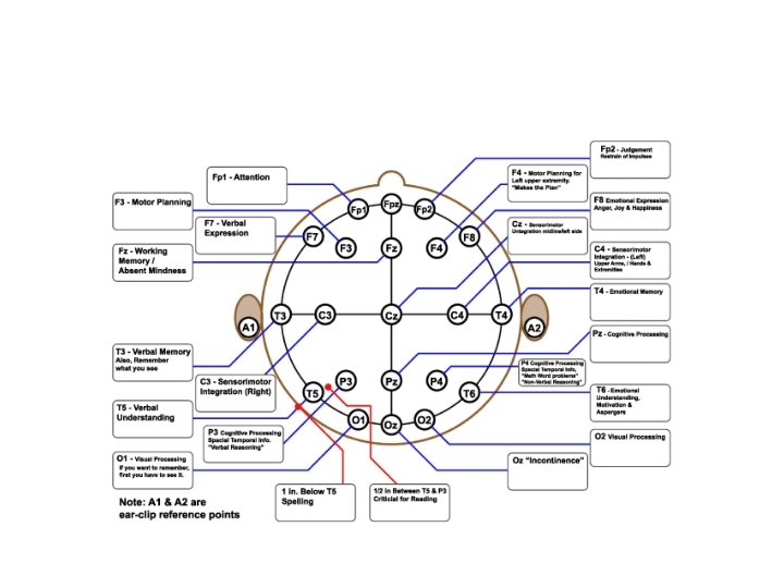
Electrode Placement Left sided leads Right sided leads
Bipolar vs Unipolar
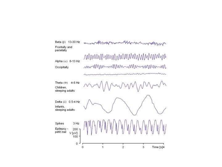
EEG waveforms • Frequency = number of complete waveforms/second (Hz) • Amplitude: size of wave measured in microvolts
EEG Waveforms cont’d
EEG frequency Ranges • • Delta: focal or diffuse Theta: central or diffuse Beta: frontal, central Alpha: occipital (alpha rhythm different from alpha frequency)
What is normal
Normal EEG
Normal Background Frequency changes with age
What are we looking for on the tracing? • Location • Symmetry – Are waveforms the same on L & R (amplitude, frequency) • Synchrony – Is there simultaneous occurrence of similar waveforms (epi on one side) • Reactivity • Morphology – How would you describe the waveform • Rhythmicity – Continuous repetition of waveforms that are similar
Abnormal EEG findings • • Generalized or focal slowing Lack of reactivity Epileptiform discharges Ictal patterns
What are these sharp waves?
Pattern Recognition
FIRDA
PLEDs
Burst Suppression
Diffuse Slowing
Focal Slowing
Breach Artifact
2 Types of Artifact
Looks like seizure…. .
Status Epilepticus
Pattern Recognition