
Introduction to Chronic Pain Definitions and Pathophysiology Rachael


- Slides: 20
Introduction to Chronic Pain Definitions and Pathophysiology Rachael Rzasa Lynn, MD 4 November 2015
Definition of Chronic Pain • Pain that persists past the normal time of healing – Variable: less than 1 month to more than 6 months • Typically use 3 months as the point of transition from acute to chronic pain – Six months is more often used in research IASP Classification of Chronic Pain, Second Edition (Revised), http: //www. iasp-pain. org/files/Content. Folders/Publications 2/Classificationof. Chronic. Pain/Introduction. pdf
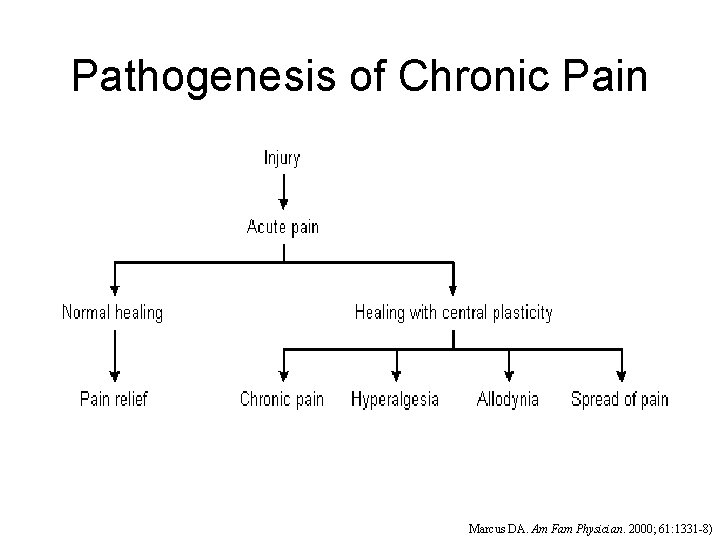
Pathogenesis of Chronic Pain Marcus DA. Am Fam Physician. 2000; 61: 1331 -8)
Nociceptive and Acute Pain • Reflects actual or imminent tissue injury • Can result in hypersensitivity as part of the normal healing process in order to promote guarding/protection of the injured tissue eg, sunburn • This pain can become more persistent and chronic via both peripheral and central mechanisms
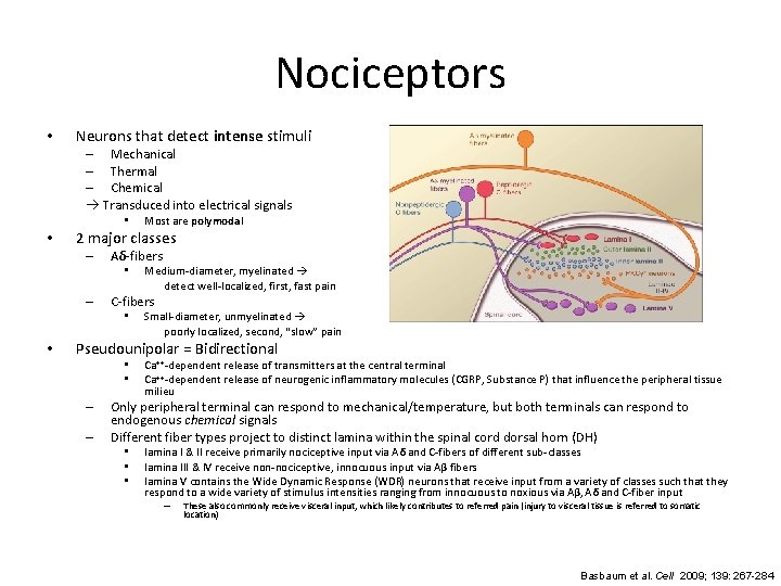
Nociceptors • Neurons that detect intense stimuli – Mechanical – Thermal – Chemical Transduced into electrical signals • • 2 major classes – Aδ-fibers • – Medium-diameter, myelinated detect well-localized, first, fast pain C-fibers • • Most are polymodal Small-diameter, unmyelinated poorly localized, second, “slow” pain Pseudounipolar = Bidirectional • • – – Ca++-dependent release of transmitters at the central terminal Ca++-dependent release of neurogenic inflammatory molecules (CGRP, Substance P) that influence the peripheral tissue milieu Only peripheral terminal can respond to mechanical/temperature, but both terminals can respond to endogenous chemical signals Different fiber types project to distinct lamina within the spinal cord dorsal horn (DH) • • • lamina I & II receive primarily nociceptive input via Aδ and C-fibers of different sub-classes lamina III & IV receive non-nociceptive, innocuous input via Aβ fibers lamina V contains the Wide Dynamic Response (WDR) neurons that receive input from a variety of classes such that they respond to a wide variety of stimulus intensities ranging from innocuous to noxious via Aβ, Aδ and C-fiber input – These also commonly receive visceral input, which likely contributes to referred pain (injury to visceral tissue is referred to somatic location) Basbaum et al. Cell 2009; 139: 267 -284
Supraspinal pathways in nociception Involves multiple levels of of brain structures: brainstem (medulla, pons, midbrain) diencephalon (thalamus, hypothalamus) primary and secondary somatosensory cortices • fronto-limbic circuits (prefrontal cortex [PFC], anterior cingulate cortex, insula, amygdala, hippocampus) • •
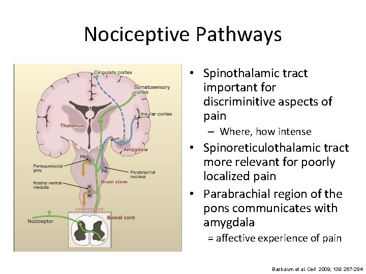
Nociceptive Pathways • Spinothalamic tract important for discriminitive aspects of pain – Where, how intense • Spinoreticulothalamic tract more relevant for poorly localized pain • Parabrachial region of the pons communicates with amygdala = affective experience of pain Basbaum et al. Cell 2009; 139: 267 -284
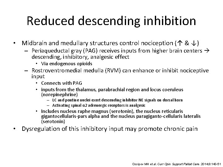
Reduced descending inhibition • Midbrain and medullary structures control nociception (↑ & ↓) – Periaqueductal gray (PAG) receives inputs from higher brain centers descending, inhibitory, analgesic effect • Via endogenous opioids – Rostroventromedial medulla (RVM) can enhance or inhibit nociceptive input • Connects with PAG • inputs from the thalamus, parabrachial region and locus coeruleus (norepinephrine) – LC and pontine nuclei exert descending inhibitor NE signals on dorsal horn – Activating spinal α 2 adrenergic receptors is analgesic • Includes nucleus raphe magnus (serotonin), the nucleus reticularis gigantocellularis-pars alpha and the nucleus paragiganto-cellularis lateralis (serotonin) • Dysregulation of this inhibitory input may promote chronic pain Ossipov MH et al. Curr Opin Support Palliat Care. 2014; 8: 143 -51
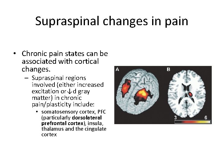
Supraspinal changes in pain • Chronic pain states can be associated with cortical changes. – Supraspinal regions involved (either increased excitation or↓d gray matter) in chronic pain/plasticity include: • somatosensory cortex, PFC (particularly dorsolateral prefrontal cortex), insula, thalamus and the cingulate cortex
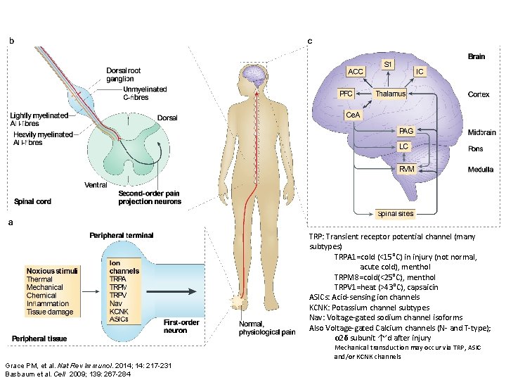
TRP: Transient receptor potential channel (many subtypes) TRPA 1=cold (<15°C) in injury (not normal, acute cold), menthol TRPM 8=cold(<25°C), menthol TRPV 1=heat (>43°C), capsaicin ASICs: Acid-sensing ion channels KCNK: Potassium channel subtypes Nav: Voltage-gated sodium channel isoforms Also Voltage-gated Calcium channels (N- and T-type); α 2δ subunit ↑’d after injury Mechanical transduction may occur via TRP, ASIC and/or KCNK channels Grace PM, et al. Nat Rev Immunol. 2014; 14: 217 -231 Basbaum et al. Cell 2009; 139: 267 -284
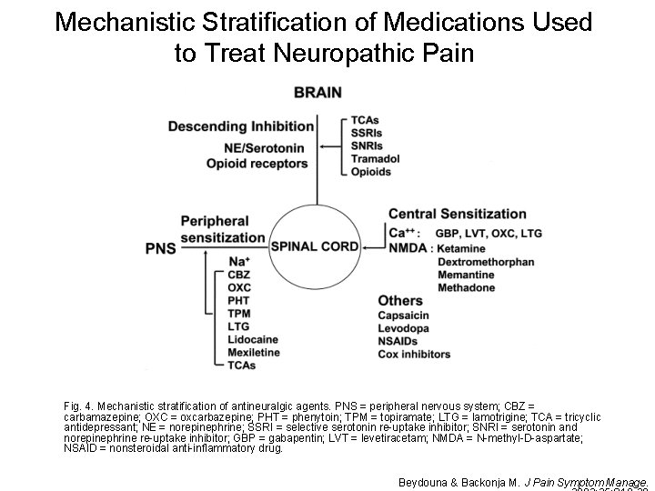
Mechanistic Stratification of Medications Used to Treat Neuropathic Pain Fig. 4. Mechanistic stratification of antineuralgic agents. PNS = peripheral nervous system; CBZ = carbamazepine; OXC = oxcarbazepine; PHT = phenytoin; TPM = topiramate; LTG = lamotrigine; TCA = tricyclic antidepressant; NE = norepinephrine; SSRI = selective serotonin re-uptake inhibitor; SNRI = serotonin and norepinephrine re-uptake inhibitor; GBP = gabapentin; LVT = levetiracetam; NMDA = N-methyl-D-aspartate; NSAID = nonsteroidal anti-inflammatory drug. Beydouna & Backonja M. J Pain Symptom Manage.
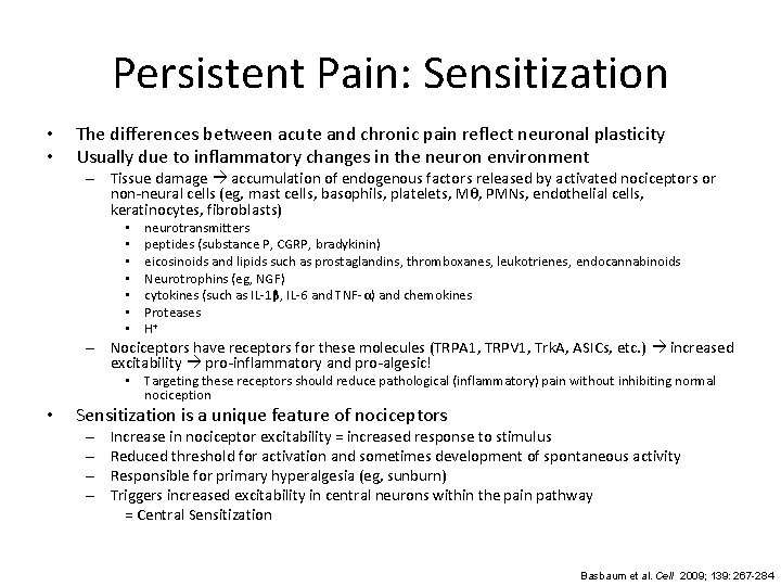
Persistent Pain: Sensitization • • The differences between acute and chronic pain reflect neuronal plasticity Usually due to inflammatory changes in the neuron environment – Tissue damage accumulation of endogenous factors released by activated nociceptors or non-neural cells (eg, mast cells, basophils, platelets, Mθ, PMNs, endothelial cells, keratinocytes, fibroblasts) • • neurotransmitters peptides (substance P, CGRP, bradykinin) eicosinoids and lipids such as prostaglandins, thromboxanes, leukotrienes, endocannabinoids Neurotrophins (eg, NGF) cytokines (such as IL-1β, IL-6 and TNF-α) and chemokines Proteases H+ – Nociceptors have receptors for these molecules (TRPA 1, TRPV 1, Trk. A, ASICs, etc. ) increased excitability pro-inflammatory and pro-algesic! • Targeting these receptors should reduce pathological (inflammatory) pain without inhibiting normal nociception • Sensitization is a unique feature of nociceptors – – Increase in nociceptor excitability = increased response to stimulus Reduced threshold for activation and sometimes development of spontaneous activity Responsible for primary hyperalgesia (eg, sunburn) Triggers increased excitability in central neurons within the pain pathway = Central Sensitization Basbaum et al. Cell 2009; 139: 267 -284
Central Sensitization • 3 major proposed pathways – Glutamate/NMDA-mediated hypersensitivity – Loss of inhibitory controls – Glia-neuron interactions • Microglia and astrocytes
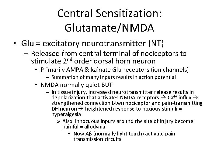
Central Sensitization: Glutamate/NMDA • Glu = excitatory neurotransmitter (NT) – Released from central terminal of nociceptors to stimulate 2 nd order dorsal horn neuron • Primarily AMPA & kainate Glu receptors (ion channels) – Summation of many inputs results in action potential • NMDA normally quiet BUT – In tissue injury, increased neurotransmitter release results in depolarization that activates NMDA receptors Ca++ influx strengthened connection btwn nociceptor and pain-transmitting DH neuron heightened response to noxious stimuli = hyperalgesia » Also, innocuous inputs around the site of injury become painful = allodynia • Now Aβ (normally light touch) activate pain transmission circuits
Central Sensitization: Loss of Inhibitory Control • GABA and glycine (gly) = inhibitory NT – Normally tonically active • When experimentally blocked hypersensitivity behavior • Decreased activity with peripheral injury thus enhanced excitation of spinal 2 nd order neurons increased activity in response to both painful (hyperalgesia) and non-painful (allodynia) stimuli
Central Sensitization: Glia-Neuron Interaction • Microglia are MΘ resident within CNS – Signal injury or infection within CNS – Immune activity involved in sickness response (lethargy, depression, anxiety) associated with illness and also chronic pain – Following peripheral nerve injury (but NOT inflammatory tissue injury) they accumulate in the superficial DH where the injured nerve terminates (and also surround and damage adjacent motorneurons) • Activated release inflammatory signals (TNF-α, IL-1β, IL-6) sensitization of central neurons and maintenance of pain Grace PM, et al. Nat Rev Immunol. 2014; 14: 217 -231
Central Sensitization: Glia-Neuron Interaction • Physical damage of peripheral afferent results in release of signals detected by microglia – ATP microglial P 2 -purinergic receptors (P 2 Xn) • May ultimately result in decreased GABA-ergic inhibition and thus increased excitability – Other neuronally released cytokines and chemokines (eg, CX 3 CL 1) that activate the microglia when their receptors (CX 3 CR 1) are bound • May be part of a positive-feedback loop » microglia produce the protein that frees CX 3 CL 1 from neuronal cell surface – Glia are activated not just in spinal cord but also in the brainstem, contributing to facilitation of pain processing at supraspinal levels Grace PM, et al. Nat Rev Immunol. 2014; 14: 217 -231
Central Sensitization: Glia-Neuron Interaction • Toll-Like Receptors (TLR 2 and TLR 4) – Innate immune receptors that respond to diverse pathogens and pathogen- or damage-associated molecular patterns (PAMPs & DAMPs) as well as endogenous signals such as IL-1 b, TNFα, IL-6 and nitric oxide – Activation of TLRs results in immune-like processes, such as the release of pro-inflammatory (and neuroexcitatory) cytokines and phagocytosis • Exposure to DAMPs may increase glial expression of P 2 X receptors increased neuronal excitability • Development of nociceptive hypersensitivity correlates temporally with increased TLR 4 expression after injury – Sensory neurons themselves may express TLRs and adaptor proteins Milligan and Watkins Nat Rev Immunol. 2009; 10: 23 -36
Central Sensitization: Glia-Neuron Interaction • Glia may become “primed” following stress, aging, illness or injury such that subsequent injury leads to unregulated activation that maintains central sensitization and leads to chronic pain • Opioids like morphine (and its “inactive” metabolites) activate TLR 4 resulting in such priming and central sensitization, which actually opposes analgesia! – This non-opioid receptor activity is also implicated in opioid reward, craving and withdrawal – As well as opioid-induced hyperalgesia
Summary • Complex changes in both pain sensation, transmission and processing lead to chronic pain – Peripheral – Central • Spinal Cord • Supraspinal structures