Introduction Restriction Endonuclease Restriction Enzyme is a bacterial
 is a bacterial enzyme that cuts ds. DNA")
Introduction : Restriction Endonuclease (Restriction Enzyme) is a bacterial enzyme that cuts ds. DNA into fragments after recognizing specific nucleotide sequence known as recognition or restriction site. and have evolved to provide a defense mechanism against invading viruses. Restriction Enzymes are believed to be evolved by bacteria to resist viral attack. Restriction Enzymes are also known as molecular scissor. 1

• • Restriction Enzymes scan the DNA sequence Find a very specific set of nucleotides Make a specific cut Restriction enzymes recognize and make a cut within specific palindromic sequences, known as restriction sites, in the DNA. This is usually a 4 - or 6 base pair sequence.

Picking a palindrome Words that read the same forwards as backwards Hannah hanna. H Level leve. L Madam mada. M

Palindromes in DNA sequences 5’ 3’ 3’ 5’ Genetic palindromes are similar to verbal palindromes. A palindromic sequence in DNA is one in which the 5’ to 3’ base pair sequence is identical on both strands.
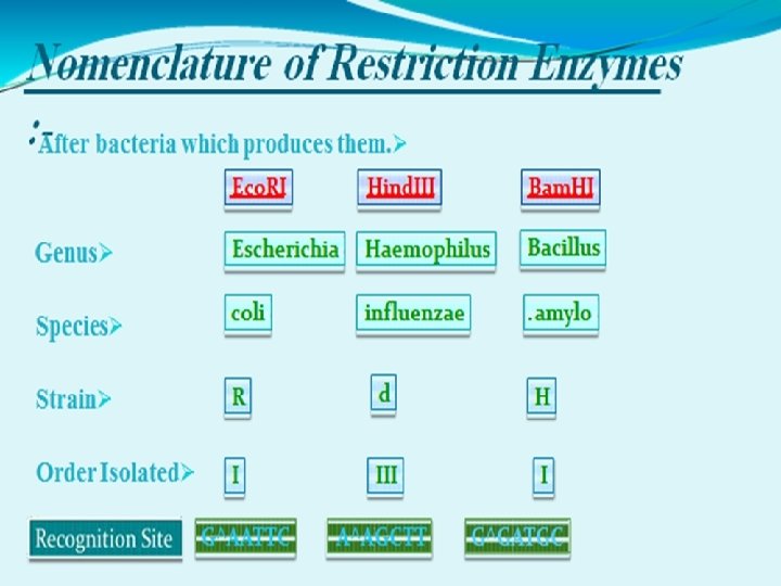

NOMENCLATURE OF RESTRICTION ENZYME • Each enzyme is named after the bacterium from which it was isolated using a naming system based on bacterial genus, species and strain. For e. g Eco. RI Derivation of the Eco. RI name Abbreviation Meaning Description E Escherichia genus co coli species R RY 13 strain I First identified order of identification in the bacterium

How Restriction Endonucleases work ? Restriction enzymes recognize a specific sequence of nucleotides, and produce a double-stranded cut in the DNA. these cuts are of two types: Blunt end Sticky end

Hae III Hae. III is a restriction enzyme that searches the DNA molecule until it finds this sequence of four nitrogen bases. 5’ TGACGGGTTCGAGGCCAG 3’ 3’ ACTGCCCAAGGTCCGGTC 5’
")
Once the recognition site was found Hae. III could go to work cutting (cleaving) the DNA 5’ TGACGGGTTCGAGGCCAG 3’ 3’ ACTGCCCAAGGTCCGGTC 5’

These cuts produce what scientists call “blunt ends” 5’ TGACGGGTTCGAGG 3’ ACTGCCCAAGGTCC CCAG 3’ GGTC 5’

“blunt ends” and “sticky ends” Remember how Hae. III produced a “blunt end”? Eco. RI, for instance, makes a staggered cut and produces a “sticky end” 5’ GAATTC 3’ 3’ CTTAAG 5’ 5’ G AATTC 3’ 3’ CTTAA G 5’

blunt end sticky end

Some more examples of restriction sites of restriction enzymes with their cut sites: Hind. III: 5’ AAGCTT 3’ 3’ TTCGAA 5’ Bam. HI: 5’ GGATCC 3’ 3’ CCTAGG 5’ Alu. I: 5’ AGCT 3’ 3’ TCGA 5’

Categorization of Restriction Enzymes on the bases of Their composition. Enzyme co-factor requirement. the nature of their target sequence. position of their DNA cleavage site relative to the target sequence.

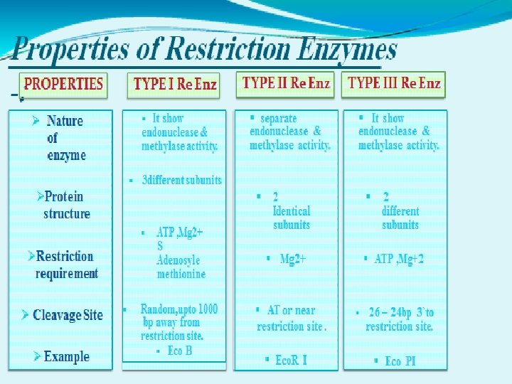
Restriction Endonuclease Types Type I- multi-subunit, both endonuclease and methylase activities, cleave at random up to 1000 bp from recognition sequence Type II- mostly single subunit, cleave DNA within recognition sequence Type III- multi-subunit, endonuclease and methylase about 25 bp from recognition sequence

Separating Restriction Fragments, I

Separating Restriction Fragments, II

Blunt ends • Some restriction enzymes cut DNA at opposite base • They leave blunt ended DNA fragments • These blunt ended fragments can be joined to any other DNA fragment with blunt ends. • Enzymes useful for certain types of DNA cloning experiments.

Sticky ends • Most restriction enzymes make staggered cuts • Staggered cuts produce single stranded “sticky-ends”

“Sticky Ends” Are Useful DNA fragments with complementary sticky ends can be combined to create new molecules which allows the creation and manipulation of DNA sequences from different sources.

ISOSCHIZOMERS & NEOSCHIZOMERS • Restriction enzymes that have the same recognition sequence as well as the same cleavage site are Isoschizomers • Restriction enzymes that have the same recognition sequence but cleave the DNA at a different site within that sequence are Neoshizomers Eg: Sma. I and Xma. I CCCGGG G C CC Xma I C C C G GG G C CC Sma I

APPLICATION OF RESTRICTION ENZYMES • They are used in gene cloning and protein expression experiments. • Restriction enzymes are used in biotechnology to cut DNA into smaller strands in order to study fragment length differences among individuals (Restriction Fragment Length Polymorphism – RFLP). • Each of these methods depends on the use of agarose gel electrophoresis for separation of the DNA fragments.

What is RFLP is a difference in homologous DNA sequences that can be detected by the presence of fragments of different lengths after digestion of the DNA samples.

Method of DNA analysis by RFLP The method of analysis of DNA by RFLP involves the following steps: 1 - In the first step fragmentation of a sample of DNA is done by a restriction enzyme, which can recognize and cut DNA wherever a specific short sequence occurs, in a process known as a restriction digest.

2 The resulting DNA fragments are then separated by length through a process known as agarose gel electrophoresis. 3 Then transferred to a membrane via the Southern blot procedure. 4 Hybridization of the membrane to a labeled DNA probe will done and then determines the length of the fragments which are complementary to the probe.

5 - Then we will observe the fragments of different length. An RFLP occurs when the length of a detected fragment varies between individuals. Each fragment length is considered an allele, and can be used in genetic analysis.
- Slides: 27