Introduction Polyps are usually asymptomatic and 90 are


 occur in two distinct")


 constitute 30 e 93%of all. BEGPs")


 or multiple hamartoma syndrome is a")


 constitute 3% of")
 Management of")

")


- Slides: 32
Introduction • Polyps are usually asymptomatic and >90% are found incidentally. • Larger polyps can present with bleeding, anaemia, abdominal pain or gastric outlet obstruction. • Although some types of polyps may have typical appearances at endoscopy the presence of dysplasia cannot be determined without histological assessment. • Therefore all types of gastric polyp must be sampled to determine the pre-malignant risk. • Some polyps are an expression of a genetic disease and may also indicate an increased risk of intestinal or extra-intestinal malignancy. • Other precancerous conditions of the stomach include chronic atrophic gastritis and intestinal metaplasia.
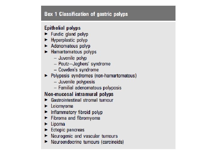
TYPES AND CLASSIFICATION OF POLYPS • The most common types of benign epithelial gastric polyps (BEGPs) are fundic gland polyps, hyperplastic polyps and adenomas. • If there are multiple gastric polyps present, these are usually of the same histological type 2. • The WHO has classified gastric polyps 3 4 but this classification is controversial. • An updated classification is suggested in box 1.
• Fundic gland polyps (also called Elster’s glandular cysts) occur in two distinct clinicopathological settings: as sporadic polyps and polyps associated with the syndrome, familial adenomatous polyposis coli. • Sporadic fundic gland polyps Fundic gland polyps (FGPs) constitute 16 e 51% of BEGPs 5 e 7 and may be observed in 0. 8 e 23% of endoscopies. • 8 e 10 They are usually multiple transparent sessile polyps, 1 e 5 mm in diameter, located in the body and fundus. • Microscopy shows cystically dilated glands lined by gastric body type mucosa. • 8 A single study 8 of 56 consecutive patients with FGPs suggested they can be diagnosed with a high degree of accuracy based on endoscopic appearance alone.
• • Sporadic FGPs are usually caused by activating mutations of the beta-catenin gene 11 12 and usually number less than ten. 13 Dysplasia occurs in <1% of sporadic FGPs. 14 e 16 FGPs are not associated with atrophic gastritis and the prevalence of Helicobacter pylori infection is very low in these patients. 17 e 20 Case reports have shown that H pylori infection may have an inhibitory effect on the development of FGPs but the cause of fundicgland polyps remains uncertain, and these polyps may regress or even disappear in time. 21
• • • • • Proton pump inhibitor-associated gastric polyps Since 1993 there have been reports of the role of long-term proton pump inhibitors (PPIs) in the genesis of fundic gland polyps. 10 22 In one study FGPs were present in 23% of patients on PPIs compared with 12% of patients not taking a PPI. 10 The polyps are usually small (<1 cm) and occur in the proximal/midgastric body. 23 These reports have been retrospective studies and show that the mean interval for polyp development was 32. 5 months and that they regress within 3 months of PPI cessation. However, other studies have not demonstrated a definitive link between long-term PPIs and FGPs. 24 e 26 Similarly, a prospective long (median of 11 years) follow-up study of 230 patients could not show any de novo FGPs. 27 Histologically, in body mucosa, long-term PPI use induces enterochromaffin cell-like (ECL) hyperplasia and typical ‘parietal cell protrusion’ and glands are lined by cells with a serrated rather than a smooth border. 23
• • • • • Fundic gland polyps in familial adenomatous polyposis FGPs are common in patients with familial adenomatous polyposis (FAP) and arise from mutation of the APC gene. 22 In this context, they are usually multiple and can ‘carpet’ the body of the stomach. Epithelial dysplasia occurs in 25 e 41% of FAP associated polyps. 13 The management of FAP is discussed later in this guideline. There is no evidence as to how to differentiate between sporadic FGP and FAP-associated FGP at endoscopy. The presence of dysplastic foci should arouse suspicion of FAP. 28 29 Two studies have shown the presence of colonic adenomas (7 e 45%) and adenocarcinomas (0 e 4. 7%) in patients with sporadic FGPs. 30 31 However, this high rate of colonic neoplasia has not been confirmed in other studies. Therefore, while the presence of numerous FGPs may be a manifestation of FAP, there is no clear evidence about the number of polyps needed to indicate further investigation iswarranted. When considering lower gastrointestinal investigation, it should be noted that almost all cases of classical FAP can be diagnosed on flexible sigmoidoscopy which is safer than colonoscopy.
• • • Recommendations for the management of FGPs < Polypectomy is not required for sporadic FGPs (moderate, net benefits, definitive). < Although FGPs have reliable endoscopic features, biopsy of probable FGPs is recommended to exclude dysplasia or adenocarcinoma (and possible FAP) and to exclude the need for polypectomy as required for other types of polyp (moderate, net benefit, qualified). < In patients with numerous FGP who are under 40 years of age, or where biopsies demonstrate dysplasia, colonic investigation should be performed to exclude FAP (low, uncertain trade offs, qualified).
• • • • • Hyperplastic polyps(HYPPs) constitute 30 e 93%of all. BEGPs 5 e 7 17 and are sessile or pedunculated polyps less than 2 cm in diameter. They can occur as single polyps usually in the antrum or as multiple polyps throughout the stomach. Multiple hyperplastic polyps are also found in Menetrier’s disease. Histologically, there is a proliferation of surface foveolar cells lining elongated, distorted pits that extend deep into the lamina propria. They may contain pyloric glands, chief cells, and parietal cells. Their histological appearance overlaps with hamartomas and inflammatory conditions. Polyp formation is strongly associated with chronic gastritis; Helicobacter-associated gastritis, pernicious anaemia, reactive or chemical gastritis when adjacent to ulcer erosions and around gastroenterostomy stomas. 32 33 Up to 80% of HYPPs have been found to regress after eradication of H pylori before endoscopic removal. 34 e 36
• • • • HYPPs rarely undergo neoplastic progression directly through neoplastic change in the polyp but are associated with an increased risk of synchronous cancer occurring elsewhere in the gastric mucosa. The prevalence of true dysplasia arising in hyperplastic polyps is debated 37 e 46 and reported rates have varied from 1. 9% to 19% and cases of adenocarcinoma have been reported ranging from 0. 6% to 2. 1%. HYPPs also denote an increased risk of neoplasia in the surrounding abnormal gastric mucosa and are associated with the occurrence of synchronous cancer elsewhere in the gastric mucosa. 40 Multiple biopsies of the intervening mucosa are therefore needed in patients with HYPPs. The risk of adenocarcinoma in the surrounding mucosa is probably higher than in the polyp itself.
• • • • • There is controversy regarding whether gastric HYPPs can be simply biopsied or whether they should be entirely removed by polypectomy because of the risk of neoplasia and because forceps biopsy sampling may miss the dysplastic foci within a hyperplastic polyp. Some authors recommend performing polypectomy for all small polyps and periodic biopsy of larger hyperplastic polyps that are too big for polypectomy. Others recommend that, as cancer usually occurs in bigger hyperplastic polyps, only large hyperplastic polyps should be removed, due to the risk of gastric polypectomy. Surveillance of hyperplastic polyps that are not removed (either because of number or size) is probably safer than multiple polypectomies but there is no randomised evidence as to which approach should be taken. A single repeat endoscopy at 1 year is reasonable, but repeated surveillance after 1 year is not recommended due to lack of evidence. One study 47 has suggested that HYPP may cause gastrointestinal blood loss and should be removed in such patients. This has not been confirmed by other studies.
• Recommendations for the management of hyperplastic polyps • < Hyperplastic polyps should be biopsied an examination of the • whole stomach should be made for mucosal abnormalities and any • abnormalities biopsied (moderate, trade offs, definite) • < Test for H pylori and eradicate when present (high, net benefit, • definitive).
• • • • Adenomatous polyps Gastric adenomas are true neoplasms and are precursors of gastric cancer. They are histological classified into tubular, villous and tubulovillous types. They constitute 3 e 26% of BEGPs, are frequently solitary and can be found anywhere in the stomach but are commonly found in the antrum. They frequently arise on a background of atrophic gastritis 48 and intestinal metaplasia, but there is no proven association with H pylori infection. Neoplastic progression is greater with polyps larger than 2 cm in diameter 49 and occurs in 28. 5 e 40% of villous adenomas and 5% of tubular adenomas. 17 41 50 e 53 There is also a strong association between gastric adenoma and synchronous or metachronous gastric adenocarcinoma. 7 44 45 The risk of association between adenomatous polyps and cancer increases with age. 45
• Recommendations for management of adenomatous polyps • < Complete removal of the adenoma should be performed when safe to • do so (high, net benefit, definitive). • < An examination of the whole stomach should be made for mucosal • abnormalities and any abnormalities biopsied (high, net benefit, • definitive). • < Endoscopic follow-up is required following resection of gastric • adenomas. Endoscopy should be repeated at 6 months for • incompletely resected polyps or those with high grade dysplasia. • Endoscopy can be repeated after 1 year for all other polyps • (moderate, trade offs, qualified).
• • • Hamartomatous polyps are rare in the stomach and include juvenile polyps, polyps of Peutze. Jeghers’ syndrome (PJS), and Cowden’s disease. The follow-up recommendations of patients with these conditions are summarised in table 1. Juvenile polyps are found in the antrum. Solitary polyps have hamartomatous or inflammatory components and do not have a neoplastic potential. Histologically, these show irregular cysts lined by normal gastric epithelium; with possible stromal haemorrhage, surface ulceration and chronic inflammation due
• • • • to torsion. Multiple polyps are associated with juvenile polyposis. Peutze. Jeghers’ syndrome PJS is a rare autosomal dominant inherited condition, characterised by the presence of hamartomatous gastrointestinal polyps and mucocutaneous pigmentation of the lips, buccal mucosa and digits. Microscopically, hyperplastic glands lined by foveolar epithelium and broad bands of smooth muscle fibres that branch out are found. PJS increases the risk of both gastrointestinal cancer through the hamartomaeadenomaecarcinoma sequence and de novo malignant change. There is a 15 -fold increase in extra-intestinal malignancies such as breast, endometrial, pancreatic and lung cancers. 54
• Recommendation for PJS gastric polyps • < Gastric polyps greater than 1 cm should be removed. As there is • a high rate of recurrence, regular endoscopic follow-up and annual • screening of other susceptible organs is recommended (moderate, • trade offs, qualified).
• • • • Cowden’s syndrome (CS) or multiple hamartoma syndrome is a rare autosomal dominant syndrome characterised by orocutaneous hamartomatous tumours, gastrointestinal polyps, abnormalities of the breast, thyroid gland genitourinary system. The gastrointestinal polyps are generally benign and malignant transformation is seen very rarely. Common benign visceral tumours and hyperplastic conditions include gastrointestinal polyps, thyroid adenoma and goitre and, fibrocystic disease of the breast in female patients, and gynaecomastia in male patients. Malignancies of the thyroid, colon, small bowel, and genitourinary tract have been reported, as well as acute myelogenous (myeloid) leukaemia, non-Hodgkin’s lymphoma and breast carcinoma. Histologically, CS polyps have elongated cystically dilated glands with papillary infoldings with a connective tissue (neural or muscular) component.
• • • Familial polyposis syndromes A summary of the cancer risk and screening recommendations for the familial polyposis syndromes is shown in table 1. Familial adenomatous polyposis (FAP) is caused by a germline mutation in the adenomatous polyposis coli APC gene on chromosome 5 q 21 and has an autosomal dominant inheritance and a high incidence of gastric cancer. Affected family members develop numerous colorectal adenomas with virtually a 100% chance of malignant progression unless the colon is removed prophylactically.
• • • • Gastric polyps occur in 30 e 100% of cases of FAP and these patients are said to have familial gastric polyposis (FGP). Most polyps are benign fundic gland polyps and gastric adenomatous polyps occur in only 5%, usually in the antrum. Duodenal adenomas and periampullary adenomas occur in 50 e 90% of patients, 55 56 are usually malignant and are the main cause of mortality after prophylactic colectomy. There is a negative association with H pylori-associated chronic gastritis. 57 Sampling of polyps is required to determine whether they are adenomas or FGPs. Endoscopic surveillance of small gastric and duodenal polyps is required every 1 e 2 years until the patient reaches the age of 50 years but large polyps/adenomas may require more frequent surveillance. Surveillance frequency can be reduced to five yearly after the age of 50 years. Partial gastrectomy and local
• • • duodenal resection or pancreaticoduodenectomy may be required in selected patients. Juvenile polyposis This condition is associated with numerous mutations (BMPR 1 A, 10 q 22. 3, SMAD 4, 18 q 21. 1) and is associated with gastric malignancy in over 50% of cases. 58 59 Juvenile polyposis is also associated with gastrointestinal bleeding 60 and proteinlosing enteropathy. Histologically, the glands are tortuous, elongated and cystically dilated and the background mucosa is oedematous and inflamed.
• • • • • • Inflammatory fibroid polyps (IFPs) constitute 3% of all BEGP 5 and are well-circumscribed lesions located in the antrum or prepyloric region, covered with a smooth surface of normal mucosa. They originate from the submucosa and should not be confused with ‘inflammatory polyps’, which are a commonly used misnomer for hyperplastic polyps. They occur in patients of all ages, but more commonly in the fifth to sixth decades of life, and more frequently in females. They do not have a malignant potential but are associated with chronic atrophic gastritis. Their aetiology remains obscure but there is a weak association with H pylori. 61 Polyps grow to 1 e 5 cm in diameter and the larger lesions often have a central depression, ulceration or a white cap. IFPs remain unchanged and asymptomatic for a long time but have the propensity to enlarge and cause gastric obstruction. Histology shows blood vessels surrounded by spindle cells (CD 34 and fascin positive) and chronic inflammatory cells, predominantly eosinophils. Multinucleated giant cells may be present, but they are not associated with a systemic allergic reaction or eosinophilia. IFPs may be amenable to complete endoscopic excision but extension into the deeper layers of the gastric wall sometimes preclude this, and for very deep polyps, forceps biopsy may be negative.
• • • • • • • Gastric neuroendocrine tumours (carcinoids) Management of gastric neuroendocrine tumours is discussed in detail in ‘Guidelines for the management of gastroenteropancreatic neuroendocrine (including carcinoid) tumours’ 62 and will therefore only be summarised here. Most gastric NETs are composed of enterochromaffin-like (ECL) cells, which be identified histopathologically by immunostaining for markers such as chromogranin A or synaptophysin. There are three main subtypes. Type 1 tumours (approximately 80% of total) are usually sessile polyps and associated with (usually autoimmune) atrophic gastritis, pernicious anaemia, achlorhydria and hypergastrinaemia, with the elevated gastrin being produced by gastric antral G cells. Type 2 gastric NETs (approximately 5% of total) occur in patients with Zollingere. Ellison syndrome, usually in the setting of multiple endocrine neoplasia type 1 (MEN 1). They are therefore associated with gastrinomas, hypergastrinaemia and gastric hyperacidity. Type 3 gastric NETs (approximately 15%) occur sporadically and are not associated with hypergastrinaemia. 63 64 Treatment and prognosis is determined by tumour type, size of polyp(s) and by the presence or absence of metastases 62 Type 1 tumours carry a very good prognosis and may need no treatment if the tumours are small (<1 cm). Larger type 1 NETs should be treated by endoscopic polypectomy or surgical antrectomy should be considered. Type 2 gastric NETs usually regress if the gastrinoma can be completely removed surgically. Type 3 gastric NETs are associated with the worst prognosis and
• • • should be treated by surgical resection. Metastatic tumours of all types are amenable to a range of palliative treatments, as discussed in more detail elsewhere. 62 Recommendations for management of gastric NET < The type of gastric NETshould be determined in each case by biopsy of the lesion and surrounding normal mucosa, and measuring fasting serum gastrin concentration (moderate, trade offs, qualified). < Tumours should be staged appropriately and treatment options should be discussed with a neuroendocrine tumour multidisciplinary team (moderate, benefit, definitive).
• • • • • Stromal tumours Management of gastrointestinal stromal tumours (GISTs) is discussed in detail in the document ‘Consensus meeting for the management of gastrointestinal stromal tumors’. 65 Therefore only a brief summary will be included here. GISTs are rare connective tissue tumours that show similar differentiation patterns to the interstitial cells of Cajal and approximately 60 e 70% arise in the stomach. Activating mutations of the KIT or PDGFRA protooncogenes are thought to be the causal molecular events. Approximately 95% of tumours are therefore positive for CD 117 (c-KIT) by immunohistochemistry. The degree of local and metastatic spread of gastric GISTs should be evaluated by CT scan and endoscopic ultrasound. If the tumour is localised to the stomach it should be surgically resected. If the tumour is unresectable or if metastases are present at diagnosis or during follow-up, treatment with the tyrosine kinase inhibitor, imatinib, should be considered and treatment should usually be continued until progression, intolerance or patient refusal occurs. NICE guidelines on the use of imatinib therapy in this tumour type are available
• ENDOSCOPIC RECOMMENDATIONS • Biopsying the gastric mucosa • Given that most polyps found at endoscopy will be FGPs and • therefore biopsies of the gastric mucosa are not indicated, it is • not cost effective to recommend biopsying the surrounding • mucosa in everyone. If biopsies are taken at the index endoscopy • they do not need to be repeated at subsequent procedures. • However, if they have not been taken, they are needed in • patients with hyperplastic and adenomatous polyps
• • • • • • Polyp sampling; forceps biopsy or polypectomy Sampling of polyps greater than 5 mm by forceps biopsy raises concern about whether it is representative of the whole polyp or whether dysplasia or cancer may be missed. A prospective multicentre study of 222 endoscopically removable polyps (excluding fundic gastric polyps and polyposis syndrome) showed complete histological agreement between initial biopsies and polypectomy in 55. 8% of cases and in 34. 7% differentiation between tumourlike lesions and neoplasia was possible. 67 However, in 2. 7% relevant differences were found, the most common being failure of biopsy to reveal foci of carcinoma in hyperplastic polyps. Thus the correct histological diagnosis can be obtained without a complete polypectomy in 97. 3% of cases. Other series which looked at whether forceps biopsy is representative of the whole polyp showed discrepancy with rates ranging from 0% to 29%. 68 e 71 Occasionally, biopsies reveal normal mucosa in a gastric polypoid lesion, which may indicate either missed sampling or a submucosal, intramural or less commonly an extra-gastric lesion. There is a higher rate of disagreement for sessile gastric polyps and flat lesions, and in one series assessing flat early gastric
• • • • • • cancers only 55% of biopsies agreed with histology of endoscopic mucosal resections (EMR). 72 There is limited data on complications following gastric polypectomy for benign polyps. Most studies in this area have evaluated the role of endoscopic therapy for treating early gastric cancers. Snare polypectomy and EMR in the stomach carry a high risk of complications. In the largest study, 7. 2% patients had post-polypectomy bleeding, 80% of whom needed endoscopic therapeutic intervention. 67 Endoscopic mucosal resection (EMR) and endoscopic mucosal dissection (EMD) of 479 flat and raised early gastric cancers in Japan had a bleeding and perforation rate of 5%. 73 Therefore, given the risks of gastric polypectomy, it may be safer to only biopsy polyps in patients with co-morbidity or at risk of haemorrhage. Polyps <1 cm probably only require two biopsies, whereas larger polyps require three to four biopsies. The risks of polypectomy also require the clinician to be confident that polypectomy is indicated. Thus, given the difficulty in confirming definite dysplasia in some polyps, an agreement by two histopathologists that dysplasia is present is ideal before embarking on polypectomy.
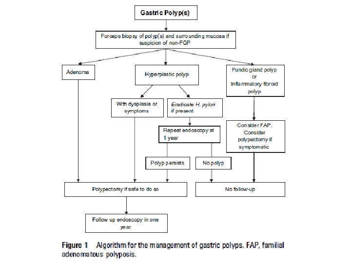
• • • • Recommendations for gastric polypectomy < Any polyp with a dysplastic focus should be completely removed when safe to do so (moderate, trade offs, qualified) < Endoscopists performing gastric polypectomy should be competent to manage the complications of bleeding (high, benefit, definitive). < The decision to perform a polypectomy must be weighed against the risk of complications especially in elderly patients with concomitant illness (high, benefit, definitive). An algorithm for the management of single and multiple gastric polyps is given in figure 1. Endoscopic follow-up All gastric polyps except FGPs and inflammatory fibroid polyps are associated with significantly increased gastric cancer risk. Table 2 summarises the characteristics of gastric polyps and their cancer risk.