Introduction Overview 8 th October 2008 Hanneke den

Introduction / Overview 8 th October 2008 Hanneke den Ouden, Justin Chumbley, Maria Joao Rosa Wellcome Trust Centre for Neuroimaging, UCL

Overview • Introduction • What’s Mf. D • Programme for 2008 • How to prepare your presentation • Where to find information and help • Experts • Overview for dummies Introduction to Mf. D 2008

Methods for Dummies 2008 Aim: to give a basic introduction to human brain imaging analysis methods, focusing on f. MRI and M/EEG Wednesdays / 13 h 00 – 14 h 00 / FIL Seminar Room Areas covered in Mf. D Introduction to Mf. D 2008 • Basic Statistics • f. MRI (BOLD) • EEG / MEG • Connectivity • VBM & DTI

PROGRAMME 2008 Autumn Introduction to Mf. D 2008

I. Basic Statistics 15 th Oct – 12 st Nov • Linear Algebra & Matrices (Nicholas Wright, Nick Henriquez) • T-tests, ANOVA’s & Regression (Nicholas Wright, Lorelei Howard) • General Linear Model (Ramiro Joly, Sinead Mullally) • Bayes for beginners (Stephen Fleming, Sharon Gilaie-Dotan) • Random Field Theory (Christian Lambert, Cirian Hill) Introduction to Mf. D 2008

II. What are we measuring? 19 th Nov – 26 st Nov • Basis of the BOLD signal (Christoph Korn, Andrea Dantas) • Basis of the M/EEG signal (Bonnie Breining, Evelyne Mercure) Introduction to Mf. D 2008

III. f. MRI Analysis 3 th Dec – 17 rd Dec • Preprocessing: – Realigning and un-warping (Mark Weyers, Hanna Marno) – Co-registration & spatial normalisation (Catherine Sebastian, Antoinette Nicolle) • Study design and efficiency (Nicholas Wright, Edoardo Zamuner) Continues after Christmas break… Introduction to Mf. D 2008

PROGRAMME 2008 Spring Introduction to Mf. D 2008
 14 th Jan – 21 st Jan •")
III. f. MRI Analysis (cont. ) 14 th Jan – 21 st Jan • 1 st & 2 nd level analysis – Design matrix contrasts and inference (Tessa Decker & Emmanuelle Volle) • Parametric modulation, temporal basis functions and correlated regressors (Mkael Symmonds, Patrick Freund) Introduction to Mf. D 2008

IV. EEG & MEG 28 th Jan – 4 th Feb • Pre-processing and experimental design (Nicolas Abreu, Mathias Gruber) • Contrasts, inference and source localisation (Maro Machizawa, Himn Sabir) Introduction to Mf. D 2008

V. Connectivity 11 th Feb – 25 th Feb • Intro to connectivity - PPI & SEM (Karine Gazarian, Carmen Tur) • DCM for f. MRI – theory & practice (Nikos Konstantinou, Stephanie Burnett) • DCM for ERP / ERF – theory & practice (Giovanna Moretto, Saloni Krishnan) Introduction to Mf. D 2008

VI. Structural MRI Analysis 4 nd Mar & 11 th Mar • Voxel Based Morphometry (Thomas Doke, Chi. Hua Chen) • Basics of DTI (Nikos Gorgoraptis, Rohit Khanna) Introduction to Mf. D 2008

How to prepare your presentation Very important!!!: Read the Presenter’s guide (available on the website) • Remember your audience are not experts… • The aim of the sessions is to – introduce the concepts and explain why they are important to imaging analysis – familiarise people with the basic theory and standard methods • Time: 45 min. + 15 min. questions – 2 presenters per session • Don’t copy last year’s slides!!!. . . • Start preparing your talk with your co-presenter at least 2 weeks in advance • Talk to the allocated expert 1 week in advance Introduction to Mf. D 2008

What if I can’t make my presentation? • If you want to change / swap your topic, try and find someone else to swap with…. • …if you still can’t find a solution, then get in touch with Maria, Justin or Hanneke as soon as possible (at least 3 weeks before the talk). Introduction to Mf. D 2008

Where to find help Mf. D Home Resources http: //www. fil. ion. ucl. ac. uk/mfd/page 2. html • Key papers • Previous years’ slides • Human Brain Function Textbook (online) • SPM course slides • Cambridge CBU homepage (Rik Henson’s slides) • Methods Group Experts • Monday Methods Meetings (4 th floor FIL, 12. 30) • SPM email List Introduction to Mf. D 2008

Experts • Will Penny – Head of Methods • John Ashburner • Stephan Kiebel • Guillaume Flandin • James Kilner • Rosalyn Moran • Carlton Chu • Andre Marreiros • Vladimir Litvak • Zoltan Nagy • Justin Chumbley • Hanneke den Ouden • Maria Joao Rosa Introduction to Mf. D 2008 Contact the expert: discuss presentation and other issues (1 week before talk) Expert will be present in the session

Website http: //www. fil. ion. ucl. ac. uk/mfd/ Where you can find all the information about Mf. D 2008: Programme Contacts Presenter’s guide Resources (Help) Etc… Introduction to Mf. D 2008
 – – Run by Christian")
Other helpful courses • Matlab for Cognitive Neuroscience (ICN) – – Run by Christian Ruff http: //www. icn. ucl. ac. uk/courses/MATLAB-Tutorials/index. htm 4. 30 pm, Thursday (not every week!) 17 Queen Square, basement seminar room • Physics lecture series – Run by FIL physics team – Details will be announced – 12 Queen Square, Seminar room Introduction to Mf. D 2008

Overview for Dummies Introduction to Mf. D 2008
 data")
Outline • Getting started with an experiment • SPM & your (f. MRI) data – Preprocessing – Analysis – Connectivity • Acronyms Introduction to Mf. D 2008

Getting started – Cogent • http: //www. vislab. ucl. ac. uk/Cogent/ – present scanner-synchronized visual stimuli, auditory stimuli, mechanical stimuli, taste and smell stimuli – monitor key presses – physiological recordings – logging stimulus & scan onset times • Try and get hold of one to modify rather than starting from scratch! People are more than happy to share scripts around. • If you need help, talk to Eric Featherstone. Introduction to Mf. D 2008

Getting started - Setting up your experiment If you need… • special equipment – Peter Aston – Physics team • special scanning sequences – Physics team • They are very happy to help, but contact them in time! Introduction to Mf. D 2008

Getting started - scanning decisions to be made • What are your scanning parameters: – how many conditions/sessions/blocks – Interstimulus interval – Scanning sequence – Scanning angle – How much brain coverage do you need • how many slices • what slice thickness – what TR • Use the physics wiki page: http: //cast. fil. ion. ucl. ac. uk/pmwiki. php Introduction to Mf. D 2008

Summary • Get you script ready & working with the scanner • Make sure it logs all the data you need for your analysis • Back up your data from the stimulus PC! You can transfer it via the network after each scanning session… • Get a scanning buddy if it’s your first scanning study • Provide the radiographers with tea, biscuits, chocolate etc. Introduction to Mf. D 2008

Use the project presentations! They are there to help you design a project that will get you data that can actually be analyzed in a meaningful way Introduction to Mf. D 2008

Hurrah! I have brain data! • So what do I do now? • This is where we get into SPM & preprocessing… • …and more decision-making! • All the processing takes a long time, so make sure you have decided in advance, and don’t need to redo your analysis Introduction to Mf. D 2008

Statistical Parametric Mapping • Mf. D 2008 will focus on the use of SPM 8 • SPM software has been designed for the analysis of brain imaging data in f. MRI, PET, SPECT, EEG & MEG • It runs in Matlab…just type SPM at the prompt and all will be revealed. • There are sample data sets available on the SPM website to play with
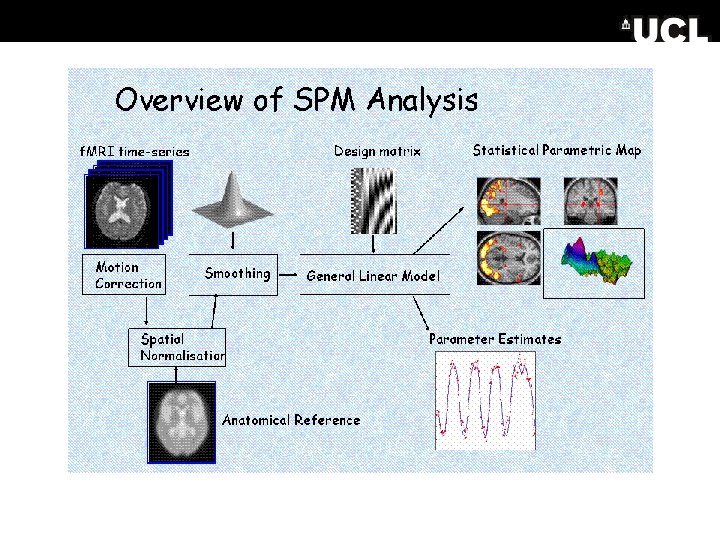

Preprocessing Possibilities… • These steps basically get your imaging data to a state where you can start your analysis – Realignment & Unwarping – Segmentation and Normalisation – Smoothing
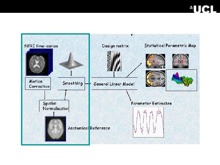

Analysis • Once you have carried out your pre-processing you can specify your design and data – The design matrix is simply a mathematical description of your experiment E. g. ‘visual stimulus on = 1’ ‘visual stimulus off = 0’

Analysis • Once you have carried out your pre-processing you can specify your design and data – The design matrix is simply a mathematical description of your experiment – E. g. • ‘visual stimulus on = 1’ ‘visual stimulus off = 0’ Our f. MRI data is a time series based on the haemodynamic response. The basis functions used in SPM are curves used to ‘describe’ or fit the haemodynamic response in relation to our model
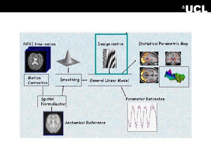

Analysis • Once you have carried out your pre-processing you can specify your design and data – The design matrix is simply a mathematical description of your experiment – E. g. ‘visual stimulus on = 1’ ‘visual stimulus off = 0’ • Our f. MRI data is a time series based on the haemodynamic response. The basis functions used in SPM are curves used to ‘describe’ or fit the haemodynamic response in relation to our model • The HRF is convolved with the design matrix, and we estimate how much variance of the BOLD response our convolved parameters can explain for each voxel, which is expressed in an SPM
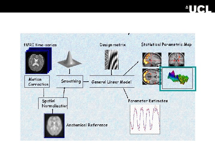

Contrasts & inference • The SPMs are then thresholded to correct for multiple comparisons • Contrasts allow us to test hypotheses about our data, using t & f tests • 1 st level analysis: activation over scans (within subject) • 2 nd level analysis: activation over subjects

Write up and publish…

Connectivity • Functional segregation – responses to an input giving a regionally specific effect • Functional integration – how one region influences another…subdivided into: – Functional connectivity: correlations among brain systems (e. g. principal component analysis) – Effective connectivity: the influence of one region over another (e. g. psycho-physiological interactions, or DCM)

Acronyms • • • DCM – dynamic causal model DTI – diffusion tensor imaging FDR – false discovery rate FFX – fixed effects analysis FIR – finite impulse response FWE – family wise error FWHM – full width half maximum GLM – general linear model GRF – gaussian random field theory HRF – haemodynamic response function ICA – independent component analysis ISI – interstimulus interval • • • PCA – principal component analysis PEB – parametric empirical bayes PPI – psychophysiological interaction PPM – posterior probability map Re. ML – restricted maximum likelihood RFT– random field theory RFX – random effects analysis ROI – region of interest SOA – stimulus onset asynchrony SPM – statistical parametric mapping VBM – voxel-based morphometry
- Slides: 39