Introduction of Abdomen and Muscles of anterior abdominal

Introduction of Abdomen and Muscles of anterior abdominal wall Dr. Tabrez

Abdomen • Lower half of trunk below the diaphragm • Divided in to two parts: i) Abdomen Proper: From Diaphragm to Pelvic brim (Pelvic inlet) ii) Pelvis: Below the pelvic brim (Pelvic inlet)

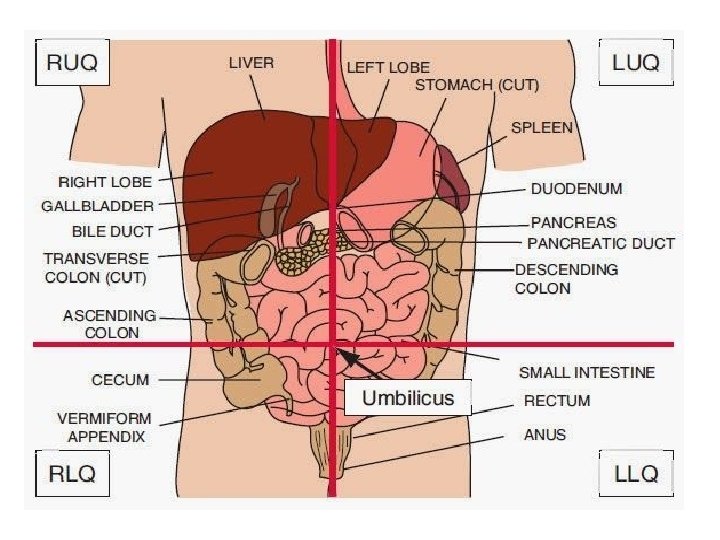
Quadrants of abdomen • • Right upper Left upper Right lower Left lower

Planes for Abdominal Regions Transpyloric plane • midway between suprasternal notch and symphysis pubis • midway between umbilicus and xiphisternal joint • a hand's breadth below the xiphisternal joint - intersects the first lumbar vertebral body near its lower border (L 1) - meets the costal margins at tips of 9 th costal cartilages. -Passes through Pylorus of stomach.

Structures found at the level of Transpyloric plane ü pylorus of stomach ü Fundus of gall bladder ü Hila of both kidneys ü Origin of superior mesenteric artery ü Lower end of spinal cord

Subcostal plane • corresponds to lowest limits of 10 th costal cartilages. • cuts front of third lumbar vertebral body nears its upper border (L 3). Transtubercular plane • level with the iliac tubercles • Cuts front of fifth lumbar vertebral body near its upper border (L 5).

Vertical planes: Right lateral and left lateral planes Indicated on the surface by vertical lines • Midway between the anterior superior iliac spines and the symphysis pubis These lines are also called 'mid-clavicular' or 'mammary' lines). •

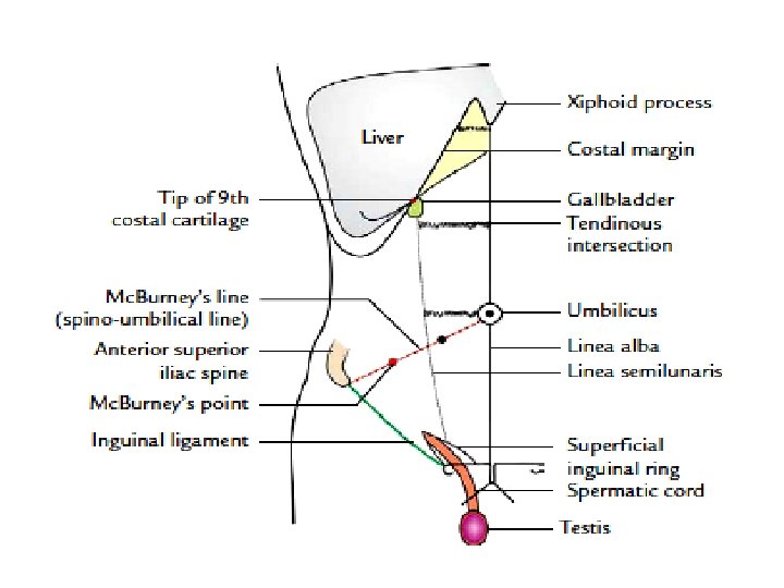
Landmarks on anterior abdominal wall

Division of Abdomen in to 9 Regions • Abdomen divided into 9 Regions- by Two Horizontal and 2 Vertical Planes. • Planes used for demarcation: – Horizontal planes : Transpyloric plane and Transtubercular planes – Vertical Planes : Right and left Lateral planes
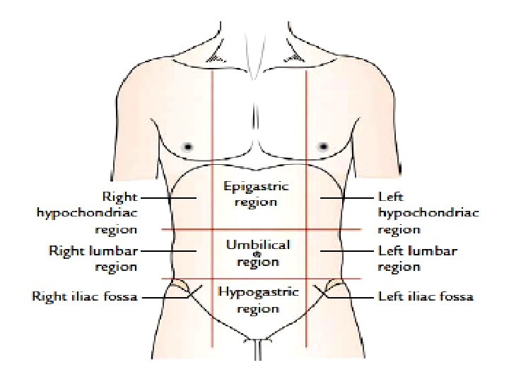

Abdominal Regions

Layers of anterior abdominal wall 1. Skin. 2. Superficial fascia. 3. External oblique muscle. 4. Internal oblique muscle. 5. Transversus abdominis muscle. 6. Fascia transversalis. 7. Extraperitoneal tissue. 8. Parietal layer of peritoneum.

1. Skin- Features 1. The skin of the anterior abdominal wall is thinner and more sensitive than the skin of the posterior abdominal wall. 2. The cleavage lines (Langer’s lines) in the anterior abdominal wall run horizontally.

3. In the multiparous women, the lower part of the anterior abdominal wall presents a number of irregularly branched white lines called striae /linea gravidarum (due to degenerative fibrosis of subcutaneous fat).

4. Umbilicus • Made up of cicatricial tissue, which represents the attachment of fetal end of the umbilical cord.

Position of umbilicus 1. In adult, it lies at the level of intervertebral disc between L 3 and L 4 vertebrae. 2. In newborn, it is slightly at a lower level due to poorly developed pelvic region. 3. In old age, it comes down to lower level due to diminished tone of the abdominal muscles.

Anatomical Significance of umbilicus 1. The level of umbilicus serves as water-shed line for venous and lymphatic drainage. • The venous blood and lymph flow upward above the level of the umbilicus and downward below the level of the umbilicus.

2. It indicates the level of T 10 dermatome, i. e. , skin around the umbilicus is supplied by the 10 th spinal segment. 3. It is one of the important sites of portacaval anastomosis.

Embryological Significance of umbilicus • It is the meeting point of four folds of embryonic plate (e. g. , two lateral folds, head fold, and tail fold).

Cutaneous nerve supply by anterior and lateral branches

Cutaneous arteries

Venous drainage

Subcutaneous venous collateral circulation

2. Superficial fascia • Above the level of the line joining the two A. S. I. S, the superficial fascia consists of a single layer but below this line it consists of two layers (a) a superficial fatty layer (Camper’s fascia) (b) a deep membranous layer (Scarpa’s fascia).

Scarpa’s fascia made up of elastic type of fibrous tissue

Significance of Holden’s line

3. External oblique muscle

4. Internal oblique muscle

5. Transversus abdominis muscle

Structures derived from flat muscles 1. Inguinal ligament E. O. M 2. Conjoint tendon(Falx inguinalis) I. O. M +T. A. M 3. Cremaster muscle I. O

Vertical muscles 1. Rectus abdominis

2. Pyramidalis muscle Absent in 20% • It is a small triangular muscle, lying anterior to the lower part of the rectus abdominis muscle within the rectus sheath. • Origin Arises by its base from the front of the pubic symphysis and pubis. • Insertion It is inserted by its apex into the linea alba midway between the umbilicus and the pubic symphysis. • Nerve Supply By the subcostal nerve (T 12). • Action It tenses the linea alba.

Functions of anterior wall muscles 1. Form strong and expandable support for the abdominal viscera and protect them from injury. 2. Compress the abdominal contents to maintain or increase the intra-abdominal pressure. 3. Move the trunk to maintain the posture.

LOVE NATURE Thank You
- Slides: 36