Introduction l l The growth and development of


 cells are formed by mitosis. Meiosis (")


 of")


- Slides: 29
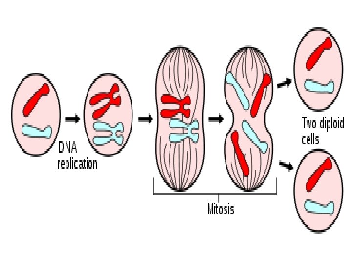
Introduction l l The growth and development of every organism depends on the precise replication of the genetic material during each cell division. Cell division is the process by which cells reproduce themselves. 2
Objectives • • Learn preparing and staining procedure to identify the stages of mitosis in onion root tip. To differentiate between the different stages of mitosis.
Types of Cell Division ü Mitosis • Mitosis is the process by which a eukaryotic cell separates the chromosomes in its cell nucleus into two identical sets, in two separate nuclei. • It is generally followed immediately by cytokinesis, • The outcome of this process: two new daughter cells with the same number and kind of chromosomes as the parent cell.
l ü l l New body (somatic) cells are formed by mitosis. Meiosis ( Reduction division ) produces progeny cells with one-half the genetic content and number of chromosomes as parent cell The formation of male and female gametes in animal cells or spores in plant cells is by meiosis. 5
Mitosis ü Purpose • Mitosis occurs in order for organisms to grow and develop. • In order to replenish dead or dying cells such as skin cells, and cells in the digestive tract. ü Karyokinesis • process of nuclear division (division of genetic material). ü Cytokinesis • Process of dividing cytoplasm/cell.
l Mitosis occurs only in eukaryotic cells and the process varies in different species. For example: l animals undergo an "open" mitosis, where the nuclear envelope breaks down before the chromosomes separate. l Fungi and yeast undergo a "closed" mitosis, where chromosomes divide within an intact cell nucleus. l Prokaryotic cells, which lack a nucleus, divide by a process called binary fission. 7
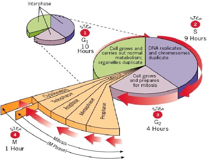
The Cell Cycle • The life of a cell is divided into three stages known as the cell cycle: 1. Interphase: cell carries out normal functions and prepares to divide. 2. Mitosis: nucleus divides splits into two. 3. Cytokinesis: cell and contents divide into two daughter cells.
Interphase • • • The cell prepares itself for cell division. This phase consist of the G 1 (first gap), S (synthesis), and G 2 (second gap) phases. The chromatin is diffuse. protein synthesis, DNA synthesis, Replication of other cellular structures.
l S phase: where each chromosome is duplicated and consists of two sister chromatids joined together by a centromere. Now, the nucleus and cell increase in l size, and chromosomes are fully extended. l 11
Mitosis • There are 4 main phases: • Prophase, • Metaphase, • Anaphase, • Telophase. • Cytokinesis (division of the cytoplasm) follows and one cell becomes two. the nucleus has to migrate into the center of the cell before mitosis can begin. •
Mitosis: Prophase Major processes during this phase: • Chromosomes condense and form visible bodies. • Chromosomes become thicker, shorter, and easily visible when stained under the light microscope. • Two “sister chromatids” join near their middle at a structure called the centromere. • The nucleolus and the nuclear membrane disappear. • The mitotic apparatus the spindle, begins to organize within the cell.
Mitosis: Metaphase • • • Chromosomes become aligned at midpoint or equator between poles of the cell. Are at their thickest and shortest structure. They are easily identified as two longitudinally double sister chromatids.
Mitosis: Anaphase • • The centromere replicates and splits The sister chromatids begin to separate and migrate to the opposite poles separate of the cell.
Mitosis: Telophase • • Ø • • Chromosomes now uncoil Nuclear envelope reappears and surrounds the chromosomes Cytokinesis The cytoplasm and all its contents are divided between the 2 daughter cells (cytoplasmic division), membrane creates between the 2 new daughter cells In plants, such as the onion root tip cells, this is seen as the formation of a cell plate
Cell Cycle
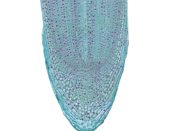
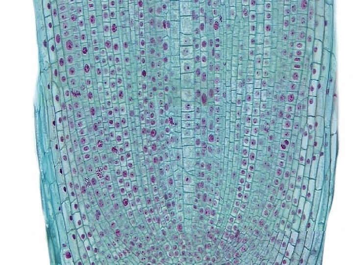
Stages of mitosis in onion root tip cells
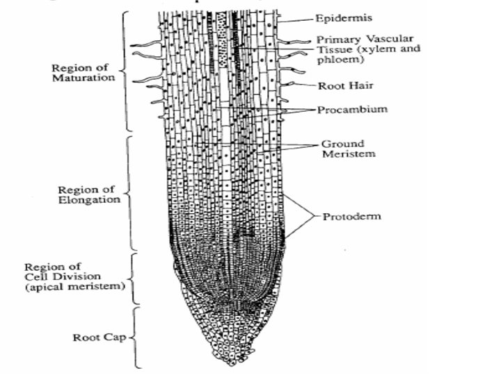
Mitosis in Root Tip • • • In a growing plant root, the cells at the tip of the root are constantly dividing to allow the root to grow. Because each cell divides independently of the others, a root tip contains cells at different stages of the cell cycle. This makes a root tip an excellent tissue to study the stages of cell division.
Materials • • • Slides & cover slips Microscope Fresh onion root tips Fixative ( methanol-acetic acid 3: 1 v/v) Forceps 1 M HCl Razor blade Stain Paper towel, or absorbent paper
Method • • Cut 2 -3 mm of onion root Use forceps to transfer an onion root tip into the cup of HCl. Leave for 4 minutes Transfer the root tip to the cup containing fixative and leave it for 4 minutes. Then place the root tip on a slide. Cover the root tip with a few drops of stain for 2 minutes Cover the root tip with one to two drops of 45% acetic acid Put a cover slip over the root, put a paper towel or other absorbent paper and with your thumb firmly press on the cover slip.
• • • Observe your preparation under the low power (X 10) of a microscope Search the slide to find cells in various stages of cell division, once you have located cells in division, change to high power (X 40) & try to observe several stages of division. Record the number of cells in each stage. Count at least three full fields of view. You should have counted over 200 cells. Record your data in the table Calculate the percentage of cells in each phase and record in the table
Interphase Prophase Anaphase Metaphase Telophase
Animation http: //www. youtube. com/watch? v=s 1 yl. UTb Xy. WU l http: //www. johnkyrk. com/mitosis. html