Introduction History of otorhinolaryngology Current achievements and problems

Introduction. History of otorhinolaryngology. Current achievements and problems of Otorhinolaryngology. Influence of occupational factors internal and external environment on the development of ENT diseases. Physiology of the auditory and vestibular analyzers. Department of ENT diseases of Tashkent Medical Akademy www. tma. uz

Otorhinolaryngology § special clinical discipline that studies the morphological and physiological features of the pathology of the ear, upper respiratory tract and adjacent areas. § Its name comes from two Greek words: otos - ear. § rhinos - the nose, § laryngos - larynx § logos - teaching.
 § Voyachek Vladimir Ignatovich (18761971)")
The founders of Otolaryngology § Simanovsky Nikolai Petrovich (18541922) § Voyachek Vladimir Ignatovich (18761971) Academician § Transfiguration of Boris Sergeevich (1892 -1970) Academician

The founders of otorhinolaryngology in Uzbekistan § § § § Professor Stein, GS (1920 1922) Shumsky Zigzmund Ignatevich (1946 1954) Affectionate Izrael Y. (1954 1967) Miraziz Kuchkar Djuraevich (1967 1997) Muminov Akram Ibrahimovic (1975 1992) Ibragimov Gairat Tulanovich (1975 1995) Dadamukhamedov Abdumalik Nazirovich

The tasks of modern otorhinolaryngology § Studies on the effect of factors external and internal environment to the ENT organs § The development of microsurgery of ear and upper respiratory tract infections; § Problems ENT oncology; § Cochlear implantation

Anatomy of the Ear

STRUCTURE OF THE BODY OF HEARING

Tympanic membrane

Tympanum

Eustachian tube

The inner ear

BLOOD SUPPLY OF INTERNAL EAR

APPEARANCE bony labyrinth 1. Canales semicirculares ossei 2. Crura ossea ampularis 3. Crus osseum simplex 4. Crus osseum commune 5. Vestibulum 6. Fenestra vestibuli 7. Fenestra cochleae 8. Cochlea 9. Canalis spiralis cochleae 10. Cupula cochlea

Structure of the membranous labyrinth 1. Ductuli semicirculare membranacea 2. Crista ampularis 3. Utriculus 4. Macula utriculi 5. Sacculus 6. Macula sacculi 7. Ductus utriculasaccularis 8. Ductus endolymphaticus 9. Saccus endolymphaticus 10. Ductus reuniens 11. Ductus cochlearis

BONE SNAIL 1. Сanalis spiralis cochleae 2. Cupula cochlea 3. Basis cochlea 4. Modiolus 5. Basis modioli 6. Lamina modioli 7. Lamina spiralis ossea 8. Scala vestibuli 9. Scala tympani

The cut bone snails 1. Сanalis spiralis cochleae 2. Cupula cochlea 3. Basis cochlea 4. Modiolus 5. Basis modioli 6. Lamina modioli 7. Lamina spiralis ossea 8. Scala vestibuli 9. Scala tympani 10. Helicotrima 11. Canalis longitudinalis modioli 12. Canalis spiralis modioli 13. Meatus acusticus internus

Scheme of the cochlea duct 1. Canalis spiralis ossea cochlea 2. Modiolis 3. Lamina spiralis ossea 5. Canalis spiralis cochlearis 6. Canalis spiralis modioli 7. Ganglion spirale 8. Paries tympanicus 9. Organum spirale 10. Paries vestibularis 11. Paries externus ductus cochlearis 12. Scala vestibuli 13. Scala tympani 14. Cavum ductus cochlearis

Auditory and vestibular analyzer § Auditory and vestibular analyzers located in the maze. § Maze is divided into three sections: § A - Snail (anterior) § B - threshold (middle section) § B - the semicircular canals (back office)

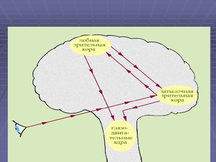
Auricular analyzer § Auditory analyzer auditory analyzer - an integrated system, originated from the outer ear and ends in the cerebral cortex. Each section of the system characterized by a certain function, which is a violation at any level leads to partial or complete hearing loss.

Auricular analyzer § Adequate stimulus of the auditory analyzer is the sound - these are the mechanical vibrations of gaseous, solid or liquid medium, which, acting on the auditory analyzer, causing it to specific physiological process, subjectively perceived as a sensation of sound.

Auricular analyzer § The air speed of sound is 332 m / s. § Hearing range of human hearing within 16 20 000 Hz. § Infrasound - sound less than 16 Hz. § Ultrasounds - the sounds of more than 20 000 Hz. § Bone-conducting tissue - ultrasounds to 225 Hz.

Auricular analyzer § Auditory analyzer is divided into conductive and sound is perceived apparatus. § By soundconducting include the outer and middle ear, peri-and endolymphatic space of the inner ear, basilar membrane plate and preddverno cochlea.

Auricular analyzer § Sound is perceived apparatus represented a peripheral receptor organ spiral. § Sound conducting apparatus - is used to deliver sound to the receptor. § Sound is perceived device converts mechanical vibrations into neural excitation process.

Methods of hearing § The study of hearing with live speech. § Whispered and colloquial speech § Kamertonal research § Elektroaudiometry(threshold and abovethreshold, speech audiometry,

Methods of hearing § Objective audiometry § Study of unconditioned reflexes auropupillyarny, auropalpebralny etc. § Investigation hearing with conditioned reflexes to sound § Play audiometry § Impedansometry § Elektrokohlegrafy § Registration of auditory evoked potentials § Otoacoustic emission study

STRUCTURE BODY BALANCE

Vestibular analyzer § Vestibular analyzer - an organ of balance, muscle tone control that supports a given position of the body and delivered to the cerebral cortex, information about the position and movement of the body in space.

Vestibular analyzer § Adequate stimulus for the semicircular canals - the angular acceleration. § Adequate stimulus for the otolith apparatus - the beginning and end of the straight-line motion, its acceleration or deceleration

Vestibular analyzer § centrifugal force, the change in head position and body in space § force of gravity, which acts on the otolithic apparatus, even during complete rest of the body.

Laws Ewald: § The movement of the endolymph in the horizontal semicircular duct of the feet to vial causes nystagmus towards the stimulated ear. The movement of the endolymph of the ampoule to the leg is in the direction of nystagmus is not stimulated ear.
 is a")
Laws Evald: § The movement of the endolymph to the ampulla (ampulopetalny) is a strong irritant horizontal semicircular duct, endolymph than the current from an ampoule (ampulofugalny). § For the vertical channels, these laws reversed.

"Iron Laws" Voyachek § The plane of the nystagmus is always coincide with the plane of rotation. § Nystagmus is always opposite to the direction "Iron Laws" Voyachek. of the shift of the endolymph § Nystagmus - a rhythmic jerking of the eyeballs, consisting of slow and fast components. § The direction of nystagmus is defined by its fast component.
, caloric, rotational, postrotational,")
Nystagmus § Depending on the stimulus, spontaneous nystagmus distinguish between (endogenous), caloric, rotational, postrotational, pressor, § electroplating. § The direction of nystagmus: right, left, up, down. § Plane-horizontal, vertical, rolling nystagmus.

Nystagmus § The strength of nystagmus - I, III degree. § - The amplitude of nystagmus - small, medium and krupnoraz-mashisty. § The frequency of nystagmus (number of shocks for to define a time interval, usually 10 seconds) - a lively, sluggish.

Position of the patient in the study of the semicircular canals: A - lateral (tilt the head forward -30 °); B - anterior (head tilt forward - 90 °); B - posterior (lateral head tilt - 90 °).

Methods of investigation of the vestibular apparatus § Caloric and rotational tests and videookulography nistagmography; § Static and dynamic stabilography; § EEG.

Methods of investigation of the vestibular apparatus § Videokulografphy using special glasses § Electronystagmography is the primary method to identify both spontaneous and experimental nystagmus.

Methods of investigation of the vestibular apparatus

Department of ENT diseases of Tashkent Medical Academy www. tma. uz
- Slides: 40