Introduction Heat cold and electricity are some of


- Slides: 84


Introduction • Heat, cold and electricity are some of the • ‘physical agents’ that can cause non-kinetic injuries to the body.

Severity of burn • ■ first degree – erythema and blistering • (vesiculation); ■ second degree – burning of the full-thickness of • the epidermis and exposure of the dermis; • ■ third degree – destruction down to subdermal • tissues, sometimes with carbonization and • exposure of muscle and bone.

First- and second-degree burns on the right thigh and scrotum of a 5 -year-old diabetic child, who had been treated by a quack, who had promised the parents to cure the child by hot baths. Death was caused by the untreated diabetes.

The extensiveness of burns on a body recovered from a fire may be varied. This individual had second and third himself with petrol before setting degree burns after dousing ﺇﺧﻤﺎﺩ himself on fire (self-immolation). Note the molten and singed hair. ﺍﻟﻤﻨﺼﻬﺮ

Dead bodies in fires pose a problem for pathologists and investigators. Death was shown to have occurred after the fire began from soot in the lungs and carboxyhaemoglobin in the blood. There were no ante-mortem burns and there was a high blood-alcohol concentration.

Heat flexures of the limbs, part way towards the ‘pugilistic attitude’ formed when the arms are raised higher. The elbows, knees and wrists are strongly flexed because muscle contraction is stronger in the flexor groups. The victim was the captain of a Russian ship who set fire to his cabin during a bout of heavy drinking.

Factors affecting degree of burn A) Extent of burnt area: is determined by rule of nine of Wallace. B) Depth of burn: The 3 rd degree burn is the most serious one. C) Site of burn. Neck, abdominal wall or genitalia are more dangerous than those of the extremities.

The ‘Rule of Nines’ for calculating the area • burned. It does not apply to infants whose body • proportions are different from adults. About 60 per cent surface burns • can be survived by children and young people up to 20 years of • age but this rapidly decreases with advancing age. •

Scalds • The general features of scalds are similar to • those of burns, with erythema and blistering, but charring of the skin is only found when the liquid is extremely hot, such as with molten metal.

Scalds or wet burns in a child accidentally left in a hot bath while her mother went to answer the telephone. This illustrates that time, as well as temperature, is a factor in causing heat damage to skin. The child died some days later from an intercurrent chest infection.

A burn on the inner sides of each upper thigh in a young woman. These are suggestive of the application of a hot-water bottle in an attempt at resuscitation after a criminal abortion. The victim died of air embolism following the use of a Higginson syringe.

Pattern of scalding from forced immersion in a hot bath. Note the clear demarcation between scalded and uninjured skin representing the fluid level of the bath. Sparing of skin on the buttocks reflects firm contact between those parts and the base of the bath.

Pattern of scalding from running water

Pathophysiological consequences of thermal injury • Burned/scalded tissue elicits an acute inflammatory • response, leading to increased capillary permeability • at the injured site; tissue fluid loss associated with • thermal injury can be severe enough to cause dehydration, • electrolyte disturbance and hypovolaemic • shock and, if the burn area exceeds 20% of the total • body surface area, the release of systemic inflammatory • mediators which may lead to acute lung injury • and multiple organ dysfunction/failure. Burned skin • provides no protection against infection, increasing • the risk of sepsis in survivors. •

Exposure to heat/hyperthermia • Hyperthermia – a condition where the core body • temperature is greater than 40°C (100°F) – occurs • when heat is no longer effectively dissipated, leading • to excessive heat retention. Its development is • more likely in those who have taken substances • (e. g. drugs, including cocaine and amphetamine) • that elevate metabolic rate/heat production or reduce • sweating, or in those with medical conditions • (e. g. hyperthyroidism). Autopsy findings in ‘heat illness’, • including ‘heat stroke’, are non-specific but • can include pulmonary and/or cerebral oedema, • visceral surface petechiae and features in keeping • with ‘shock’ and multiple organ failure in those who • survive for a short period. •

Charred body at the scene of a fire showing the ‘pugilist attitude’ and post-mortem skin splits on the chest. Extreme care must be taken to preserve the teeth in such cases, in order to assist identification of the deceased.

Post-mortem burn on the arm and chest caused by a hot-water bottle being left over the heart in an attempt at resuscitation. The lesion is sharp-edged, there is no erythematous margin, and the surface is brown and leathery from post-mortem drying.

Larynx of a house-fire victim showing heat blanching of the epiglottis and glottal entrance from breathing hot gas. The lower cut end of the trachea shows copious soot-containing mucus, also indicating respiration during the fire.

A small bronchus from a fire victim, showing histological evidence of soot deposition, together with desquamation of the epithelium and plugging with cellular debris. (Van Gieson; original magnification 10. )

Soot in the tracheal mucus, a sign of breathing while the fire was in progress. Though soot can passively reach the glottis after death if the mouth is open, it cannot reach the lower air passages in any significant quantity. Histology of the smaller bronchi offers complete proof of active respiration.

Pathological investigation of bodies recovered • from fires: When a fire results in a fatality, there should be a • low threshold for treating the death with caution, • in view of the potential for there to have been an • attempt made to conceal a homicide by ‘burning • the evidence’. The need for safety of • investigators after events such as gas explosions, is a very • important consideration in the examination of • fire scene.

The finding of a body in a burnt-out car should always be treated with suspicion. Carboxyhaemoglobin levels may be low in rapid fl ash petrol fires leading to difficulties in assessing vitality at the time of the fire.

The pathological investigation of bodies recovered • from fires should attempt to: • ■ confirm the identity of the deceased; • ■ determine whether the deceased was alive at some • time during the fire (or was dead before it started); • ■ determine why the deceased was in the fire (and • why they could not get out of it); • ■ determine the cause of death; and • ■ determine (or give an opinion as to) the manner • of death. •

Visual identification from facial features may be • possible if there has been limited fire damage to the • body, but heat damage can cause major distortion of • such features even in the absence of direct burns. • Personal effects may assist identification, as • will unique medical features and factors such as • the presence of scars and tattoos, but where there • is severe charring of the body more robust means • of identification must be relied on such as dental • examination and comparison of the dentition with • available ante-mortem records or DNA analysis. •

Post-mortem radiography should usually be • performed before dissection, with particular emphasis on radiographs to assist identification (dentition, surgical prostheses, etc. ), to identify fractures • (including healing fractures with callus) and to exclude projectiles such as bullets and shrapnel.

Determination of ‘vitality’ (the fact that someone • was alive) during the fire at post-mortem examination • may be possible by the finding of soot • in the airways, oesophagus and/or stomach – the • implication that respiration was required to inhale • the soot. The presence of soot below the level of • the vocal cords, often accompanied by thermal injury • of the epithelial lining of the airways, is particularly • useful, and may be confirmed under the • microscope •

Blood samples can be taken for a rapid • assessment of carboxyhaemoglobin, as a convenient marker of the inhalation of the combustion products of fire i. e. the inhalation of carbon monoxide, a product of incomplete combustion. While carboxyhaemoglobin levels are • commonly elevated in fire deaths (a level of over 50 per cent

Failure to escape from fire Deceased was already dead before the start of the fi re • ● Deceased was intoxicated (alcohol and/or drugs) • ● Deceased was elderly and/or disabled • ● Deceased was immobile • ● Deceased was rapidly overcome by fumes/smoke because of ‘poor • physiological reserve’ (e. g. ischaemic heart disease or chronic • obstructive airways disease) • ● Deceased had insufficient time to escape the fire owing to the nature • of the fire itself (an explosion or ‘fl ash fire’) • ● There was panic/confusion • ● Escape routes were obstructed (deliberately or accidentally) • ● Deceased was in an unfamiliar environment (and did not know where • the escape route was) •

Soot staining can be seen in the oesophagus in this body recovered from a house fire. Such staining indicates that the deceased was alive – and able to swallow – at the time of the fire.

Deaths occurring during a fire • The majority of burnt bodies recovered from • fires will not have died from the direct effect of burns, but from exposure to the products of combustion (smoke, carbon monoxide, cyanide and a cocktail of toxic combustion byproducts) and/or the inhalation of hot air/gases

Interference with respiration (owing to a • reduction in environmental oxygen and/or the production of carbon monoxide and other toxic substances) ● Inhalation heat injury leading to • laryngospasm, bronchospasm and so-called ‘vagal inhibition’ and cardiac arrest ● Exposure to extreme heat and shock • ● Trauma • ● Exacerbation of pre-existing natural disease • or burns

Post-mortem fire-related skull fractures in a severely charred body. There is a reddish-brown heat haematoma/extradural haemorrhage on the inner surface of the carbonized cranial vault.

Post-mortem artefactual skin splitting in charred skin. No haemorrhage can be seen in the depths of the splits and they should not be confused with ante-mortem injuries.

Cold injury (hypothermia) • Cold injury (hypothermia) has both clinical and • forensic aspects, as many people suffer from and • die of hypothermia even in temperate climates in • winter. In marine disasters, hypothermia may be • as common a cause of death as drowning. •

Mild cases • – shivering • – feeling cold • – lethargy • – cold, pale skin • ● Moderate cases • – violent, uncontrollable shivering • – cognitive impairment • – confusion • – loss of judgment and reasoning • – loss of coordination, including difficulty moving around or stumbling – memory loss • – drowsiness • – slurred speech • – apathy • – slow, shallow breathing • – weak pulse • •

Pinkish discoloration over the large joints in fatal hypothermia.

Numerous superficial haemorrhagic gastric erosions of the lining of the stomach in hypothermia. These are often called ‘Wischnewsky’ spots.

Frostbite of the knuckles.

● Severe cases • – loss of control of hands, feet and limbs • – uncontrollable shivering that suddenly stops • – unconsciousness • – shallow or no breathing, weak • – irregular or no pulse • – stiff muscles • – dilated pupils •

The body may rapidly lose temperature when • immersed in cold water, as water has a cooling effect that is 20– 30 times that of dry air. Hypothermia may confer a protective effect on survival following cold water immersion, but survival may be accompanied by severe hypoxic brain injury.

Generally, the elderly, children and trauma • patients are susceptible to hypothermia. • Hypothermia can be classified into mild (core temperature 32– 35°C compared with a normal of 37. 5°C), moderate (30– 32°C), or severe (< 30°C).

Other gastrointestinal lesions sometimes • found in deaths caused by hypothermia include haemorrhagic erosions and infarction in the small bowel (because of red blood cell ‘sludging’ and submucosal thrombosis), and haemorrhagic pancreatitis with fat necrosis. Cold injury to the extremities may be severe enough to cause frost bite, which reflects tissue injury that varies in severity from erythema to infarction and necrosis following microvascular injury and thrombosis

The phenomenon of ‘hide and die syndrome’ • describes the finding of a body that appears to • be hidden, for example under furniture or in the corner of a room, etc. ,

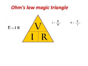
Electrical injury Injury and death from the passage of an electric • current through the body is common in both industrial and domestic circumstances. The essential factor in • causing harm is the current (i. e. an electron flow) • which is measured in milliamperes (m. A). This in • turn is determined by the resistance of the tissues • in ohms () and the voltage of the power supply in • volts (V). •

Almost all cases of electrocution, fatal or • otherwise, originate from the public power supply, which is delivered throughout the world at either 110 V or 240 V. It is rare for death to occur at less than 100 V.

Usually, the entry point is a hand that touches • an electrical appliance or live conductor, and the exit is to earth (or ‘ground’), often via the other hand or the feet.

In either case, the current will cross the • thorax, which is the most dangerous area for a shock because of the risks of cardiac arrest or respiratory paralysis.

Firm contact electric marks on a fingertip. Two blisters have been formed by the heat of local skin resistance; they have collapsed on cooling.

Firm contact blister and adjacent spark burn from 240 V mains supply. The victim was holding a faulty electric drill. There were no other marks on the body.

Collapsed blisters and a spark burn from holding a faulty power tool.

Multiple electrical burns on a hand, with blisters showing the typical pale raised margins and areas of peeling epidermis. There is a blackening from metallization as the current was passing for several hours; the victim was an electrician who fell into an air-conditioning plant.

Homicidal electrocution in the bath. An electric heater was dropped into the water with the wire to the earth terminal deliberately disconnected. The 240 V supply entered the body diffusely through the water and completed the circuit by earthing through contact with the breasts against the metal taps, leaving electrical marks on the skin.

Suicide by electricity. Both wrists were wired in this fashion to a 240 V supply socket, the circuit being completed from arm to arm across the chest.

When a live metal conductor is gripped by • the hand, pain and muscle twitching will occur if • the current reaches about 10 m. A. If the current in the • arm exceeds about 30 m. A, the muscles will go into • spasm, which cannot be voluntarily released because • the flexor muscles are stronger than the extensors: • the result is for the hand to grip or to hold on. This • ‘hold-on’ effect is very dangerous as it may allow the • circuit to be maintained for long enough to cause • cardiac arrhythmia, whereas the normal response would have been to let go so as to stop the pain.

If the current across the chest is 50 m. A or • more, even for only a few seconds, fatal ventricular fibrillation is likely to occur, and alternating current (AC, common in domestic supplies) is much more dangerous than direct current (DC) at precipitating cardiac arrhythmia •

The tissue resistance is important. Thick dry • skin, such as the palm of the hand or sole of the foot, may have a resistance of 1 million ohms, but when wet, this may fall to a few hundred ohms and the current, given a fixed supply voltage, will be markedly

The mode of death in most cases of electrocution • is ventricular fibrillation caused by the direct effects • of the current on the myocardium and cardiac conducting • system. These changes can be reversed • when the current ceases, which may explain some • of the remarkable recoveries following prolonged • cardiac massage after receipt of an electric shock. • The victims of such an arrhythmia will be pale, • whereas those who die as a result of peripheral respiratory • paralysis are usually cyanosed. Even rarer • are the instances in which the current has entered • the head and caused primary brain-stemparalysis, • which has resulted in failure of respiration. •

The electrical lesion • Unless the circumstances are accurately • known, it can be difficult to know whether a dead victim has been in contact with electricity

Usually, however, there is a discrete focal • point of entry and, as the electrical current is concentrated at that point, enough energy can be released to cause a thermal lesion. The entry points may be multiple and obvious, or they may be single and very inconspicuous.

The focal electrical lesion is usually a blister, • which occurs when the conductor is in firm • contact with the skin and which usually collapses soon after infliction, forming a raised rim with a concave centre. Characteristically, the skin is pale, often white, and there is an areola of pallor

local vasoconstriction), sometimes accompanied by • a hyperaemic rim. The blister may vary from a few • millimetres to several centimetres. The skin often • peels off the large blisters leaving a red base. The • other type of electrical mark is a ‘spark burn’, where • there is an air gap between metal and skin. Here, a • central nodule of fused keratin, brown or yellow in • colour, is surrounded by the typical areola of pale • skin. Both types of lesion often lie adjacent to each • other. In high-voltage burns, multiple sparks may • crackle onto the victim and cause large areas of • damage, sometimes called ‘crocodile skin’ because • of its appearance •

Multiple burns from high-voltage (multikilovolt) electrical supply lines. The ‘crocodile skin’ is caused by arcing of the current over a considerable distance.

Death from lightning • Hundreds of deaths occur each year from atmospheric • lightning, especially in tropical countries. • A lightning strike from cloud to earth may involve • property, animals or humans. Huge electrical forces • are involved, producing millions of amperes and • phenomenal voltages. Some of the lesions caused • to those who are struck directly or simply caught • close to the lightning strike are electrical, but other • will be from burns and yet others result from the • ‘explosive effects’ of a compression wave of heated • air leading to ‘burst eardrums’, pulmonary blast injury • and muscle necrosis/myoglobinuria. •

All kinds of bizarre appearances may be found, • especially partial or complete stripping of clothing from the victim, which may arouse suspicions of foul play. Severe burns, fractures and gross lacerations • can occur, along with the well-known magnetization or even fusion of metallic objects in the clothing. The usual textbook description is of ‘fern or branch-like’ patterns on the skin – the so-called Lichtenberg

The ‘Lichtenberg figure’ and lightening fatalities. Note the fern-like branching pattern of skin discoloration on the chest

Thermal injury • It's injury caused by application of exposure to extreme • heat or cold � Classification of thermal injury • 1. Due to heat • A- General effects of exposure to heat • 1 - Heat stroke (Heat hyperpyrexia) • 2 - Heat exhaustion (Heat collapse) • 3 - Heat cramps (Miner's cramps) • B- Local application of heat • 1 -Dry heat >>> Burn • 2 - Moist heat >>> Scald •

2. Due to cold • A- General effects of exposure to cold • 1 - Hypothermia • B- Local application of cold • 1 - Dry cold >>> Frost bite • 2 - Moist cold >>> Trench foot (Immersion foot or hand) • � General effects of exposure to cold • Hypothermia • � Manifestation • Mild degree: -CNS depression, increased metabolic rate, increased pulse, • Shivering, Dysarthria • Moderate degree: -Further CNS and vital sign depression, Loss of shivering, • Arrhythmias, Inability to rewarm spontaneously • Severe degree: -Comatose and areflexes, profoundly depressed vitals •

MEDICOLEGAL IMPORTANTS OF HYPOTHERMIA Death due to cold exposure in most of cases are accidental in nature 2 - Suicidal and Homicidal death due to cold • exposure NOT common 3 - Cold water more penetrating than cold air SO more fatality 4 - Alcoholic, ill health or malnutrition condition reduce resistance of person to cold • • •

Frost bite • It occur due to exposure to dry cold, as in case of • mountaineers Usually affect distal part (fingers, toes more • affect than tip of nose & ear lobule). Atmospheric temperature below zero • Trench foot OR immersion foot or hand • Necrosis of finger, toe or hand & feet may occur • at low temperature below 5Ċ, if the limbs are wet due to immersion •

General effects of exposure to heat • A- HEAT EXHAUSION • It's due to salt-deficiency & dehydration • Signs & symptoms • 1 - Rapid weak pulse & low BP • 2 - Pale cold skin with profuse sweating (temperature • normal or slightly rise) 3 - Vomiting • 4 - Cramps • 5 - NO loss of consciousness, NO rise of temperature, NO • convulsion 6 - Hypokalemia, high hemoglobulin, high blood urea • 7 - Oligouria with high specific gravity • TTT with Intravenous fluid •

B- Heat cramps • This is condition characterized by several painful paroxysmal cramps in the • muscle of extremities and abdomen, due to exposure to hard work in high • temperature lead to rapid dehydration of the body by excessive loss of salt • and water (this may occur alone or in the presence of heat exhaustion) • TTT with Intravenous fluid • C- HEAT HYPERPYREXIA (HEAT STROKE) • Occur when head is exposed directly to sunlight or heat, this will affect the • heat regulation center, SO the mechanisms which control the body • temperature become disrupted (SWEATING), SO temperature rise • Heat stroke is a medical emergency and death rate is high • Signs & symptoms • 1 - Sudden rise of temperature • 2 - Flushed dry skin (NO sweating) • 3 - No vomiting, No cramps, No dehydration • 4 - Convulsion, Coma • TTT >>> cooling of the body • •

B- Local application of heat • BURN • � Classification according to depth • 1. First degree • **Affect superficial layer of skin • **Painfull • **Healing of this burn dose not leave scar • **It appears as blister surrounded by red, hyperemic skin. • 2. Second degree • **There is destruction of full thickness of skin • **Most Painfull • **Healing of this burn leave scar, which produce disfigurement or impaired • function according to site & size • **It appear as shriveled area of coagulated tissue surrounded by burn of first • degree • 3. Third degree • Characterized by gross destruction of subcutaneous tissue, muscles • Painless due to destruction of nerve endings • Healing of this burn leave scar which produce disfigurement or impaired • function according to site & size •

� FACTORS AFFECTING THE PROGNOSIS OF • BURNS 1 - THE EXTENT of burn (MOST important factor) • Can be estimated by (RULE OF 9) • A- Head & Neck = 9% • B- Chest (Front=9%, Back= 9%) • C- Abdomen (Front=9%, Back= 9%) • D- Lower limb • - Right (Front=9%, Back= 9%) • - Left (Front=9%, Back= 9%) • E- Upper limb (Right=9%, Left= 9%) • F- Perineum and external genitalia = 1% •

2 - THE DEGREE (DEPTH) of burn • 3 - THE SITE of burn • Burn of the face, neck or abdomen more dangerous than burn of • the limb 4 - THE AGE of victim • Infant and old persons more affected than young adult • 5 - GENERAL HEALTH of victim • Persons with chronic disease more affected than healthy persons • AGE OF THE BURN • 1 - 2 -3 hrs vesication • 2 - 36 -72 hrs disappearance of inflammatory zone, presence of pus • 3 - 1 week separation of superficial slough • 4 - 2 week separation of deeper slough • 5 - after 2 week appearance of red granulation surface without • slough

� CAUSES OF DEATH FROM BURNS • � IMMEDIATE CAUSES • 1 - Asphyxia • 2 - Primary (Neurogenic) shock • 3 - Associated mechanical injuries • � DELAYED CAUSES • 1 - Secondary (Hypovolumic) shock • 2 - Toxemia • 3 - Infection • 4 - Acute edema glottis • � LATE CAUSES • 1 - Jaundice • 2 - Infection • 3 - Deep vein thrombosis • 4 -Curling's ulcer, Marjolin's ulcer • Curling's ulcer is an acute peptic ulcer of the duodenum resulting as a complication from severe burns (occur after 7 -10 days of burn) • Marjolin's ulcer: - these are the ulcer malignant changes in burnt area. • •

POSTMORTEM PICTURE OF DEATH DUE TO • BURN 1 - The body show ante-mortem burn of different • degree, and may be take pugilistic (boxer's) attitude • 2 - Generalized redness of the body due to • carboxy-hemoglobulin in the blood 3 - Soot in the airway • 4 - Hemoconcentration • 5 - Generalized visceral congestion • 6 -Skull fracture due to severe burn •

Thermal fracture Traumatic fracture • The scalp Show ante-mortem burn Show ante- • mortem wounds The brain Small and shrunken • Edematous The extradural hematoma Dose not fill space between skull & brain • Fill space between skull & brain • The fracture No displacement of bone May show displacement of bone • •

SUREST SIGNS OF ANTEMORTEM BURNS • INJURY AND DEATH 1 - Soot or carbon practicles in respiratory tract • 2 - In absence of soot, concentration of • carboxy hemoglobin in blood beyond 5%, antemortem vital reaction IN and AROUND • burnt margin.

DIFFERENCE BETWEEN ANTEMORTEM & POSTMORTEM BUR Antemortem burn Postmortem burn Hyperemia Present IN and around the burn Absent Vesicles Tense, filled with serum rich by albumin, chloride & polymorphs If present, NOT tense, filled with Fluid poor in protein & NO chloride or polymorphs Floor of ruptured vesicles Red Yellow Signs of inflammation & repair Present Absent Soot in airway May be present Absent CO in the blood May be present Absent Hemoconcentration Present Absent Healing May be present Absent Sepsis May be present Absent Other causes of death Absent Present • • •

DIFFERENCE BETWEEN DRY BURNS, SCALDS & CORROSIV BURN DRY BURN SCALDS CORROSIVE Causes Dry heat E. g. flame Moist heat E. g. hot liquid or vapour Corrosive Direction of spread From below to upward From above to downward Degree All degree may seen 1 st degree only All degree Vesicles Around the burnt area After sometime All over burnt area within few minute Absent Cloths Burnt Not affected Eaten up and discolored Healing According to degree of burn No permanent scar Heal with thick scar Singeing of hair Present Absent Soot in airway May be present Absent Co in blood May be present Absent Nature of affection Usually accidental • • • •

BURN DUE TO ELECTROCAUSION • � NATURE • Accidental death is the most common • Homicidal & Suicidal very rare • � FACTORS AFFECTING THE SEVERITY OF BURN OF • ELECTROCUTION • 1 -STRENGHT OF CURRENT (Most important factor) • The severity of the injury is directly proportional to the strenght of current • 2 - TYPE OF CURRENT • Alternating current more dangerous than the continued one • 3 - DURATION OF CONTACT • The severity of the injury is directly proportional to the duration of the contact 4 - DIRECTION OF ELECTRICAL BURN • In upper parts of the body more dangerous than the lower parts of body • 5 - RESISTANCE OF THE BODY • • In high voltage burn, multiple sparks may crackle onto the victim and cause • multifocal spark lesion called CROCODILE SKIN APPEARANCE • • •

CAUSE OF DEATH FROM ELECTROCUTION • 1 - Ventricular fibrillation (main cause of death) • 2 - Peripheral asphyxia • Due to muscular spasm, this restricts respiratory movement • 3 -Central asphyxia • Occur when current pass through the respiratory center • 4 - Cerebral anoxia • Due to prolonged ventricular fibrillation, this may cause • brain damage due to inadequate blood supply •

Lightning • It's electric discharge coming down from the clouds to • earth. Its direct current It also produce wound of entry (usually head) and • wound of exit (usually foot). It passes through blood vessels and skin in tree like • fashion (called lightning print OR ARBORESCENT MARKINGS) • sometimes can be absent IN general lightning cause any type of injury. • •
 Insidan region jh
 Static electricity and current electricity
 Static electricity and current electricity
 How are static electricity and current electricity alike
 What is a conductor
 Concept map
 Welcome 1 unit 10 lesson 1
 Late postmortem changes
 Heat and cold
 What is contact force
 Some trust in horses
 Chapter 23.1 performing range of motion exercises
 What is the formula for latent heat
 They say it only takes a little faith to move a mountain
 They say it only takes a little faith to move a mountain
 Cakecountable or uncountable
 Some say the world will end in fire some say in ice
 Some say the world will end in fire some say in ice
 Introduction to statistics and some basic concepts
 Properties of heat
 Dry cooking method
 4 forces of nature
 Ib physics topic 5 questions and answers
 Pron and cons
 Electricity and magnetism lecture notes
 Basic electricity and optics
 Electricity and magnetism jeopardy
 Difference between charge and electric charge
 Advanced automotive electricity and electronics
 Sph3u electricity and magnetism
 Relationship between electricity and magnetism
 Induced magnetic moment
 Magnetism grade 5
 Electricity and magnetism
 Electricity and magnetism
 Fuse symbol igcse
 Ampere
 Supply and use tables
 Magnet current
 Public authority for electricity and water
 Electricity and magnetism
 Electricity and magnetism
 Compare and contrast cold wave and wind chill factor
 Gabby and sydney bought some pens and pencils
 Some prepositions show time and place and others add detail
 Van der graaf generator fysica
 What material does not conduct electricity
 Weather merit badge answers
 Particle model of electricity
 The buildup of electric charges on an object
 How does a photocopier use static electricity
 Snc1d electricity
 Substance
 Effects of electric current on human body
 Physics circuits symbols
 Cheap electricity pasadena
 Ohms law triangle
 Current
 Tariff systems grade 10
 What is missing
 Water conducts electricity
 Salt water electricity generator
 Coulomb symbol
 Electricity merit badge powerpoint
 Energy slogan
 Elements that are shiny and conduct heat are called ______.
 Electricity is all around us
 Bill nye current electricity
 What is electricity
 Electricity explained
 Is static electricity ac or dc
 Electricity powerpoint
 Electricity merit badge
 Electricity merit badge
 Where does electricity come from
 How electricity works
 The path of least resistance electricity
 Rubbing amber
 Iec 61968 5
 Chapter 6 section 1 electric charge worksheet answers
 Chapter 20 static electricity answers
 Chapter 13 basics of electricity
 Electrical merit badge
 Biomass electricity generation
 Kirchhoff's current law examples

