
INTRODUCTION AND EPIDEMIOLOGY Bullets or fragments Severity Wound
INTRODUCTION AND EPIDEMIOLOGY Bullets or fragments Severity Wound entirely different Management Preparation in our hospital
– these cause wounds, Assessment Treatment
TYPES LOW VELOCITY AND HIGH VELOCITY MISSILE WOUNDS
1. Low velocity missile wounds From hand guns Heavy bullets 150 -350 mps Crushed and forced apart Drilling a simple hole o Vital structures Comparable to knifes No energy transmitted o No hidden injuries
2. High velocity missile wounds l From rifles and fragments of explosives l Velocity greater than speed of sound l Impact l KE=1/2 mv* l Severity -Part, Length and Energy l Energy transferred -density and stability.
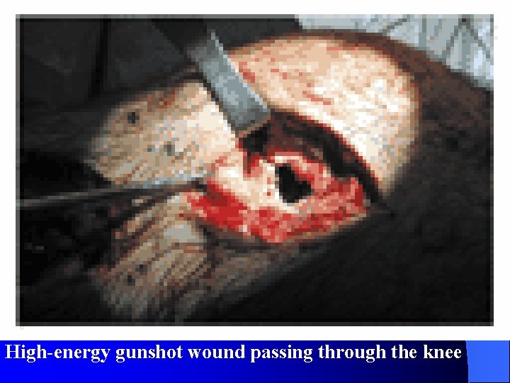
High-energy gunshot wound passing through the knee
Severity lies in two: 1. Shock Waves – Compressed – Moves away-1500 mps – Millionth of a second – Extremely high pressures – Damage tissues at distance
2. Temporary Cavitation – Transfer energy – Forwards and outwards violently. Cavity 30 to 40 times diameter of missile. Subatmospheric pressure Entry and exit Collapse down- actively sucking About 500 ml totally destroyed. v
Dead Muscles l Plum red colour, doesn’t contract and doesn’t bleed. l Directly proportional to density Homogenous very susceptible Lung, resistant.
l Inversely proportional to elastic fibres Skin and lung resistant. l Hits bone fragments as secondary missile Large exit wounds
Instability of the missile Stable on impact, § Only 10 -20% Unstable, 60 -70% Fragments on impact § All energy Liver, spleen and brain Pulped tissue with debris and bacteria
MANAGEMENT OF OPEN WOUND FIRST AID – Pressure bandage – Very occasionally tourniquet – Bony damage splinting
Wound excision 1) Exploration 2) Removal of dead, damaged contaminated 3) Foreign bodies 4) Arrest bleeding 5) Restoration of normality
TECHNIQUE Tourniquet shouldn't be used - Live and dead difficult But in massive bleeding Preparation B) Irrigation C) Enlargement A)
D) Excision: Principle a. Skin b. Fat c. Fascia d. Muscle –be radical e. Haematomas E) Haemostasis - By packing -Haemostats and ligatures
Management of specialized tissues 1. Nerve: – identify with a non-absorbable suture 2. Tendon: – Cover it – Trimmed and repair deferred 3. Bone: - – Cover it – Restore anatomy
4. Joints Exception for delayed closure • Infection and adhesion • Antibiotics 5. Blood vessels Should be repaired FASCIOTOMY - In the whole length of compartment
Hallmarks of modern war injury Multiple injuries to different body systems To maim not to kill Fragments No Characteristic Variety of injuries
Features of missile injuries Low energy- limited injury Cavitation In bone creates secondary missiles Muscles creates the ideal culture medium.
MANAGEMENTOF MISSILE INJURIES Missile wounds of soft tissue - Complete exploration - Delayed primary closure
Stages of operation: 1. Cleaning, incision 2. Deep fascia exposed and incised 3. Neurovascular bundle 4. Removal of foreign matter
5. Dead Muscle excised; 4 C’s for muscle excision: a. Colour b. Contractility c. Consistency d. Capillary bleeding 6. Tendon ends trimmed
7. Major artery and vein, trimmed and sutured 8. Bone fragments must not be discarded 9. Injured joints 10. At the end, wound irrigated left open 11. Immobilization 12. Antibiotic cover
Principles of missile injury surgery Preserve skin Divide Fascia Repair vessels not nerves Remove dead tissue Stabilize bone with external fixation Clean and close joint cavities Leave wounds open
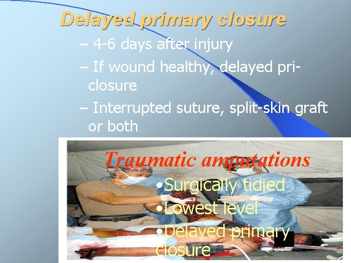
Delayed primary closure – 4 -6 days after injury – If wound healthy, delayed priclosure – Interrupted suture, split-skin graft or both Traumatic amputations • Surgically tidied • Lowest level • Delayed primary closure
Missile wounds of the abdomen l Nasogastric tube and urinary catheter l Digital rectal l Timing vary l Blood in realistic quantities
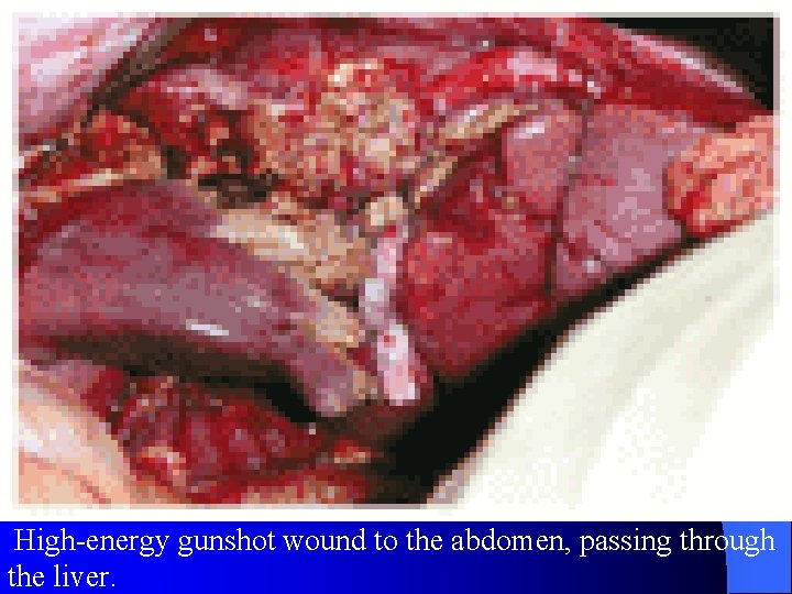
High-energy gunshot wound to the abdomen, passing through the liver.
STEPS OF OPERATION: 1. Full midline incision 2. Source of bleeding Ø Stomach -Inspect posterior gastric wall - opening lesser sac Ø Retroperitoneal haematoma in duodenal region -Kocher’s method
ØRetropertoneal haematomas of ascending and descending colon -Exploration Ø Non-expanding retroperitoneal haematoma over the kidneys -left undisturbed
Wounds of colon and rectum Right side: v Pri repair or pri. resection v Vented ileo tranverse anastomosis
Left side; One stage procedure • If high risk resected -Proximal colostomy and distal mucous fistula • Hartman procedure • Subsequent restoration •
Extra peritoneal rectal injuries: -Sigmoid end colostomy Good dependant drainage -Tip of coccyx and anus Severe Ligature of internal iliac artery
Renal Injury • Conservatively • Ureter to surface or ‘ a pig tail stent’ Bladder and urethral injuries • Suprapubic cystostomy Liver injury • Compression and packing • Drainage of surrounding
Damage to spleen and pancreas • Spleen and tail of pancreas -Resection • Head of pancreas – fatal Peritoneal toilet • Warm saline closure
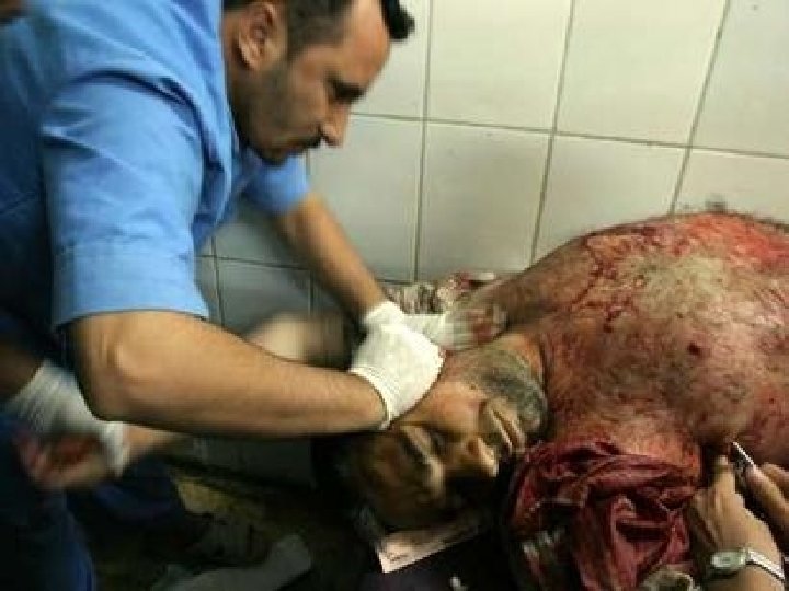
MISSILE WOUNDS OF THE CHEST High mortality Airtight seal Ø To prevent open pneumothorax Tube thoracostomy Ø Prevent accumulation of blood or air Ø By chest radiography
Entry and exit excised Opening sealed Delayed pri. Closure 20% require formal thoracotomy
Indications (thoracotomy) More than 1. 5 liters initial blood loss Continuing loss of >200 ml/h Cardiac tamponade Other mediastinal injuries Persistent air leak Retained foreign bodies >1. 5 c. m. in diameter
MISSILE WOUNDS OF THE HEAD Ø High energy - lethal Ø Low energy and tangential - air way, ventilation & maintenance of BP & perfusion pressure Ø Localise FB and bone fragments Ø CT Ø Excision
Ø Irrigation and suction Ø Temporalis fascia and fascia lata - Close dura Ø Head and face - Exception of delayed pri. closure Ø IPPV - Reduces intracranial pressure Ø Transducers - To monitor ICP
SHOT GUN INJURIES Excision Indriven wadding and plugs Laparotomy Retention of lead shot High lead concentration Will fall
DO’S AND DONTS OF MISSILE INJURIES DO ü Incise skin generously ü Incise fascia widely ü Identify neurovascular bundle
ü Excise all devitalized tissue ü Remove ü Leave ü Dress all indriven clothing wound open wound ü Record all injuries
Dont’s Excise too much skin – Practice keyhole surgery – Repair tendons or nerves –
– Remove attached pieces of bone – Close the deep fascia – Insert synthetic prostheses – Pack the wound – Close the skin
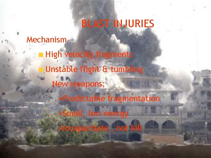
BLAST INJURIES Mechanism High velocity fragments Unstable flight & tumbling New weapons; • Predictable fragmentation • Small, low energy • Incapacitate , not kill
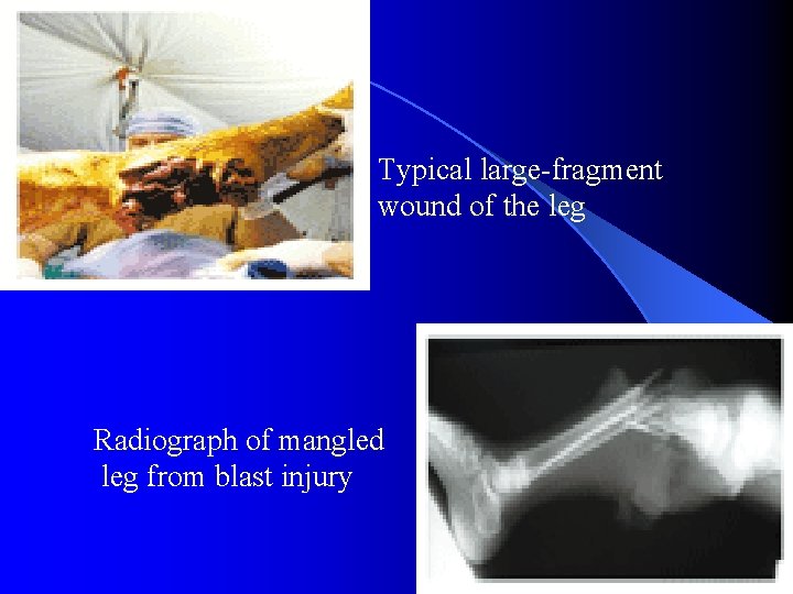
Typical large-fragment wound of the leg Radiograph of mangled leg from blast injury
Two components 1. Blast pressure wave = dynamic over pressure Ø +ve and - ve phase 2. Mass movement of air = Blast wind
BLAST PRESSURE WAVE l Positve pressure phase; Few milli seconds Upto 7000 KN/m* Over & around an obstruction l Incident pressure – 90* to the direction l Reflected pressure
l Speed of sound l In water – greater l Affect air – fluid interfaces
Mass movement of air l Rapid expansion - Super sonic displacements ‘ Fresh air moving very fast’ l Disrupts environment, hurling debris and people
General management l Primary blast wave - Middle ear, lungs & bowel l Combined injury l Heavy contamination l Larger & deeper – priority l Left open
Regional Management Auditory system 3 ways: 1. TM rupture 2. Dislocation of ossicles 3. Disruption of inner ear Depends on angle
Respiratory system Rapid inward movement - pulmonary contusion Stress waves Alveolar – capillary memb. & leakage - Spreading haemorrhage Ø post blast respiratory insufficiency(PBRI)
PBRI Clinical picture Ø Cough with frothy blood – stained sputum Ø Dyspnoea & a feeling of apprehension Oxygen saturation well below 90%
PBRI Guide lines v. ABCDE v Avoid over hydration v. Administer high- flow oxygen v. Arterial blood analysis
v. Mechanical ventilation v Positive en-expiratory pressure (PEEP) v Corticosteroids avoided
GIT l Common in under water l Perforation of stomach, si & caecum
Clinical presentation l Increasing abdominal pain l Signs of peritonitis l Gas under diaphragm
Diagnosis l USG. l CT l DPL l Laparoscopy
The Eye l Conjunctival haemorrhage l Pupil l Distortion of iris l Hyphema
Other factors l Chemical & thermal burns l Inhalation of toxic gases & smoke

- Slides: 64