Introduction ABDOMEN Dr Kumar Satish Ravi M B
,")
Introduction ABDOMEN Dr. Kumar Satish Ravi M. B. B. S. , M. D. (JIPMER), MAMS

Xiphoid process Costal margin Posterior superior iliac spine Iliac crest Iliac tubercle Anterior superior iliac spine Pubic crest Pubic tubercle Symphysis pubis
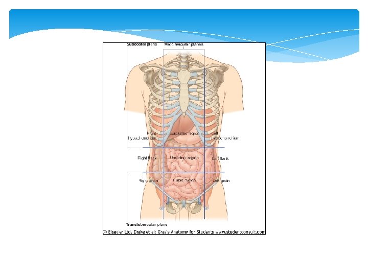

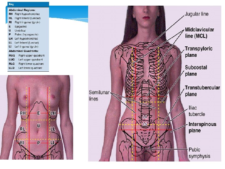
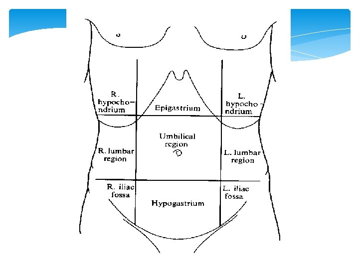
Body Regions Imaginarily divided into 9 regions Midline sections: Epigastric = above stomach Umbilical = umbilicus or navel Hypogastric = below the stomach
 Lateral sections: Right and left hypochondriac Positioned near ribs, specifically cartilages")
Body Regions (con’t) Lateral sections: Right and left hypochondriac Positioned near ribs, specifically cartilages Right and left lumbar Positioned near small of back (lumbar region) Right and left iliac Named for upper bone of hip (ilium) Also called inguinal region (referring to groin)

Body Positions Anatomical Standing erect, facing forward, arms at sides, palms forward, toes pointed forward Prone Lying face down Supine Lying face up
, scapular tip (posterior) Lower border= inferior costal")
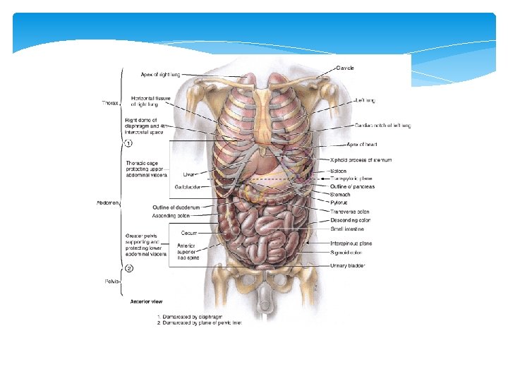
Related anatomy Thoraco-abdomen: Upper border= nipple (anterior), scapular tip (posterior) Lower border= inferior costal margin

Related anatomy Anterior abdomen: Upper border=anterior costal margin Lower border=inguinal crease Lateral border=anterior axillary's line

Related anatomy Back: Upper border=scapular tip Lower border=iliac crest Lateral border= posterior axillary line
")
Soft tissue land marks ( Snells’)

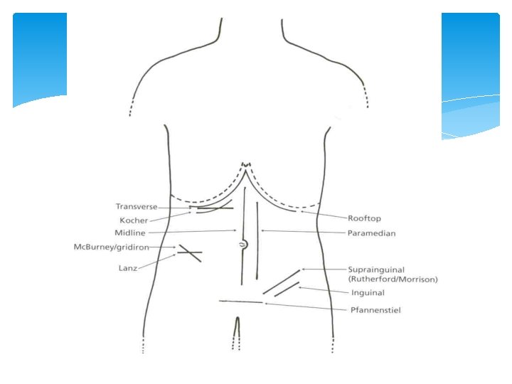
THE ANATOMY of abdominal incision Definition: incision defined as cut made with knife for surgical purposes. Types of abdominal incision: The abdominal incisions are classified into: Vertical abdominal incision Transverse abdominal incision Oblique abdominal incision Abdomino thoracic incision

The vertical incisions: Midline incision Para median The transverse abdominal incisions: Upper and lower transverse incision Pfannenstiel incision LANZ incision for appendicectomy oblique abdominal incisions The subcostal or Kocher’s incision Rutherford Morison incision Mc. Burney incision for appendicectomy

19

THE APPLIED ANATOMY of abdominal incision Abdominal cavity can be approached through numerous incisions but only correct diagnosis will enable surgeon to choose correct incision. When the pre-operative diagnosis is reasonably certain incision may be chosen with confidence. Laparotomy for undiagnosed abdominal disease is most usefully approached through vertical incision equidistant above and below the umbilicus and once the diagnosis confirmed, incision may be enlarged in an upward or downward direction.

Choice of incision depends on many factors these includes: - The organs to be investigated The type of surgery to be preformed Whether speed is an essential consideration The build of the patient The degree of obesity The presence of previous abdominal incisions Closure of the abdominal incision: The ideal method of abdominal wound closure has not been discovered. However it should be free from complications such as: 21

Burst abdomen Incisional hernia Persistent sinuses It should be comfortable to the patient Should leave reasonably good scar Incisional Hernia: - defined as protrusion of omentum or organ through surgical incision. However if the muscles and aponeurotic layers of the abdomen doesn’t heal properly an incisional hernia can result, infection, bowel obstruction and obesity are predisposing factors to incisional hernia 22
 and")
Structure of the Anterior Abdominal Wall: 1. Skin 2. Fascia: superficial Fatty Layer(camper) and deep Membranous layer (scarpa) 3. Muscles 4. Extraperitoneal fascia 5. Parietal peritoneum

Transpyloric plane Hilum of the kidneys Pylorus of the stomach Body of pancreas Fundus of the gall bladder
 Deep membranous layer")
LAYERS OF ANTEROLATERAL ABDOMINAL WALL skin Superficial fatty layer (camper’s fascia) Deep membranous layer (scarpa’s fascia) External oblique Internal oblique Transverse abdominis Fascia transversalis Extraperitoneal fascia Parietal peritoneum
 Deep membranous layer (scarpa’s fascia) External oblique")
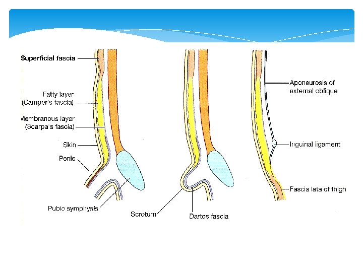
Superficial fascia Superficial fatty layer (camper’s fascia) Deep membranous layer (scarpa’s fascia) External oblique Deep membranous layer (scarpa’s fascia) Superficial penile fascia Attachment of fascia lata Ischiopubic ramus Colles’ fascia Dartos fascia

Muscles

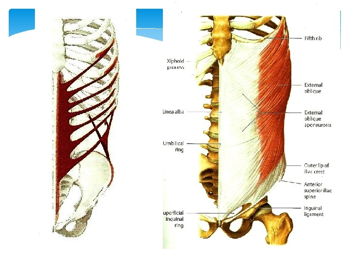
2. 2. 2 Muscles External oblique Origin Muscular slips from the outer surfaces of the lower eight ribs (ribs 5 -12) Insertion Innervation Function Ant. 2/3 outer Anterior rami of lip of iliac crest; lower six thoracic aponeurosis Table spinal nerves (T 7 ending in to T 12) midline raphe (linea alba) Compress abdominal contents; both muscles flex trunk; each muscle bends trunk to same side, turning anterior part of abdomen to opposite side

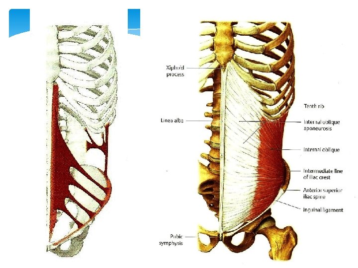
2. 2. 2 Muscles Internal oblique Origin Insertion Thoracolumbar fascia; iliac crest between origins of external and transversus; lateral twothirds of inguinal ligament Inferior border of the lower three or four ribs; aponeurosis ending in linea alba; pubic crest and pectineal line Innervation Anterior rami of lower six thoracic spinal nerves (T 7 to T 12) and L 1 Function See External Oblique

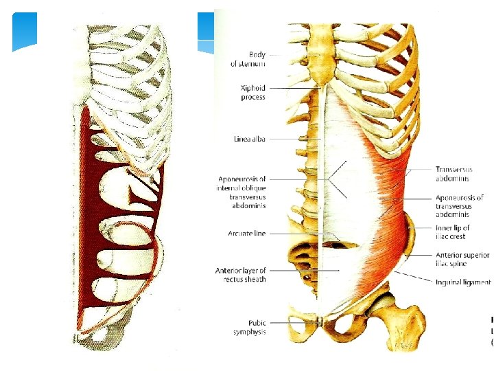
2. 2. 2 Muscles Transverse abdominis Origin Insertion Thoracolumbar fascia; medial lip of iliac crest; lateral one-third of inguinal ligament; costal cartilages lower six ribs (ribs 712) Aponeurosis ending in linea alba; pubic crest and pectineal line Innervation Anterior rami of lower six thoracic spinal nerves (T 7 to T 12) and L 1 Function Compress abdominal contents

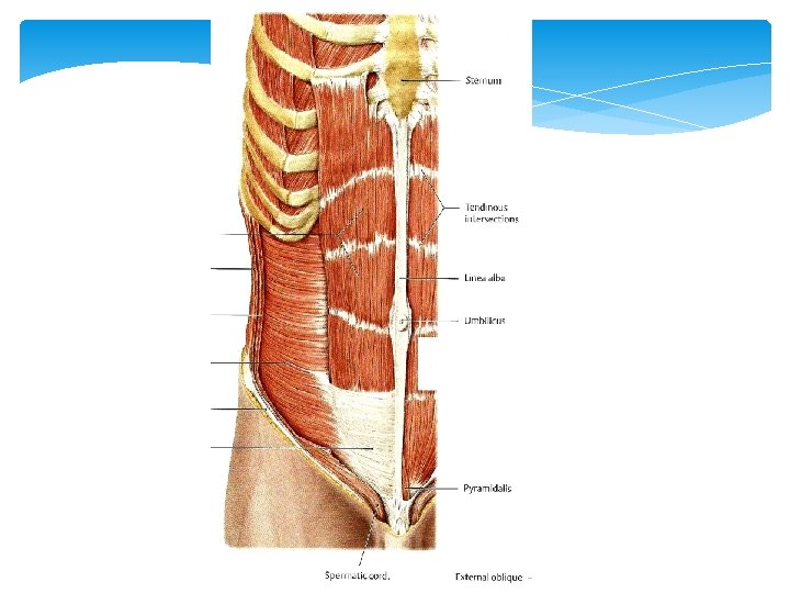
2. 2. 5 Rectus sheath Rectus abdominis Origin Pubic crest, pubic tubercle, and pubic symphysis Insertion Innervation Function Costal cartilages of ribs 5 -7; xiphoid process Anterior rami of lower seven thoracic spinal nerves (T 7 to T 12) Compress abdominal contents; flex vertebral column; tense abdominal wall

PYRAMIDALIS : Pyramidalis is a triangular muscle that lies in front of the lower part of rectus abdominis within the rectus sheath. O- Ant surface of pubis I- Linea Alba Innervation: Terminal branches of the subcostal nerve, which is the ventral ramus of the twelfth thoracic spinal nerve. Actions: Pyramidalis contributes to tensing the lower linea alba, but is of doubtful physiological significance.

Cremaster muscle: Loosely arranged muscle fasciculi lying along spermatic cord. May form incomplete coating around cordcremasteric fascia. Arises mainly from the inferomedial border of internal oblique / transversus abdominis. Medial portion –pubic tubercle & lateral pubic crest. Innervation: Genital branch of the genitofemoral nerve, L 1 & L 2 Actions: Cremaster pulls the testis up towards the superficial inguinal ring.

CONJOINT TENDON The conjoint tendon is formed from the lower fibres of internal oblique and the lower part of the aponeurosis of transversus abdominis. It is attached to the pubic crest and pectineal line. It descends behind the superficial inguinal ring and acts to strengthen the medial portion of the posterior wall of the inguinal canal.

Cutaneous nerves

Deep nerves of the ant. Abd. Wall: Lower six thoracic nerves Fig. from Snells’

Cutaneous vessels
 of the anterolateral abdominal wall : Ø")
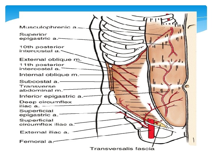
The primary blood vessels (arteries and veins) of the anterolateral abdominal wall : Ø Superior epigastric vessels and branches of the musculophrenic vessels from the internal thoracic vessels. Ø Inferior epigastric and deep circumflex iliac vessels from the external iliac vessels. Ø Superficial circumflex iliac and superficial epigastric vessels from the femoral artery and greater saphenous vein, respectively. ØPosterior intercostal vessels of the 11 th intercostal space and the anterior branches of subcostal vessels

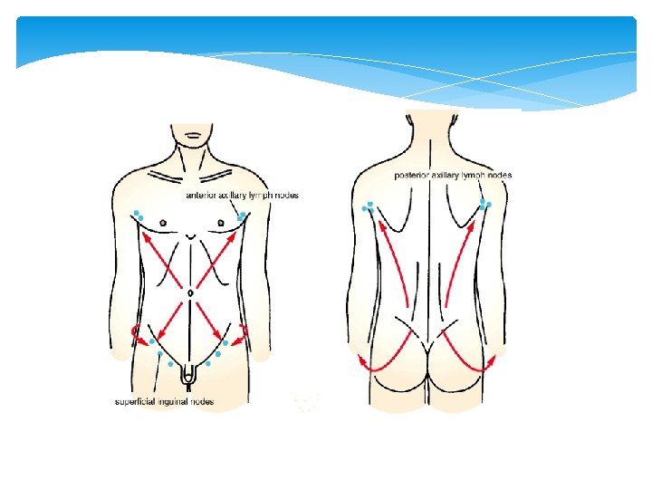
Veins The venous drainage passes above mainly into the axillary vein via the lateral thoracic vein and below into the femoral vein via the superficial epigastric & the great saphenous veins.

Axillary lymph nodes Water shed line Superficial inguinal lymph nodes

Photograph shows a CAPUT MEDUSAE accentuated by a large amount of ascites in a patient being prepared for liver transplantation Henseler K P et al. Radiographics 2001; 21: 691 -704 © 2001 by Radiological Society of North America

RECTUS SHEATH
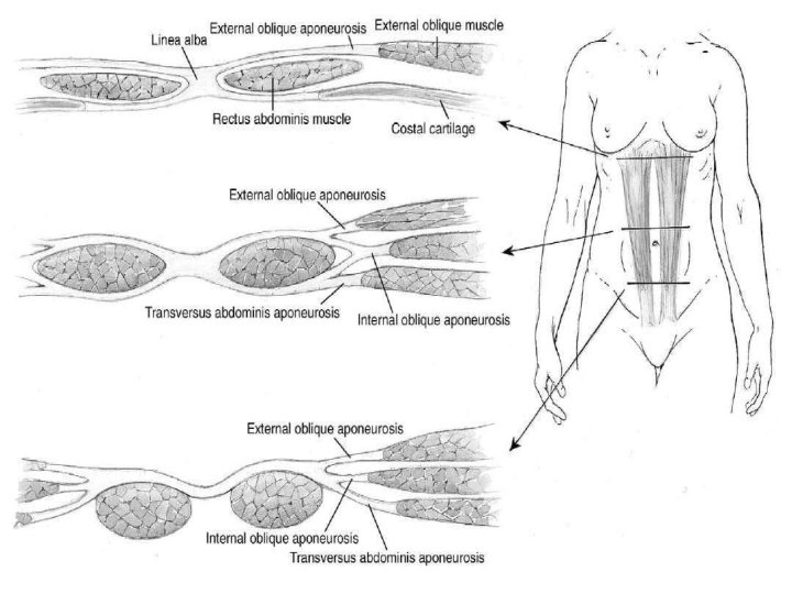
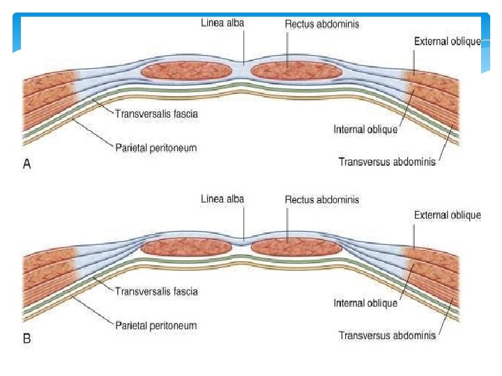
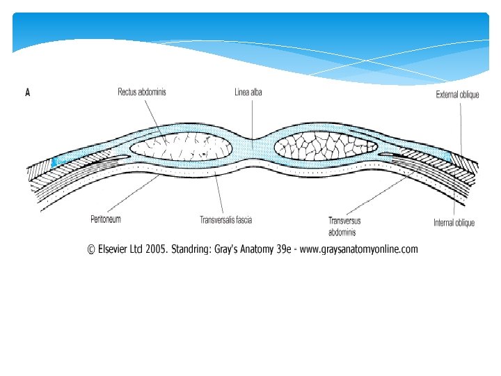

Transverse sections of the Rectus sheath seen at three levels. A. Above the costal margin. B. Between the costal margin and the level of the anterior superior iliac spine. C. Below the level of the anterior superior iliac spine and above the pubis. (Snells’) ANT. LAT MED POST.

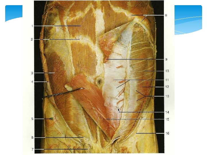
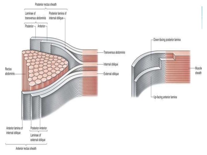
Left: The anterior wall of the sheath has been partly removed, revealing the rectus muscle with its tendinous intersections. Right: The posterior wall of the rectus sheath is shown. The edge of the arcuate line is shown at the level of the anterior superior iliac spine.

Contents of Rectus sheath: 1. Muscles: Rectus abdominis & Pyramidalis 2. Arteries: Sup & Inf epigastric; 3. veins: Sup & Inf. Epigastric; 4. Nvs: Terminal brs of lower six thoracic nerves

RECTUS SHEATH Aponeurotic sheath covering rectus abdominis muscle. Features Formation Contents

Divarication of recti

2. 2. 2 Muscles Neurovascular plane Ant Post

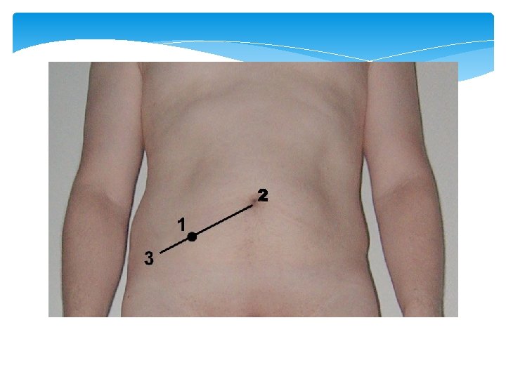
Appendix Mc Burneys point junction between the medial two third and the lateral one third on the line joining the ASIS and umbilicus

Umbilicus Ø Site of attachment of umbilical cord Ø Water-shed: in reference to Lymphatic and venous drainage

Umbilicus ØSkin around it supplied by T 10 segment Nerve.

Umbilicus Ø Site of Porto-caval anastomoses: Portal hypertension causes, Caput medusae

Umbilicus v. Rasberry red/Cherry red tumor: Vitellointestinal duct remnant – tumor. v. Weeping of umbilicus: Persistence of urachus v. Exomphalos: Non return of the physiological herniation of the intestinal loop. v. Ectopia vesicae: Failure of development of infraumbilical part of ant. Abd. Wall. v Umbilical hernia

A. Omphalocele showing failure of the intestinal loops to return to the body cavity after physiological herniation. The herniated loops are covered by amnion. B. Omphalocele in a newborn. C. Newborn with gastroschisis. Loops of bowel return to the body cavity but herniate again through the body wall, usually to the right of the umbilicus in the region of the regressing right umbilical vein. Unlike omphalocele, the defect is not covered by amnion.

Remnants of the vitelline duct. A. Meckel's, or ileal diverticulum combined with fibrous cord (vitelline ligament). B. Vitelline cyst attached to the umbilicus and wall of the ileum by vitelline ligaments. C. Vitelline fistula connecting the lumen of the ileum with the umbilicus. ( Langman’s)

A. Arrangement of the fatty layer and the membranous layer of the superficial fascia in the lower part of the anterior abdominal wall. Note the line of fusion between the membranous layer and the deep fascia of the thigh (fascia lata). B. Note the attachment of the membranous layer to the posterior margin of the perineal membrane. Arrows indicate paths taken by urine in cases of ruptured urethra.

Clinical importance: The membranous layer of the superficial fascia is important clinically because beneath it is a potential closed space that does not open into the thigh but is continuous with the superficial perineal pouch via the penis and scrotum. Rupture of the penile urethra may be followed by Extravasation of urine into the scrotum, perineum, and penis and then up into the lower part of the anterior abdominal wall deep to the membranous layer of fascia. The urine is excluded from the thigh because of the attachment of the fascia to the fascia lata.

Clinical Significance of Fascia and Fascial Spaces of Abdominal Wall Ø Liposuction is a surgical method for removing unwanted subcutaneous fat using a percutaneously placed suction tube and high vacuum pressure. The tubes are inserted subdermally through small skin incisions. (REF: SNELL’S)

- Slides: 72