Intro to Radiologic Technology RADT A RTEC A
")


 Crookes tube – With electrical supply 2) Screen coated with barium")

 • Joint Review Committee on Education in Diagnostic Medical")

 • American Registry")


 • Nuclear Medicine Technology (NM) • Radiation Therapy")
 • Computed Tomography(CT) • Magnetic Resonance Imaging (MR)")
 Can get formal education 1)")

 Mina Colunga Registered Technologist in")
 – Specializing")


 Angiography")
 Angiography")

 = $ 23 -$40 per hour")
 – Post primary certification 1) Must have primary certification in radiography,")


 --uses high frequency sound waves")

 “real time” images 82")


- Primary certification Mike Smith, RT (R) (CI) 1) Must have")
- Post primary certification JOE CAR, RT (R) (VI) 1) Must")


 Commercial Radiologist Assistant = RA • Sales •")
 • Still not widely accepted • Must have a primary certification")

 • Registry (out of state) • X rays")

 • LLU • PART OF RADIOLOGIST GROUP •")

- Slides: 136
Intro to Radiologic Technology (RADT A)
RTEC A INSTRUCTOR • MINA COLUNGA, B. S. , RT. , C. R. T. Instructor, minacolunga@yahoo. com or mcolunga@elcamino. edu WEB page: www. elcamino. edu/faculty/mcolunga 2
WHY CHOOSE RADIOGRAPHY? 3
Is this a safe profession? Why do you want to do this? Why are you taking this class? 4
Preconceived Ideas regarding the X-ray field
What is x-rays? X-rays are electromagnetic radiation with extremely short wavelengths. They can pass through many materials.
What is radiation? Radiation – transfer of energy through space or a material away from the source
• Radiology- Medical specialty in which x-rays, radium, and radioactive substances are applied in the diagnosis and treatment of the patient • Diagnostic Imaging- Medical specialty in which xrays, radium, radioactive substances, sound waves, and radio frequencies are applied in the diagnosis and treatment of the patient • Radiologist- Physician who applies any form of radiation in the diagnosis and treatment of disease.
• Radiographer- Skilled person qualified by education to provide patient services using imaging modalities as directed by a physician qualified to order and/or perform radiographic procedures. (X-ray Technologist) • Radiograph- a photographic record produced by xrays through an object.
Types of Radiation Non-ionized ex: radio Ionized ex: x-rays, gamma
Electromagnetic Spectrum
History of Radiology 14
– November 8, 1895: Historical Perspectives • Wilhelm Conrad Röntgen discovered x-rays – German Physicist – University of Wurtzburg 15
• Wilhelm Röntgen in 1895 - discovered xrays • Working with Crooke’s vacuum tube – He found invisible rays were produced. – These new rays could go through skin and flesh – Give a picture of a person's bones. 16
17
X-rays – the Basic Radiological Tool Röntgen’s experimental apparatus Crookes tube Taken 22 Dec. 1895 18
First Radiograph • Anna Bertha Röntgen • 30 minute exposure. 19
Collaborative Events • Crookes tube – Air evacuated glass tube – Cathode side – Anode side – Electrical supply • Screen or board painted with barium platinocyanide • Low light work area 20
21
“Willie Röntgen” • Honored in 1901 with the first Nobel prize in physics for his efforts. 22
In the beginning…. . 23
Early years in Radiologic Technology • Nurses or nurses aides taught how to “take an x-ray” • NO special education • Only “ON THE JOB” training • Experience the best teacher • The first Technologist is credited to be EDWARD C. JERMAN. 24
An early therapy session 25
26
In 30 years • Developed from a technical trade to one of a professionalism • Once thought that anyone could be trained to quickly = “push the buttons’ • To now where it is considered a profession that requires analytical thinking and problem solving 27
28
29
• X rays began to be used in industry and medicine • Years later, they noticed it can be harmful • They could be harmful to: – living tissue – even cause cancer if the exposures were too great or too prolonged 30
Early signs of possible damage from Radiation exposure • Skin dryness • Erythema • Ulcers formed 31
Acute: Ulceration 32
33
Radiologic Technologists Practices RADIATION SAFETY TO SELF AND OTHERS 34
35
36
37
38
HISTORY REVIEW Who is this? 39
HISTORY REVIEW Wilhelm Conrad Röntgen 40
HISTORY REVIEW What did he discover? 41
HISTORY REVIEW He discovered x-rays 42
HISTORY REVIEW What were the series of events that led to the discovery? 43
HISTORY REVIEW 1) Crookes tube – With electrical supply 2) Screen coated with barium platinocyanide 3) Low light area 44
Accreditation, Certification, Registration, Licensing? ? ? What is all that? 45
Accrediting Agencies for Schools (JRC’s) • Joint Review Committee on Education in Diagnostic Medical Sonography (JRCDMS) • Joint Review Committee on Education in Nuclear Medicine Technology (JRCNMT) • Joint Review Committee on Education in Radiologic Technology 46
Individual Certification • Take an exam • Pay a fee • You then get registered • Nearly all hospitals require appropriate certifciation as a condition of employment. 47
National: Registry Agencies • American Registty of Diagnostic Medical Sonographers (ARDMS) • American Registry of Radiologic Technologists • Nuclear Medicine Certification Board 48
State Licensing Agencies • Vary from state to state • List of individual state requirement can be obtained at www. arrt. org • • Must provide proof of certification Fill out paperwork Pay a fee Sometimes take an exam 49
Certification vs. License • ARRT – National certification • R. T. – Must take an exam • Pass with 75% – Can take this after completing program • CRT – State Licensing – Must pass ARRT or other equivalent national exam to get this – Pay fee to get radiography license (R) – Take fluoroscopy exam and pay a fee for (F) license 50
RADIOLOGIC TECHNOLOGY It covers all of our individual disciplines. 51
RADIOLOGIC TECHNOLOGY Radiography Mammography Computed Tomography Magnetic Resonance Imaging • Quality Management • Sonography • Radiation Therapy • • Bone Densitometry Vascular Sonography Breast Sonography Cardiac Interventional Radiography • Vascular Interventional radiography • Radiologist Assistant • Nuclear Medicine • • 52
5 Primary Certifications • Radiography (R) • Nuclear Medicine Technology (NM) • Radiation Therapy (T) • Sonography (US) (RDMS) • Magnetic Resonance Imaging (MR) 53
Post Primary Certifications • Mammography (M) • Computed Tomography(CT) • Magnetic Resonance Imaging (MR) or (MRI) – Note: Both a primary and postprimary track • Quality Management (QM) • Cardiac-Interventional Radiography (CI) • Vascular-Interventional Radiography (VI) • Sonography (US) or (RDMS) – Note: Both a primary and postprimary track • Vascular Sonography (VS) • Breast Sonography (BS) • Bone Densitometry (BD) • Registered Radiologist Assistant (RA) 54
MRI and Sonography are PRIMARY and POST PRIMARY 1) Can get formal education 1) On the job training 1) if you have a primary certification in radiography, nuclear medicine or radiation therapy 2) meet clinical requirements. 55
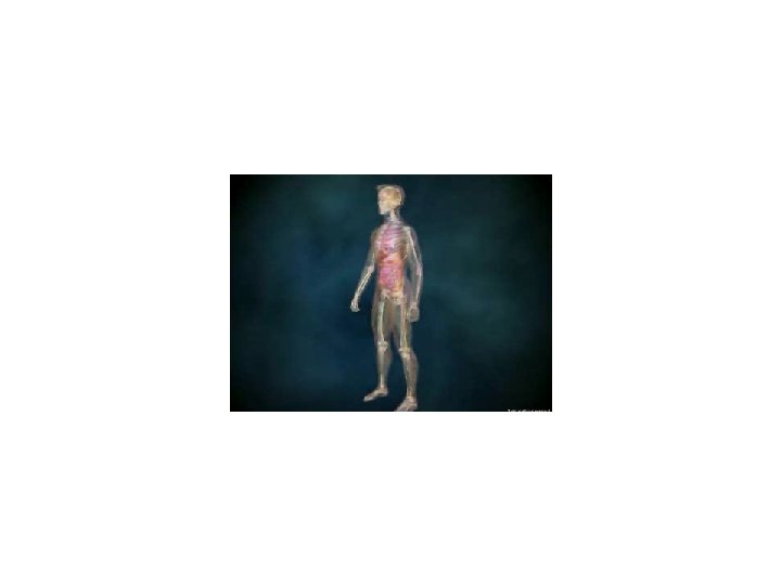
Individual Disciplines of Radiology 56
Radiography : Primary Certification Mina Colunga R. T. (R) Mina Colunga Registered Technologist in the specialty of Radiography 57
RADIOGRAPHY • Diagnostic Radiology – Technologist – Radiographer – Technician (Limited Licensure) – Specializing in the use of x-rays to create images of the body including the skeletal system, chest and abdomen 58
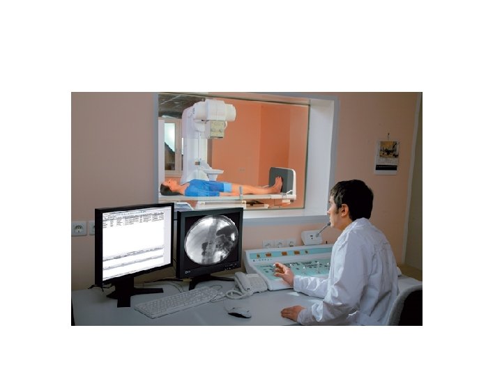
Two Types of x-ray examinations Radiography Fluoroscopy
Fluoroscope • 1898 by Thomas Edison 60
Types of Diagnostic Exams • • • Chest Extremities Skull/ Facial Spine Gastrointestinal Interventional 62
All types of EXAMS & PEOPLE • • • Head to toes Trauma Special procedures Critical patients Walk ins Surgery • • • Infants Elderly All classes All ethnicity All backgrounds 63
Uses Ionizing Radiation to create images of the human body 64
65
66
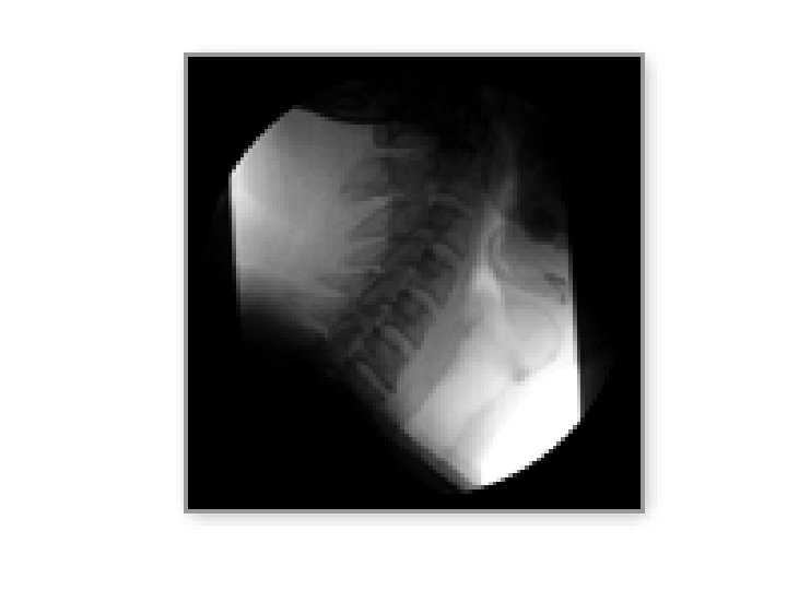
Flouroscopyxrays in motion 67
Fluoroscopy 68
69
70
71
Beyond Diagnostic Radiography 1. 2. 3. 4. 5. 6. 7. 8. Ultrasound (sonography) Angiography Computerized tomography (CT) Magnetic Resonance Imaging (MRI) Positron Emission Tomography (PET) Nuclear Medicine Mammography Radiation Therapy
Beyond Diagnostic Radiography 1. 2. 3. 4. 5. 6. 7. 8. Ultrasound (sonography) Angiography Computerized tomography (CT) Magnetic Resonance Imaging (MRI) Positron Emission Tomography (PET) Nuclear Medicine Mammography Radiation Therapy
SALARY RANGES RT’s • New R. T. (R) = $ 23 -$40 per hour – ON-CALL + O. T. $48, 000 – $83, 000 YR • Advance disciplines • R. T. (CT), (NM), (S), (M), etc – $ 30 - $50 PER HOUR 76
Bone Densitometry (BD) – Post primary certification 1) Must have primary certification in radiography, nuclear medicine or radiation therapy 2) Meet clinical requirements 77
Bone Densitometry- measures mineral content and density of bones 78
Low Doses of Radiation 79
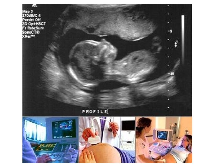
Career in Radiography Ultrasound (sonography) --uses high frequency sound waves
Ultrasound beam is transmitted and reflected – as special crystal at the end of the transducer can determine the type of tissue Determines depth 81
Uses SOUND WAVES (NOT X-RAYS) “real time” images 82
ULTRASOUND uses a technique similar to Navy SONAR to produce diagnostic images. 83
84
85
86
U/S & the “real thing” 87
Vascular Sonography 88
90
Angiography 92
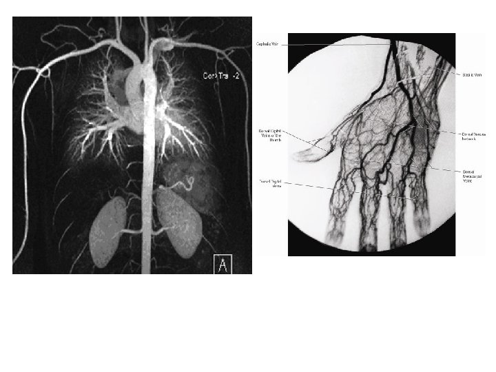
ANGIOGRAPHY is a specialized radiographic examination where the images of the blood vessels of the body are demonstrated by injection of contrast media 93
94
SUB SPECIALITY IN ANGIOGRAPHY • Cardiovascular Interventional Technology • Vascular Interventional Technology Must have certification in diagnostic radiography in order to be trained and certified in these special procedures.
Cardiac Interventional Radiography (CI)- Primary certification Mike Smith, RT (R) (CI) 1) Must have primary certification in radiography 2) Meet clinical requirements 96
Vascular Interventional Radiography (VI)- Post primary certification JOE CAR, RT (R) (VI) 1) Must have primary certification in radiography 2) Meet clinical requirements 97
98
99 99
Angiogram A medical imaging technique using xray and contrast agent to visualize the inside of blood vessels and organs of the body.
Computed Tomography Also known as CT, Cat Scans
Computed Tomography Uses ionized radiation to obtain cross sectional images Designated by CT Jennifer Smith, R. T. (R) (CT) 1) Must have primary certification in radiography, nuclear medicine or radiation therapy 2) Meet clinical requirements 104
Computed Tomography • Able to do 3 D reconstruction
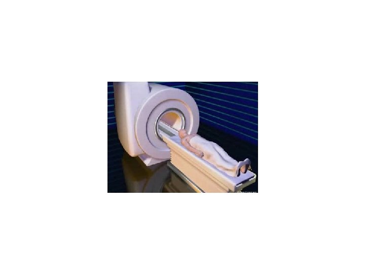
MRI • Magnetic Resonance Imaging
MRI SIGNAL PRODUCTION • Uses – Magnet field – radio waves 108
• MRI – Uses magnetic and radio waves to create images – Can be whole body or cross sectional – Designated by MRI • Jeremy Assef, R. T. , CRT, (MRI)
112
Which one is MRI? CT? 113
Look for the signs…. 114
What are the differences between MRI and CT? CT • Uses ionizing radiation • Can be used on any patient • Fast MRI • Uses magnets and radiowaves • Cannot be used on patients who have metal in their body • Slow 115
Which is better? 116
What are the similarities between CT and MRI? 117
Nuclear Medicine
• Nuclear Medicine – Uses radioactive isotopes to produce images – Radiation comes from within the patient • Primary or Post primary certification
PET scan
Mammography
• Mammography – Breast imaging using ionized radiation
Radiation Therapy • Medical dosimetrists are involved in treatment planning and dose calculations • 1 -4 year program 124
125
126
Radiation therapy
• Radiation Therapy – Involved the treatment of diseases – Use high level of ionized radiation (megavolt) to kill cancerous cells • Primary certification
Additional Opportunities Education Administration Management (QM) Commercial Radiologist Assistant = RA • Sales • Application specialist • • • 130
Radiologist Assistant (RA) • Still not widely accepted • Must have a primary certification in radiography • Must meet clinical requirements 131
TRAVELING TECHNOLOGIST = SEE THE WORLD AND GET $$$ 132
Other working opportunities… • Registry (local) • Registry (out of state) • X rays taken around the world !! 133
Variety of Work Settings • physicians offices, • clinical outpatient facilities, • free standing imaging centers, • mobile imaging centers • portable services to rehabs • Mammo’s to under privileged areas • Urgent care 134
RA • Radiology Assistant (Like PA) • LLU • PART OF RADIOLOGIST GROUP • Still not widely accepted 135
Questions ? • Diagnostic Imaging Modalities 136