Intrauterine fetal death stillbirth Intrauterine fetal death stillbirth

Intrauterine fetal death/ stillbirth

Intrauterine fetal death �stillbirth is defined as a baby delivered after 24 completed weeks of pregnancy , which does not breath or show any signs of life at any time after being completely expelled from mother.

Causes of IUFD � Fetal ◦ ◦ ◦ Cord accidents (knots, thrombosis, strangulation and torsion) Feto–fetal transfusion Feto–maternal haemorrhage Chromosomal and genetic disease Structural abnormality Infection –bacterial, viral ( TORCH) ◦ Anaemias of fetal origin e. g. alpha-thalassaemia � Direct maternal effects ◦ Obstetric cholestasis ◦ diabetes ◦ Antibody production e. g. rhesus disease, platelet alloimmunisation, congenital heart block ◦ Hypertension ◦ Any disorder causing septicemia with hypoperfusion

�placental causes ◦ Placental dysfunction causing hypoxemia i-e FGR ◦ Placental abruption ◦ Placental infarction ◦

Diagnosis of the death � Clinical ◦ ◦ ◦ feature Decreased/absent fetal movement--- 50 per cent of cases Disapperance or reduction of pregnancy symptom Failure of fundal height to rise Fetal heart not audible on pinard scope antenatal visit signs of an acute event such as abruption, ruptured membranes or the onset of labour � Doppler apparatus ◦ CTG and auscultation can be very misleading. ◦ Inability to detect fetal heart on sonicaid

ultrasound � Fetal death must be diagnosed by ultrasound. � must be performed by a trained practitioner � Colour-flow mapping can be very useful, especially in the obese woman. � features. : ◦ Absent fetal heart activity ◦ Spalding’s sign (overlapping of the fetal skull bones when the fetus has been dead for some time); ◦ Retraction of brain tissue ◦ oligohydramnios; ◦ signs of fetal hydrops; ◦ Empty fetal bladder ◦ intrafetal gas.

X-RAY �Spalding’s sign �Robert’s sign ◦ Presence of gas shadows in fetal circulatory system.
 isoimmunisation � If the woman is Rh. D negative-- blood")
Prevention of rhesus (d) isoimmunisation � If the woman is Rh. D negative-- blood for Kleihauer testing Anti-Rh. D immunoglobulin should be given as soon as possible after presentation and not delayed. � Delivery may not occur until after the 72 -hour watershed beyond which immunoprophylaxis is less effective. � A further dose of anti-Rh. D immunoglobulin might be necessary once the Kleihauer result is known and clearance of fetal cells should be rechecked at 48 hours where a large fetomaternal haemorrhage has been encountered. �

Management �Breaking the news �History �Examination �Investigation

HOW TO DELIVER �sympathetic approach �vaginal delivery �CS ◦ ◦ ◦ ◦ major placenta praevia Severe cephalopelvic disproportion Previous classical CS 2 or more previous CS Neglected /obstructed labour Uterine rupture Transverse /shoulder presentation

TIMING OF DELIVERY � over 85 per cent of women will deliver spontaneously within 3 weeks � The risk of coagulation problems is 30 % if the fetus has been retained for 4 weeks. � Conditions that pose a risk to maternal safety ◦ (abruption, pre-eclampsia, infection) ◦ then advice on expediting delivery needs. � conservative management can be offered ◦ testing for DIC twice weekly.

INDUCTION OF LABOUR If the woman appears to be physically well, her membranes intact, no evidence of infection or bleeding--choice of immediate IOL or expectant management. � If there is ruptured membranes, infection or bleeding, immediate IOL. � third-trimester induction ---prostaglandin E 2 preparations. � Now, the combination of the anti-progesterone mifepristone and the prostaglandin analogue misoprostol is recommended. � A standard protocol for mifepristone/misoprostol induction is shown below. � ◦ Mifepristone: 200 mg 24– 48 hours before induction. ◦ Misoprostol: 50– 100 mcg pv 6 hourly for 24 hours. membranes should be left intact for as long as possible, as ascending infection can rapidly develop. � previous CS, the risk of uterine rupture is increased in IOL. -- careful surveillance -- Mechanical methods �

Immediate postnatal care �PPH ◦ pre-eclampsia, abruption, prolonged fetal death or infection. ◦ retained placenta ---Prolonged chorioamnionitis and repeated small abruptions �Risk of thrombosis risk ◦ (pre-eclampsia, infection, immobility) ◦ LMWH

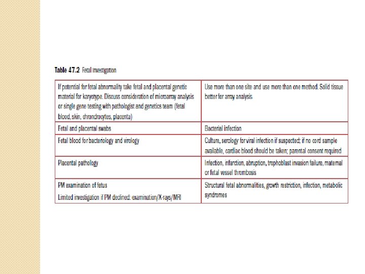
Evaluation of still born baby � Photographs ◦ Gross structural abnormality � Radiology ◦ Grossly malformed baby � Karyotyping ◦ ◦ ◦ Obvious fetal anamoly IUGR Family H/O abnormal children Recurrent preg loss Non immune hydrops fetalis Potter syndrome � Autopsy examination ◦ Congenital malformation ◦ Actual cause of death

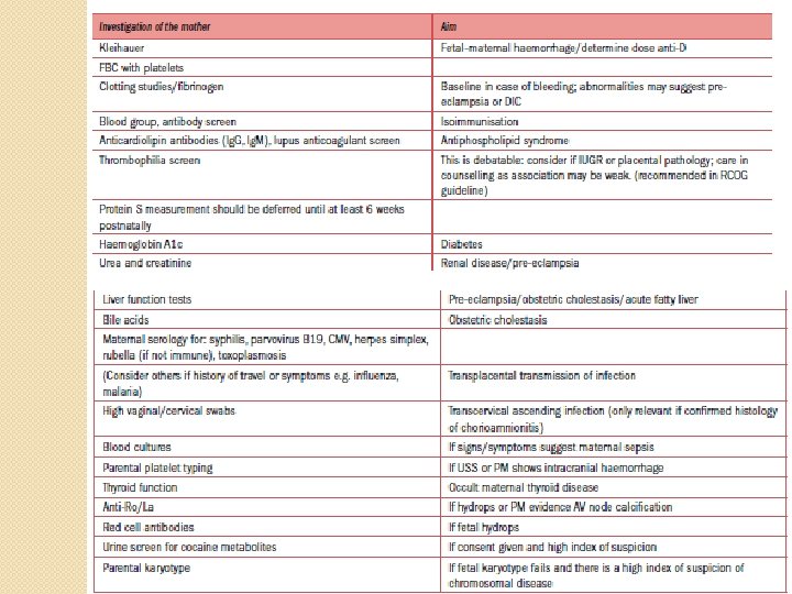
Investigation of the fetus Postmortem examination �written and verbal information ◦ aim of PM examination, ◦ the extent of the examination, ◦ how samples of tissue are dealt with afterwards ◦ whether samples are kept for research or audit. �They also need to know how long the process will take and whethere will be any delay in releasing the baby for burial or cremation.

What is postmortem examination and how it is performed? Postmortem examinations may take up to 5– 7 working days to complete when the brain needs to be fixed for proper examination. � Examinations should be completed within 3 days after receipt of the body if no special examination is required. � There will then be additional time for histopathological analysis, microbiology, metabolic studies, etc. , which can add significant delays to the final report. � It is recommended that at least 60 per cent of reports are issued within 42 days and 90 per cent within 56 days. � The baby is always treated with dignity and respect. � The incisions are closed and do not involve the face or limbs. � In most cases the baby when dressed will look no different from the way it did before the PM. The exception may be the very small macerated fetus whose skin is so thin that suturing is not possible. In these cases, the baby is wrapped. �
 �SUPPRESSION OF LACTATION ◦")
AFTER DELIVERY �Depression, which can become posttraumatic stress disorder (PTSD) �SUPPRESSION OF LACTATION ◦ good supportive bra, NSAIDs if there is discomfort, and time will be enough. ◦ single dose (1 mg) of cabergoline, a longacting dopamine agonist, is highly effective. �CONTRACEPTION ◦ leaflet on contraception

FOLLOW-UP schedule a visit once all information is available. � encourage parents to talk spontaneously and to be honest in replies. � When an underlying cause for the fetal death has been identified, this must be clearly explained. � a clear plan of management for the next pregnancy should be outlined. � Early conception may be associated with very marginally higher risks of preterm birth and SGA babies, but these risk increases are not great enough to recommended delayed conception � Finally, parents must be given the opportunity to come back if they are unclear about certain aspects and, for some, a preconceptual visit is helpful. �

Management of Subsequent Pregnancy after Stillbirth � Preconceptional or Initial Prenatal Visit Detailed medical and obstetrical history � Review evaluation of prior stillbirth � Determination of recurrence risk � Discuss recurrence of comorbid obstetric complications � Smoking cessation � Preconceptional weight loss in obese women � Genetic counseling if family genetic condition exists � Diabetes screen � Thrombophilia screen: antiphospholipid antibodies (only if history indicates) � Support and reassurance �

� First Trimester ◦ Dating sonography ◦ First-trimester screen: PAPP -A, h. CG, NT ◦ Support and reassurance � Second Trimester ◦ Fetal sonographic anatomical survey at 18– 20 weeks’ gestation ◦ Maternal serum screening (quadruple) or single-marker alpha fetoprotein if firsttrimester ◦ Possible uterine artery Doppler studies at 22– 24 weeks’ gestationa ◦ Support and reassurance � Third Trimester ◦ Sonographic screening for fetal-growth restriction, starting at 28 weeks ◦ Kick counts starting at 28 weeks ◦ Antepartum fetal surveillance starting at 32 weeks or 1– 2 weeks earlier than prior stillbirth ◦ Support and reassurance � Delivery ◦ Elective induction at 39 weeks ◦ Delivery before 39 weeks only with documented fetal lung maturity by amniocentesis

Thank you
- Slides: 23