
intra partum fetal monitoring DEFINITION To assess the


- Slides: 85
intra partum fetal monitoring
DEFINITION To assess the fetal behavior in labor. OBJECTIVE To detect hypoxia and acidosis in labor as they can cause organ damage. METHODS OF FETAL MONITORING 1. Clinical methods 2. Biophysical 3. Biochemical
• CLINICAL • Intermittent auscultation -using fetoscope or doppler ultrasound -Fetal heart rate recorded every 15 min in the first stage labour and 5 min in the second stage of labour -Normal findings: F. H. R between 120 bpm and 160 bpm -NON REASSURING FINDINGS Increase F. H. R. >160 bpm Decrease F. H. R. <120 bpm Irregularity
BIOPHYSICAL METHODS • • External (indirect)F. H. R Monitoring Internal(direct) F. H. R Monitoring External (indirect)uterine contraction monitoring Internal(direct)uterine contraction monitoring Scalp stimulation Vibro acoustic stimulation Fetal ECG
cardiotocography A procedure of graphically recording fetal heart activity and uterine contractions. USES OF CARDIOTOCOGRAM 1. To diagnose fetal distress early that is before acidosis. 2. Typical pattern helps to identify cord compression/utero placental insufficiency/head compression. 3. Can diagnose abnormal uterine contraction by internal tocometry. 4. Fetal ECG can be obtained by internal electronic fetal monitoring.
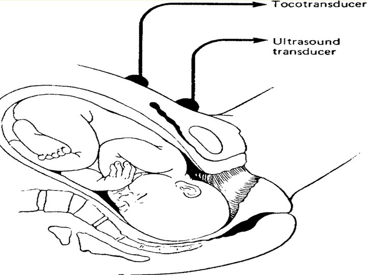
METHODS TO DETECT FETAL HEART ACTIVITY EXTERNAL METHODS 1. Ultra sound mode(most common method) INTERNAL METHODS 1. Direct fetal E. C. G mode ULTRA SOUND MODE /INDIRECT ELECTRONIC FETAL MONITRING Principle: -Ultrasonic waves undergo shift in frequency as they are reflected from moving fetal heart valves and blood is ejected in pulsatile fashion during systole. -It picks up the mechanical activity of fetal heart.
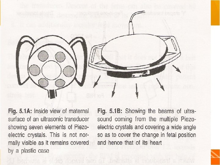
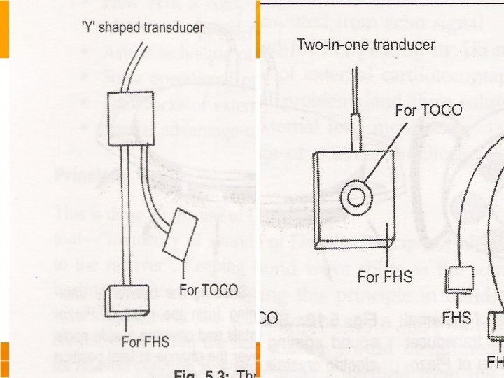
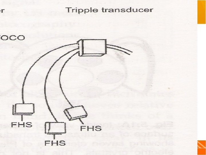
STRUCTURE OF ELECTRONIC TRANSDUCER Made of seven piezo electric crystals Centre crystal emits ultra sound continuously and surrounding six receives it. Operates at a frequency of 1. 5 MHz SPECIAL TYPES OF TRANSDUCERS Y-shaped Two-in One transducer Triple transducer Beltless transducer FHR is calculated by measuring the time interval between two consecutive beats and converting into beats per min.
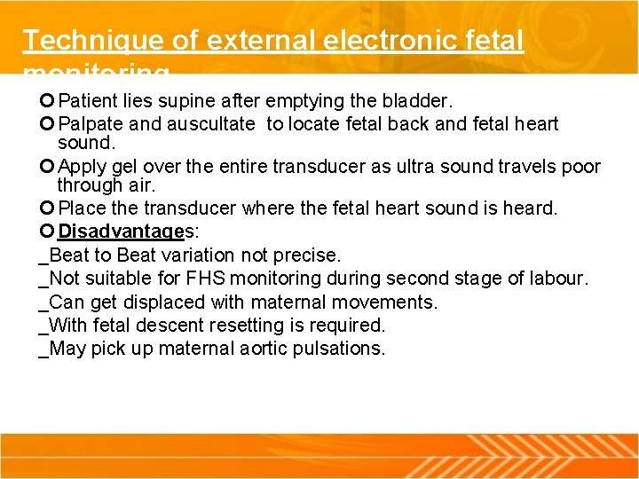
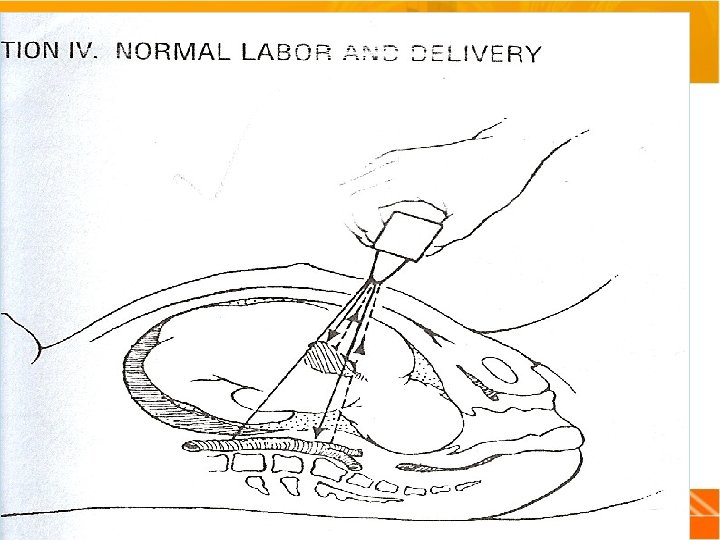
Technique of external electronic fetal monitoring Patient lies supine after emptying the bladder. Palpate and auscultate to locate fetal back and fetal heart sound. Apply gel over the entire transducer as ultra sound travels poor through air. Place the transducer where the fetal heart sound is heard. Disadvantages: _Beat to Beat variation not precise. _Not suitable for FHS monitoring during second stage of labour. _Can get displaced with maternal movements. _With fetal descent resetting is required. _May pick up maternal aortic pulsations.
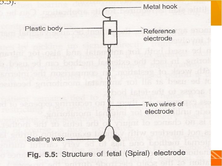
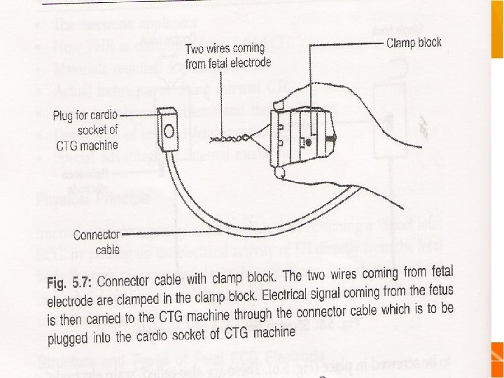
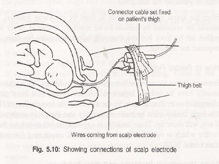
INTERNAL ELECTRONIC FETAL MONITORING Principle: Obtaining a direct E. C. G. by picking the electrical activity of fetal heart directly from the fetal body by the placement of of electrode on it. Structure: 1. Metal hook: to penetrate and fix the electrode to the skin of presenting part of the fetus and it also act as one of the electrical poles of the E. C. G. 2. Small plastic body: second pole of the E. . C. G. also known as the reference electrode. 3. Two wires of electrode. Electrode wire is connected to clamp block that is connected to connector cable and that will be connected to CTG.
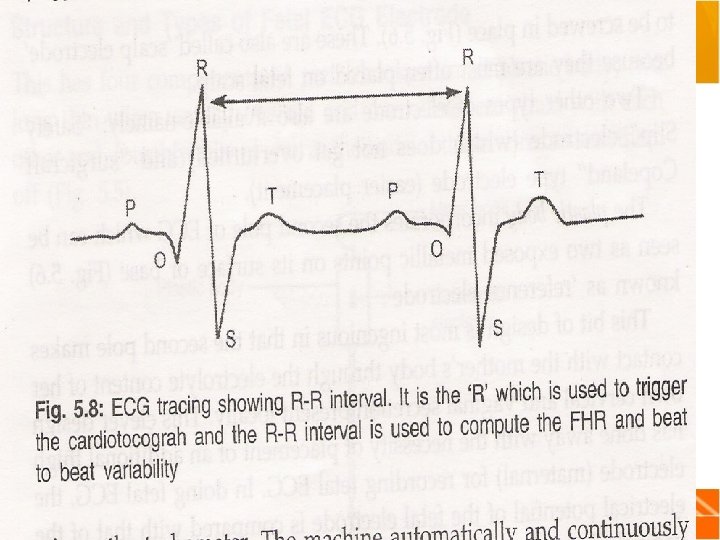
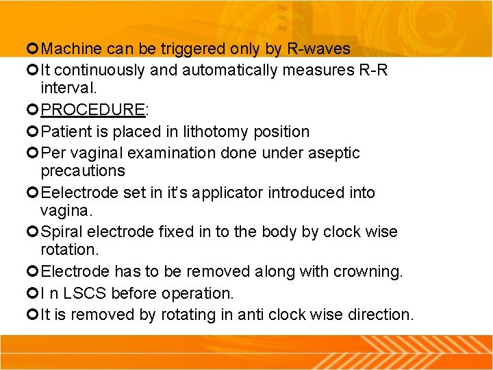
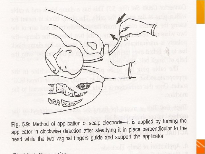
Machine can be triggered only by R-waves It continuously and automatically measures R-R interval. PROCEDURE: Patient is placed in lithotomy position Per vaginal examination done under aseptic precautions Eelectrode set in it’s applicator introduced into vagina. Spiral electrode fixed in to the body by clock wise rotation. Electrode has to be removed along with crowning. I n LSCS before operation. It is removed by rotating in anti clock wise direction.
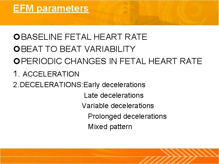
EFM parameters BASELINE FETAL HEART RATE BEAT TO BEAT VARIABILITY PERIODIC CHANGES IN FETAL HEART RATE 1. ACCELERATION 2. DECELERATIONS: Early decelerations Late decelerations Variable decelerations Prolonged decelerations Mixed pattern
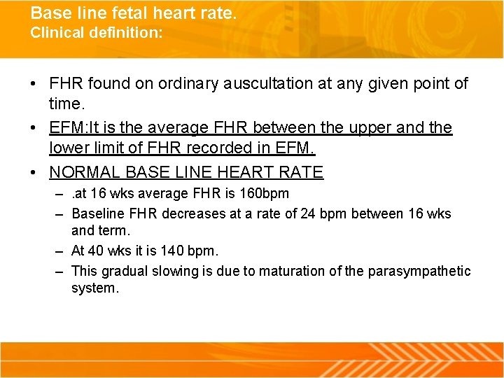
Base line fetal heart rate. Clinical definition: • FHR found on ordinary auscultation at any given point of time. • EFM: It is the average FHR between the upper and the lower limit of FHR recorded in EFM. • NORMAL BASE LINE HEART RATE –. at 16 wks average FHR is 160 bpm – Baseline FHR decreases at a rate of 24 bpm between 16 wks and term. – At 40 wks it is 140 bpm. – This gradual slowing is due to maturation of the parasympathetic system.
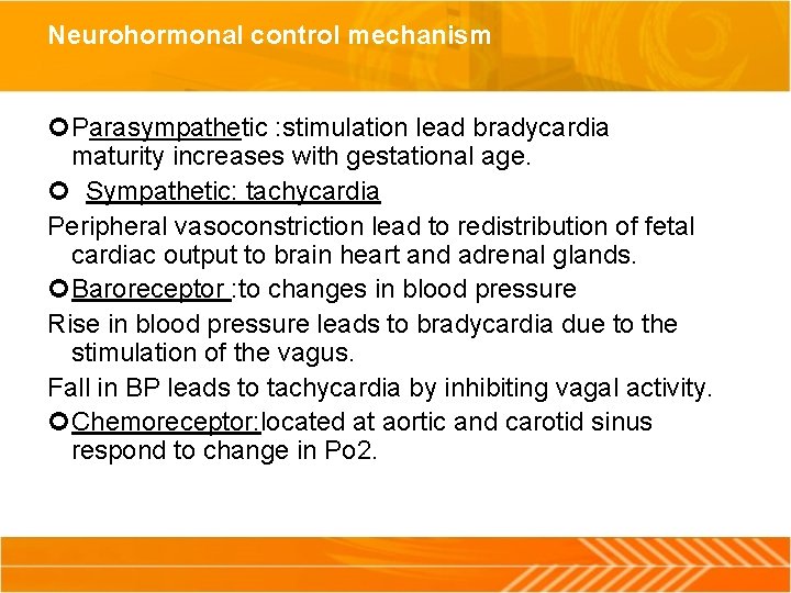
Neurohormonal control mechanism Parasympathetic : stimulation lead bradycardia maturity increases with gestational age. Sympathetic: tachycardia Peripheral vasoconstriction lead to redistribution of fetal cardiac output to brain heart and adrenal glands. Baroreceptor : to changes in blood pressure Rise in blood pressure leads to bradycardia due to the stimulation of the vagus. Fall in BP leads to tachycardia by inhibiting vagal activity. Chemoreceptor: located at aortic and carotid sinus respond to change in Po 2.
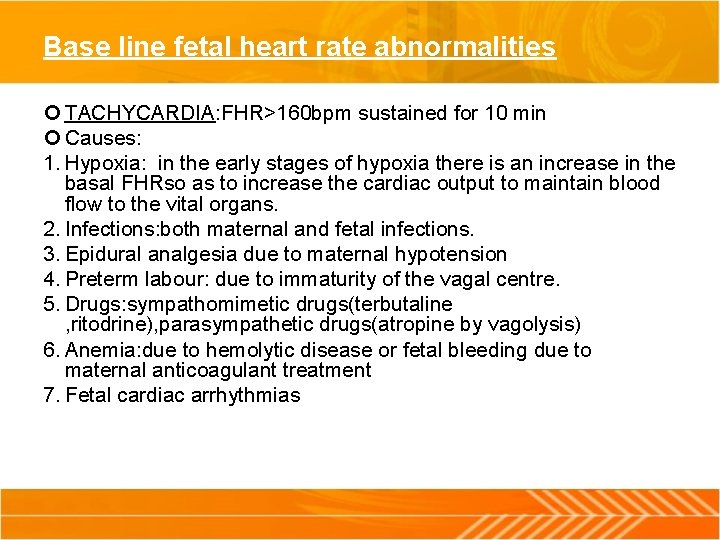
Base line fetal heart rate abnormalities TACHYCARDIA: FHR>160 bpm sustained for 10 min Causes: 1. Hypoxia: in the early stages of hypoxia there is an increase in the basal FHRso as to increase the cardiac output to maintain blood flow to the vital organs. 2. Infections: both maternal and fetal infections. 3. Epidural analgesia due to maternal hypotension 4. Preterm labour: due to immaturity of the vagal centre. 5. Drugs: sympathomimetic drugs(terbutaline , ritodrine), parasympathetic drugs(atropine by vagolysis) 6. Anemia: due to hemolytic disease or fetal bleeding due to maternal anticoagulant treatment 7. Fetal cardiac arrhythmias
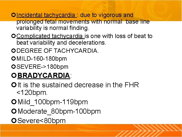
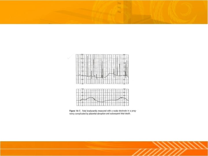
Incidental tachycardia : due to vigorous and prolonged fetal movements with normal base line variability is normal finding. Complicated tachycardia is one with loss of beat to beat variability and decelerations. DEGREE OF TACHYCARDIA. MILD-160 -180 bpm SEVERE->180 bpm BRADYCARDIA: It is the sustained decrease in the FHR <120 bpm. Mild_100 bpm-119 bpm Moderate_80 bpm-100 bpm Severe<80 bpm
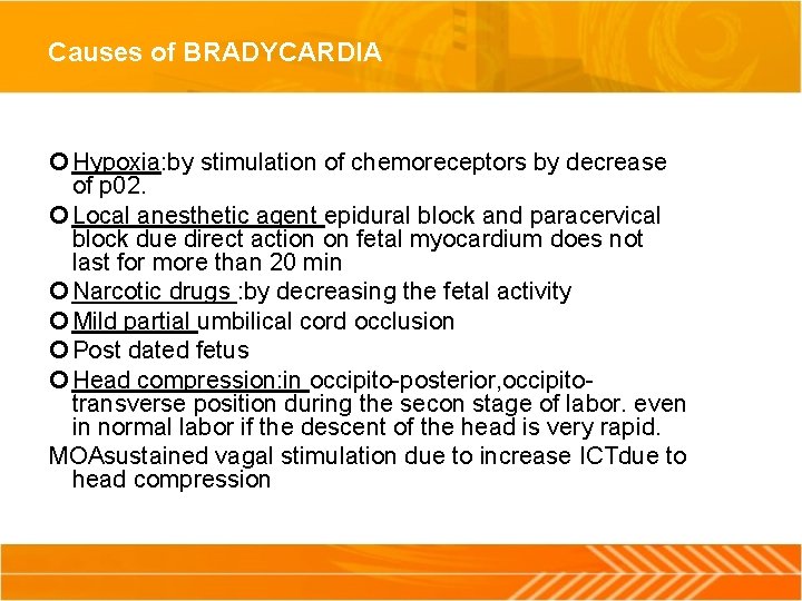
Causes of BRADYCARDIA Hypoxia: by stimulation of chemoreceptors by decrease of p 02. Local anesthetic agent epidural block and paracervical block due direct action on fetal myocardium does not last for more than 20 min Narcotic drugs : by decreasing the fetal activity Mild partial umbilical cord occlusion Post dated fetus Head compression: in occipito-posterior, occipitotransverse position during the secon stage of labor. even in normal labor if the descent of the head is very rapid. MOAsustained vagal stimulation due to increase ICTdue to head compression
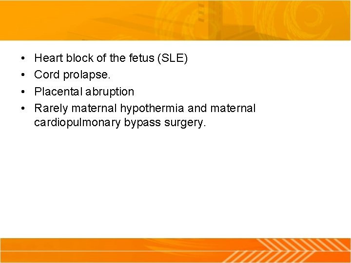
• • Heart block of the fetus (SLE) Cord prolapse. Placental abruption Rarely maternal hypothermia and maternal cardiopulmonary bypass surgery.
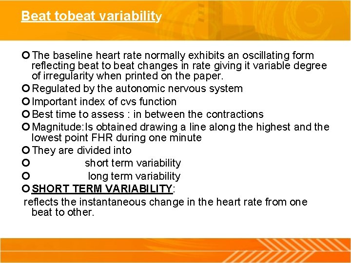
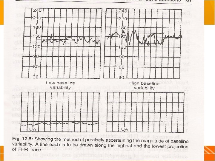
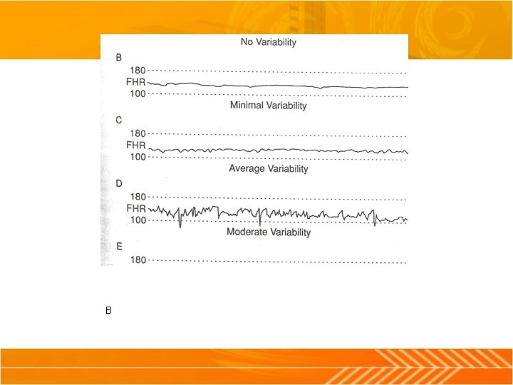
Beat tobeat variability The baseline heart rate normally exhibits an oscillating form reflecting beat to beat changes in rate giving it variable degree of irregularity when printed on the paper. Regulated by the autonomic nervous system Important index of cvs function Best time to assess : in between the contractions Magnitude: Is obtained drawing a line along the highest and the lowest point FHR during one minute They are divided into short term variability long term variability SHORT TERM VARIABILITY: reflects the instantaneous change in the heart rate from one beat to other.
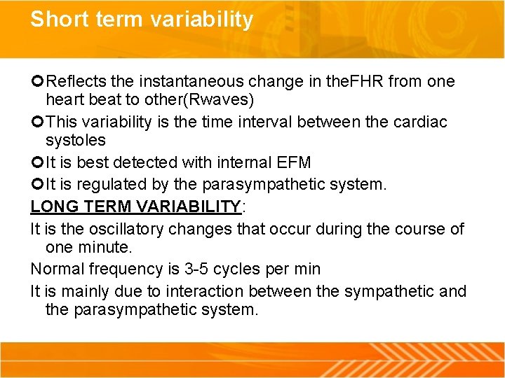
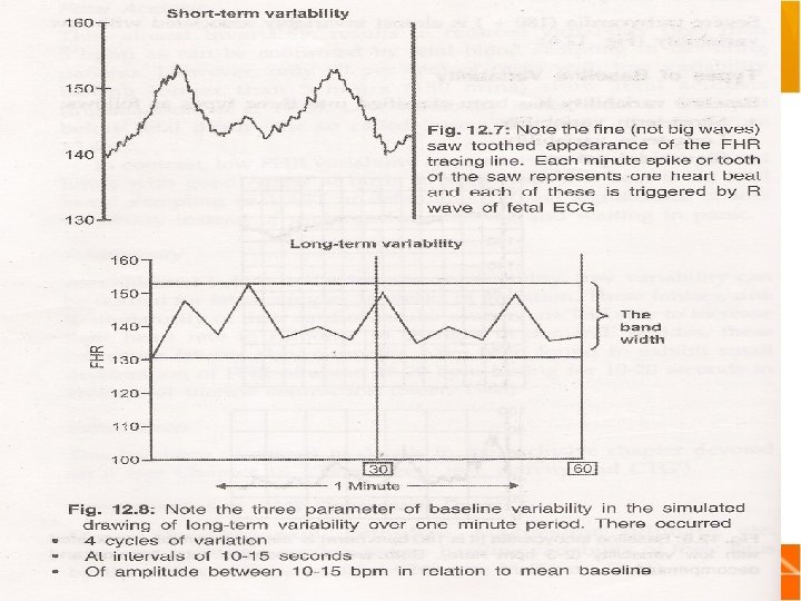
Short term variability Reflects the instantaneous change in the. FHR from one heart beat to other(Rwaves) This variability is the time interval between the cardiac systoles It is best detected with internal EFM It is regulated by the parasympathetic system. LONG TERM VARIABILITY: It is the oscillatory changes that occur during the course of one minute. Normal frequency is 3 -5 cycles per min It is mainly due to interaction between the sympathetic and the parasympathetic system.
B
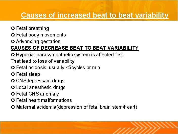
Causes of increased beat to beat variability Fetal breathing Fetal body movements Advancing gestation CAUSES OF DECREASE BEAT TO BEAT VARIABILITY Hypoxia: parasympathetic system is affected first That lead to loss of variability Fetal acidosis: usually <5 cycles pr min Fetal sleep CNSdepressant drugs Local anesthetic drugs Fetal CNS anomaly Fetal heart malformations Maternal acidemia(depression of fetal brain stem/heart)
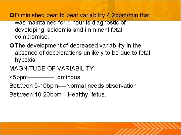
Diminished beat to beat variability, 4. 2 bpm/min that was maintained for 1 hour is diagnostic of developing acidemia and imminent fetal compromise. The development of decreased variability in the absence of decelerations unlikely to be due to fetal hypoxia MAGNITUDE OF VARIABILITY <5 bpm------- ominous Between 5 -10 bpm----Normal needs observation Between 10 -20 bpm---Healthy fetus.
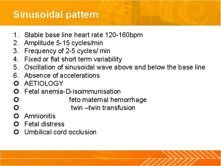
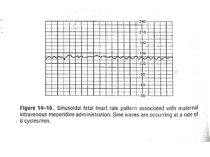
Sinusoidal pattern 1. 2. 3. 4. 5. 6. Stable base line heart rate 120 -160 bpm Amplitude 5 -15 cycles/min Frequency of 2 -5 cycles/ min Fixed or flat short term variability Oscillation of sinusoidal wave above and below the base line Absence of accelerations AETIOLOGY Fetal anemia-D-isoimmunisation feto maternal hemorrhage twin –twin transfusion Amnionitis Fetal distress Umbilical cord occlusion
Mild amplitude-5 -15 bpm mediate 16 -24 bpm major>25 bpm • PERIODIC FETAL HEART RATE CHANGES • ACCELERATIONS • DECELERATIONS: Early decelerations Late decelerations Variable decelerations Prolonged decelerations Mixed pattern
• MANAGEMENT WHEN FHR VARIABILITY DECREASES • Shift the mother to the other [position • O 2 by mask • Maintain normal BP
PERIODIC CHANGES in the FETAL HEART RATE It is the deviation from the baseline in response to fetal stimulation , movement , uterine contraction ACCELERATION: I t is the increase in the FHR>15 bpm from the base line and lasting for 15 -20 sec Primary cause: 1. fetal movement 2. uterine contraction 3. fetal stimulation 4. umbilical cord occlusion partial 5. fetal blood sampling
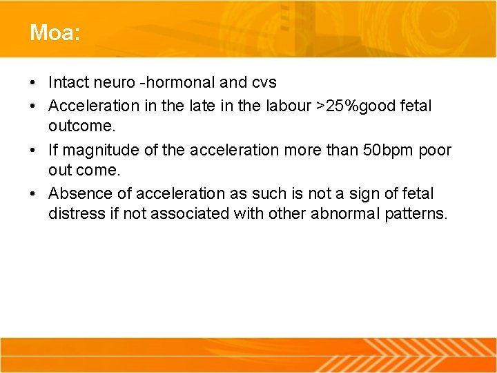
Moa: • Intact neuro -hormonal and cvs • Acceleration in the late in the labour >25%good fetal outcome. • If magnitude of the acceleration more than 50 bpm poor out come. • Absence of acceleration as such is not a sign of fetal distress if not associated with other abnormal patterns.
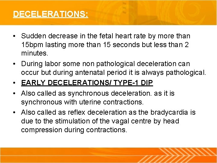
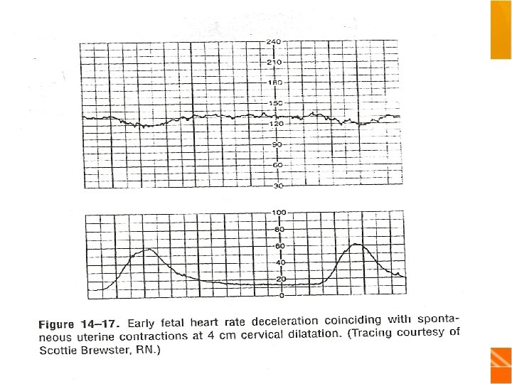
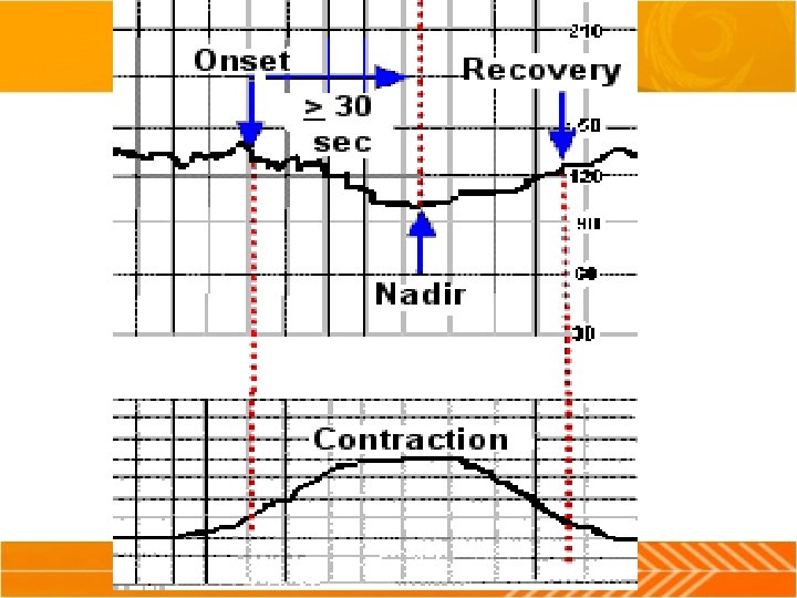
DECELERATIONS: • Sudden decrease in the fetal heart rate by more than 15 bpm lasting more than 15 seconds but less than 2 minutes. • During labor some non pathological deceleration can occur but during antenatal period it is always pathological. • EARLY DECELERATIONS/ TYPE-1 DIP • Also called as synchronous deceleration. as it is synchronous with uterine contractions. • Also called as reflex deceleration as the bradycardia is due to the stimulation of the vagal centre by head compression during contractions.
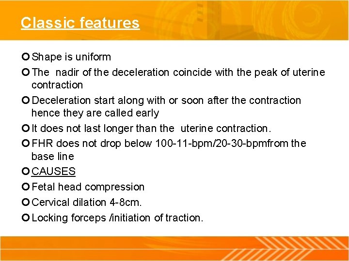
Classic features Shape is uniform The nadir of the deceleration coincide with the peak of uterine contraction Deceleration start along with or soon after the contraction hence they are called early It does not last longer than the uterine contraction. FHR does not drop below 100 -11 -bpm/20 -30 -bpmfrom the base line CAUSES Fetal head compression Cervical dilation 4 -8 cm. Locking forceps /initiation of traction.
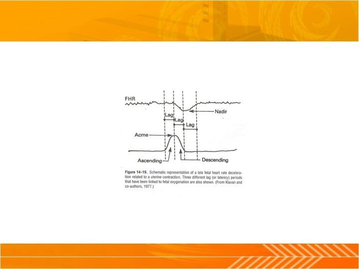
Management of early deceleration Per vaginal examination for dilatation and descent of head. To exclude any obstruction in the passage. To exclude cord prolapse. LATE DECELERATION /TYPE 11 DIP At least 3 or more late decelerations following consecutive contractions for the diagnosis of late deceleration CLASSIC FEATURES Symmetrical decrease in the fetal heart rate beginning at or after peak of uterine contractions Nadir of late deceleration comes after a time lag of 15 seconds after peak of uterine contraction. Deceleration outlasts contraction.
mechanism • Due to hypoxia produced by the uterine contraction there will be decreased blood flow to the inter villous space. • Hypoxia depends on the duration and the amplitude of uterine contraction. deceleration also related with the po 2 that is if it is below 10 mm. Hg • Deceleration is bought by the action of chemoreceptor mediated. • Hypoxic depression of the myocardium is other mechanism.
Degree of deceleration • • Degree of drop in rate: Moderate: upto 100 bpm Severe: below 100 bpm Magnitude of beat lost: Less than 15 bpm Between 15 -45 bpm Greater than 45 bpm
Clinical causes PREUTERINE: Maternal hypotension 1. Supine hypotension 2. Following epidural block Severe anemia without hypovolaemia UTERINE Uterine hyper activity INTRA UTERINE 1. Chronic placental insufficiency 2. Placental abruption cause acute and severe late deceleration.
Preuterine and uterine causes has good prognosis if the offending condition is detected and removed. Intrauterine cause due to placental insufficiency lead to true hypoxia which is not reversible and will be associated with base line tachycardia and loss of beat to beat variability. » MANAGEMENT Patient in the left lateral position If uterine contraction frequent Terbutaline 0. 25 mg oppose the increased vagal activity brought by hypoxia Oxygen inhalation Hypovolaemia should be corrected.
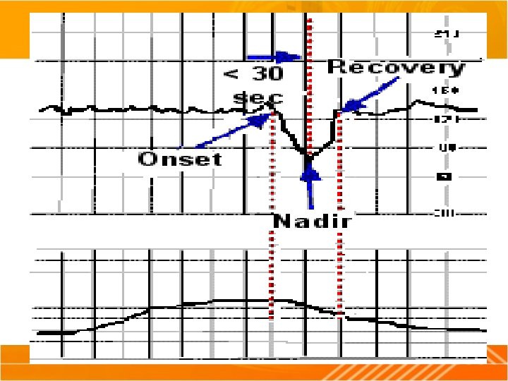
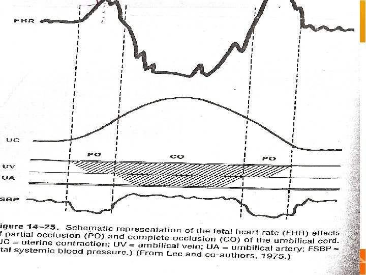
VARIABLE DECELERATIONS /TYPE 111 They are variable in relation to uterine contraction CAUSES 1. PARTIAL CORD OCCLUSION umbilical vein is occluded as it is thin walled. Lead to decrease venous return to the heart Heart compensates to this by tachycardia This is responsible for the acceleration before the deceleration. COMPLETE CORD OCCLUSION Along with the venous blood flow arterial blood flow is also obstructed.
Complete cord occlusion Along with the arterial blood flow venous blood flow is also obstructed That leads to increase in the peripheral resistance Which in turn leads to increase in the blood pressure This stimulates the aortic stretch receptors Vagal nerve will be stimulated leads to bradycardia Acceleration after deceleration is due to the reflex tachycardia Rapid descent of the fetal head.
SIGNIFICANT VARIABLE DECELERATIONS The decelerations decreasing to less than 70 bpm and lasting for more than 60 sec Other predisposing factors Short cord (35 cm) and long cord(80 cm) Volume of liquor Breech Occipito- posterior Face PROGNOSIS Frequency Duration Amplitude Presence of initial acceleration Rebound tachycardia
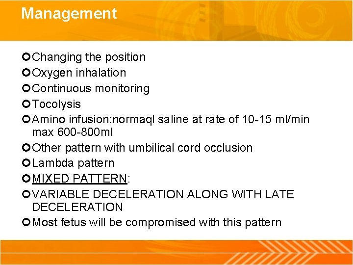
Management Changing the position Oxygen inhalation Continuous monitoring Tocolysis Amino infusion: normaql saline at rate of 10 -15 ml/min max 600 -800 ml Other pattern with umbilical cord occlusion Lambda pattern MIXED PATTERN: VARIABLE DECELERATION ALONG WITH LATE DECELERATION Most fetus will be compromised with this pattern
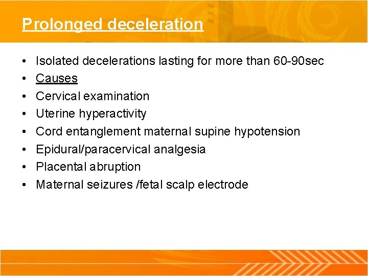
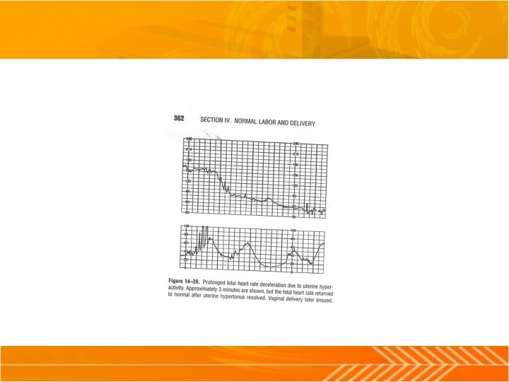
Prolonged deceleration • • Isolated decelerations lasting for more than 60 -90 sec Causes Cervical examination Uterine hyperactivity Cord entanglement maternal supine hypotension Epidural/paracervical analgesia Placental abruption Maternal seizures /fetal scalp electrode
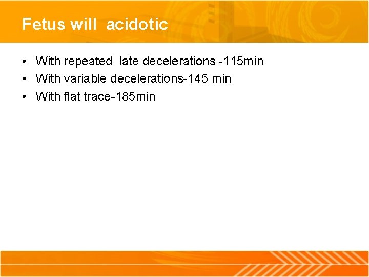
Fetus will acidotic • With repeated late decelerations -115 min • With variable decelerations-145 min • With flat trace-185 min
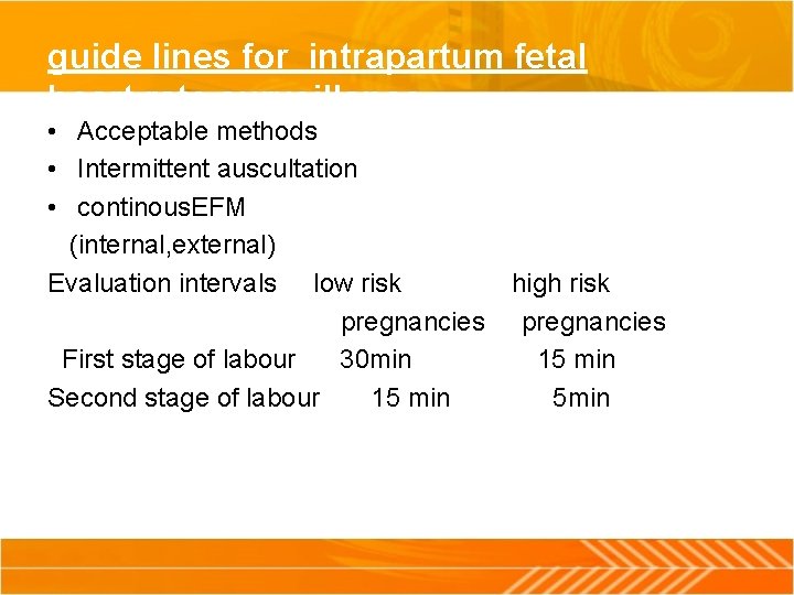
guide lines for intrapartum fetal heart rate surveillance • Acceptable methods • Intermittent auscultation • continous. EFM (internal, external) Evaluation intervals low risk high risk pregnancies First stage of labour 30 min 15 min Second stage of labour 15 min 5 min
Intra partum surveillance of uterine activity • EXTERNAL MONITORING • Transducer(tocodynamometer) held against the abdominal wall • Uterine contractions are measured by the displacement of the transducer placed on the abdomen close to the fundus. • Displacement of the transducer is proportional to the strength of the uterine contraction. • Disadvantage is exact intensity of the uterine contraction is not measured • Gives good indication of onset peak and end of uterine contraction.
Internal • Aim: • Is to measure intra uterine pressure that is the change in pressure of amniotic fluid during contractions. • METHODS AVALIABLE • Fluid filled catheter • Catheter tip pressure transducer • Catheter cum pressure transducer method
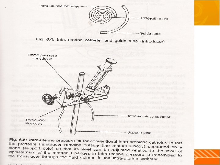
Method of insertion of catheter • Do an aseptic vaginal examination • Pass two fingers between the presenting part and the posterior lip of the cervix. • Insert the guide tube along the fingers following the curvature of the pelvis so that the tip lies just behind the presenting parts in the hind waters • Since the guide lies in the hind waters liquor starts draining through the guide as soon as inserted. • With the guide insitu insert the perforated end of catheter in to the guide tube and gently advance until 18 inch is reached
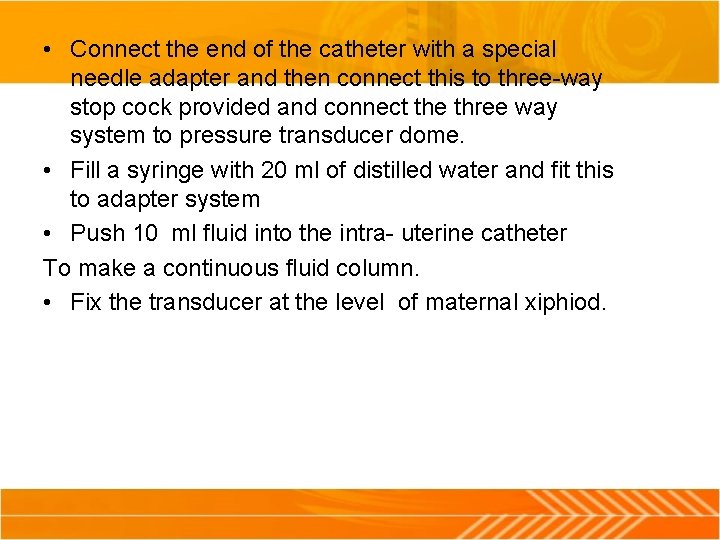
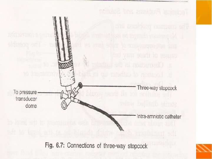
• Connect the end of the catheter with a special needle adapter and then connect this to three-way stop cock provided and connect the three way system to pressure transducer dome. • Fill a syringe with 20 ml of distilled water and fit this to adapter system • Push 10 ml fluid into the intra- uterine catheter To make a continuous fluid column. • Fix the transducer at the level of maternal xiphiod.
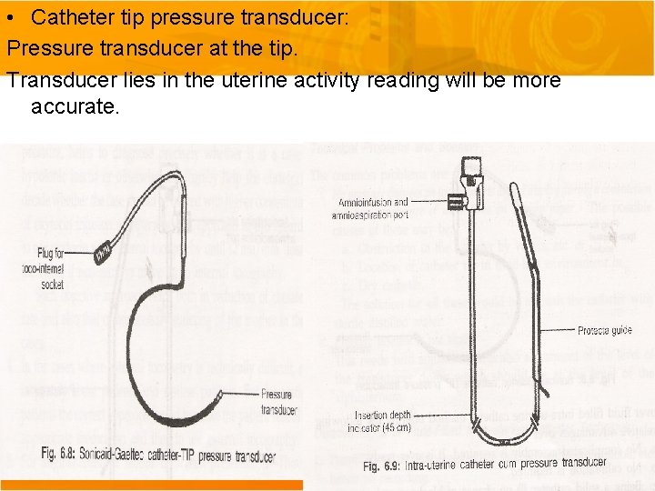
• Catheter tip pressure transducer: Pressure transducer at the tip. Transducer lies in the uterine activity reading will be more accurate.
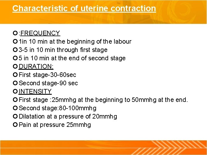
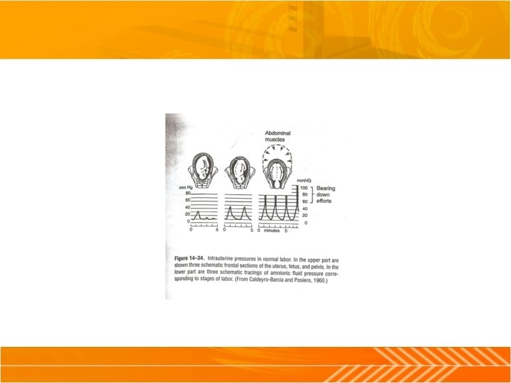
Characteristic of uterine contraction : FREQUENCY 1 in 10 min at the beginning of the labour 3 -5 in 10 min through first stage 5 in 10 min at the end of second stage DURATION: First stage-30 -60 sec Second stage-90 sec INTENSITY First stage : 25 mmhg at the beginning to 50 mmhg at the end. Second stage: 80 -100 mmhg Dilatation at a pressure of 20 mmhg Pain at pressure 25 mmhg
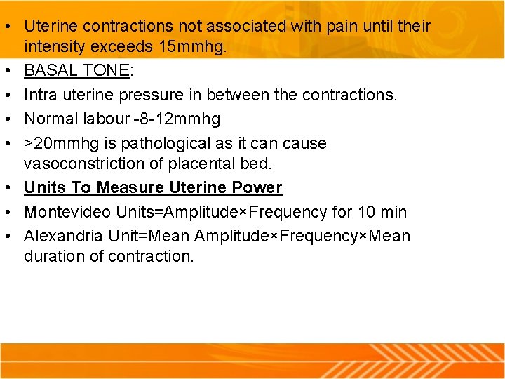
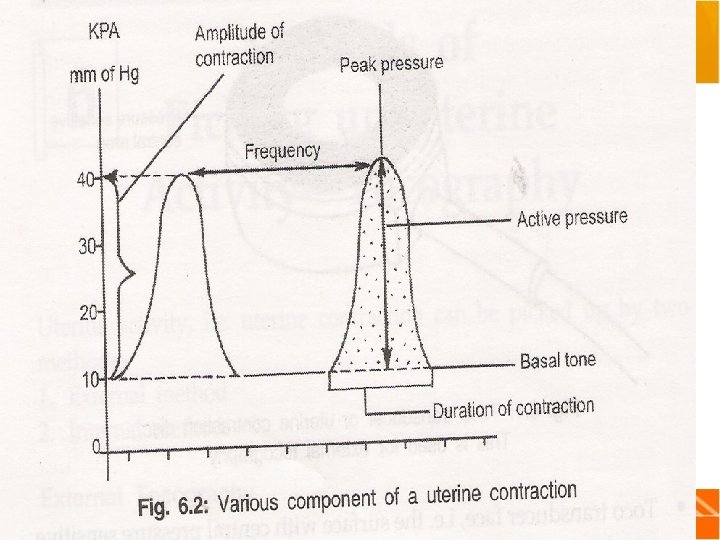
• Uterine contractions not associated with pain until their intensity exceeds 15 mmhg. • BASAL TONE: • Intra uterine pressure in between the contractions. • Normal labour -8 -12 mmhg • >20 mmhg is pathological as it can cause vasoconstriction of placental bed. • Units To Measure Uterine Power • Montevideo Units=Amplitude×Frequency for 10 min • Alexandria Unit=Mean Amplitude×Frequency×Mean duration of contraction.
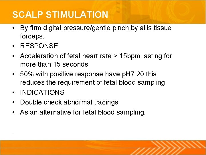
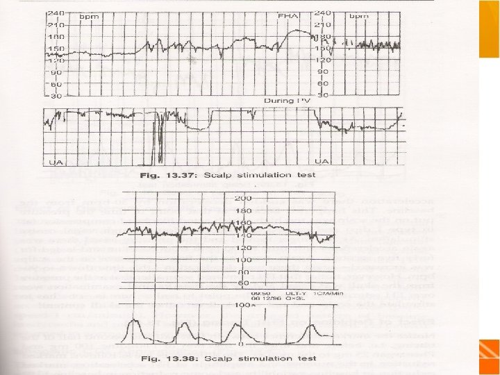
SCALP STIMULATION • By firm digital pressure/gentle pinch by allis tissue forceps. • RESPONSE • Acceleration of fetal heart rate > 15 bpm lasting for more than 15 seconds. • 50% with positive response have p. H 7. 20 this reduces the requirement of fetal blood sampling. • INDICATIONS • Double check abnormal tracings • As an alternative for fetal blood sampling. .
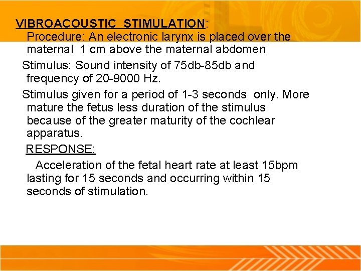
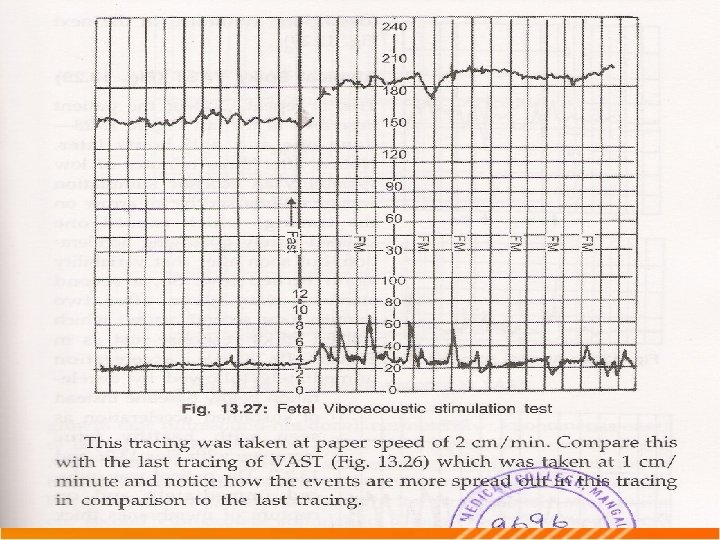
VIBROACOUSTIC STIMULATION: Procedure: An electronic larynx is placed over the maternal 1 cm above the maternal abdomen Stimulus: Sound intensity of 75 db-85 db and frequency of 20 -9000 Hz. Stimulus given for a period of 1 -3 seconds only. More mature the fetus less duration of the stimulus because of the greater maturity of the cochlear apparatus. RESPONSE: Acceleration of the fetal heart rate at least 15 bpm lasting for 15 seconds and occurring within 15 seconds of stimulation.
FETAL PULSE OXIMETER • Assess fetal oxy Hb saturation once membranes ruptured. • Procedure; . • A pad like sensor inserted through cervix and positioned against fetal face and held in place by uterine wall • Normally in 50 -80% saturation is 70 -90% • Low limit 30% persist >2 min • Nellcor-N-400 fetal oxygen monitoring system.
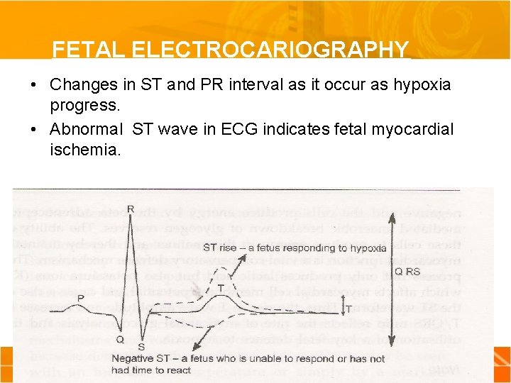
FETAL ELECTROCARIOGRAPHY • Changes in ST and PR interval as it occur as hypoxia progress. • Abnormal ST wave in ECG indicates fetal myocardial ischemia.
• The rise in the T wave amplitude and increase in T/QRS complex reflects myocardial glycogenolysis that occurs during hypoxia. • Increase in the T wave height occur in early hypoxia. • T-wave depression occur in late hypoxia.
Fetal blood sampling • Objective: Measurement of the Ph of the capillary blood helps to identify fetus in distress. • Procedure: • Illuminated endoscope inserted through the sufficiently dilated cervix • Press firmly against fetal scalp. • Skin wiped with a clean cotton swab and silicone gel is coated so that blood will accumulate as discrete globules. • Incision of 2 mm depth is made with special blade and blood is collected in a heparin zed capillary tube.
• >7. 25 p. H: Observe cardio tocogram. • 7. 20 -7. 25 p. H: Repeat again within 30 minutes. • <7. 20: Repeat again immediately and expedite delivery • Base excess gives information about the buffering available if continues to produce lactic acid due to glycolysis. • Base excess negative p. H falls rapidly. • Interruption of placental blood flow leads to respiratory acidosis first if the interruption is brief. • If hypoxia is prolonged then anaerobic metabolism which inturn produces metabolic acidosis.
spectroscopy • It is based on the spectroscopic observance of returning laser beams of different wave lengths in relation to time. • It is the change in the optical density and absorbance of light that produces the effect. • Laser diode transmits waves of 4 pre set wave lengths. 775, 805, 845, 904. • Placement of the patient: sensor is placed on the mothers abdomen over the fetal head.
FETAL OXYGEN PARAMETERS THAT WILL BE DISPLAYED: 1. Oxyhaemoglobin level 2. Deoxyhaemoglobin level 3. Total haemoglobin 4. Cytochrome aa 3 level 5. Relative change in local blood volume. The organ targeted is fetal brain.
THANK YOU