
Intra Aortic Balloon Pump IABP Yan Wing Wa
Intra Aortic Balloon Pump (IABP) Yan Wing Wa Department of Intensive Care Pamela Youde Nethersole Eastern Hospital Spring 2009 1

What is an IABP? • The Intra-Aortic Balloon Counterpulsation system is a volume displacement device. • It should be positioned so that the tip is approximately 1 to 2 cm below the origin of the left subclavian artery and above the renal arteries. • On chest x-ray the tip should be visible in the 2 nd or 3 rd intercostal space. 2
Purpose of IABP • Hemodynamic support in medical conditions • Prophylactic hemodynamic support • Cardiac support for hemodynamically challenged patients with mechanical defects PRIOR to correction 3
Surgical Indications • Post surgical myocardial dysfunction • Support for weaning from cardiopulmonary bypass (CPB) • Cardiac support following correction of anatomical defects • Maintenance of graft patency post CABG • Pulsatile flow during CPB 4
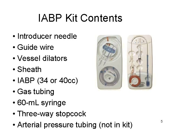
IABP Kit Contents • Introducer needle • Guide wire • Vessel dilators • Sheath • IABP (34 or 40 cc) • Gas tubing • 60 -m. L syringe • Three-way stopcock • Arterial pressure tubing (not in kit) 5

IAB Sizing Chart • The IAB Should be selected according to the following chart (chart located on every box). Note: The 50 cc balloons are no longer made by Datascope 6
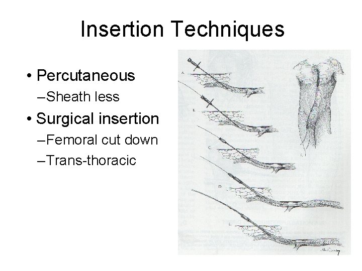
Insertion Techniques • Percutaneous – Sheath less • Surgical insertion – Femoral cut down – Trans-thoracic 7
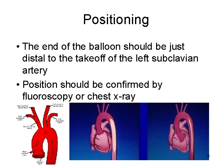
Positioning • The end of the balloon should be just distal to the takeoff of the left subclavian artery • Position should be confirmed by fluoroscopy or chest x-ray 8
Insertion Position • On chest x-ray the tip should be visible in the 2 nd or 3 rd intercostal space 9
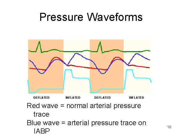
Pressure Waveforms DEFLATED INFLATED Red wave = normal arterial pressure trace Blue wave = arterial pressure trace on IABP 10
Desired Outcome • Appropriately timed blood volume displacement (30 – 50 m. L) in the aorta by the rapid shuttling of helium gas in and out of the balloon chamber, resulting in changes in inflation and deflation hemodynamics 11
Inflation • The goal of inflation is to increase or augment perfusion 12
Inflation of IABP Results In: • • • Increased coronary perfusion pressure Increased systemic perfusion pressure Increased O 2 supply to both the coronary and peripheral tissue Increased baroreceptor response Decreased sympathetic stimulation causing decreased HR, decreased SVR, and increased LV function 13
Deflation • The goal of deflation is to reduce LV workload • Deflation creates a "potential space" in the aorta, reducing aortic volume and pressure 14
Deflation of the IABP Results In: • • Afterload reduction and therefore a reduction in MVO 2 Reduction in peak systolic pressure, therefore a reduction in LV work Increased cardiac output Improved ejection fraction and forward flow 15
How do you Know? • Afterload reduction? – Change in diastolic pressures – Unassisted and assisted • Decreased workload? – Change in systolic pressures – Unassisted and assisted • Improved cardiac output? – Increase in pulse pressures 16
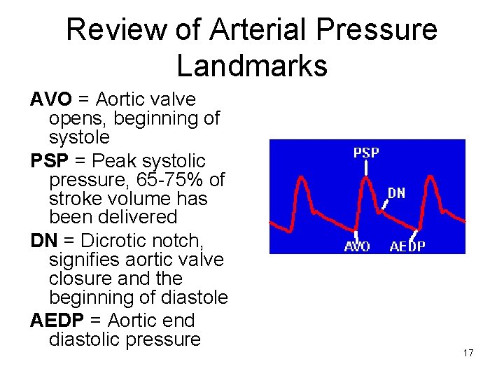
Review of Arterial Pressure Landmarks AVO = Aortic valve opens, beginning of systole PSP = Peak systolic pressure, 65 -75% of stroke volume has been delivered DN = Dicrotic notch, signifies aortic valve closure and the beginning of diastole AEDP = Aortic end diastolic pressure 17
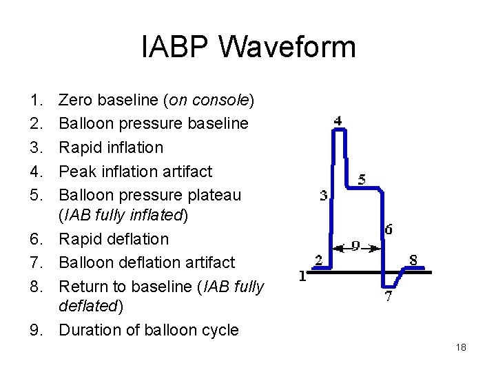
IABP Waveform 1. 2. 3. 4. 5. 6. 7. 8. 9. Zero baseline (on console) Balloon pressure baseline Rapid inflation Peak inflation artifact Balloon pressure plateau (IAB fully inflated) Rapid deflation Balloon deflation artifact Return to baseline (IAB fully deflated) Duration of balloon cycle 18
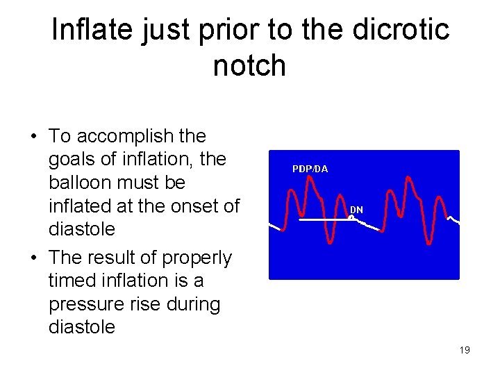
Inflate just prior to the dicrotic notch • To accomplish the goals of inflation, the balloon must be inflated at the onset of diastole • The result of properly timed inflation is a pressure rise during diastole 19
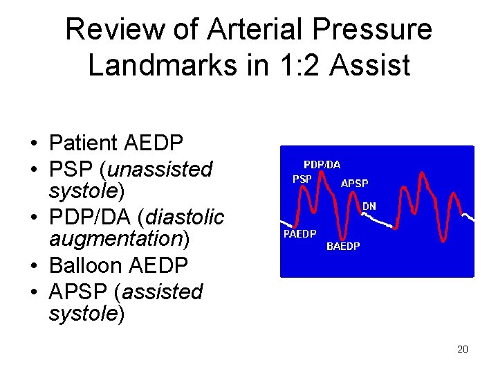
Review of Arterial Pressure Landmarks in 1: 2 Assist • Patient AEDP • PSP (unassisted systole) • PDP/DA (diastolic augmentation) • Balloon AEDP • APSP (assisted systole) 20
Inflation Hemodynamics • Coronary artery blood flow and pressure are increased – Increased renal and cerebral blood flow • Increased diastolic pressure increases perfusion to distal organs and tissues 21
Inflation Hemodynamics • Coronary collateral circulation is potentially increased from the increase in coronary perfusion pressure (CPP) • Systemic perfusion pressure is increased 22
Coronary Perfusion Pressure • The pressure at which blood perfuses through the coronary circulation, mainly in diastole • Blood flow to the left ventricle occurs principally during diastole and is largely determined by the driving pressure (diastolic pressure minus left ventricular diastolic pressure), the diastolic interval and the state of coronary vasodilatation 23
Deflation Hemodynamics • The pressure that the LV must generate (afterload) is less throughout the systolic phase – Decreases myocardial O 2 demands • Isovolumetric contraction (IVC) phase of systole is shortened – Decreases myocardial O 2 demands 24
Deflation Hemodynamics • Reduced afterload allows the LV to empty more effectively so stroke volume (SV) is increased • Enhanced forward cardiac output decreases the amount of blood shunted from left to right in cases of intraventricular septal defects and incompetent mitral valve 25
Triggering • It is necessary to establish a reliable trigger signal before balloon pumping can begin • The computer in the IAB console needs a stimulus to cycle the pneumatic system, which inflates and deflates the balloon • The trigger signal tells the computer that another cardiac cycle has begun 26
Triggering • In most cases it is preferable to use the R wave of the ECG as the trigger signal • There are other trigger options for instances when the R wave cannot be used or is not appropriate 27
Trigger Loss • The console MUST see a trigger to initiate an inflate/deflate cycle • If no trigger is seen when the clinician attempts to start pumping, no pumping will occur and an alarm will be sounded • If the trigger is lost after pumping starts, no further pumping will occur until a trigger is re-established 28
Trigger Loss • If the current trigger is lost the clinician choose an alternate, available trigger to resume pumping • For example, if the ECG lead becomes disconnected the arterial pressure (AP) trigger may be selected until the ECG is re -established 29
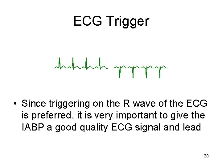
ECG Trigger • Since triggering on the R wave of the ECG is preferred, it is very important to give the IABP a good quality ECG signal and lead 30
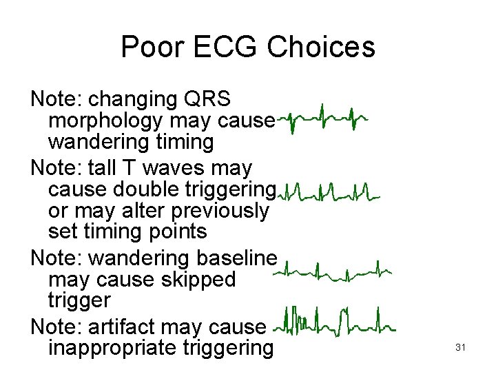
Poor ECG Choices Note: changing QRS morphology may cause wandering timing Note: tall T waves may cause double triggering or may alter previously set timing points Note: wandering baseline may cause skipped trigger Note: artifact may cause inappropriate triggering 31
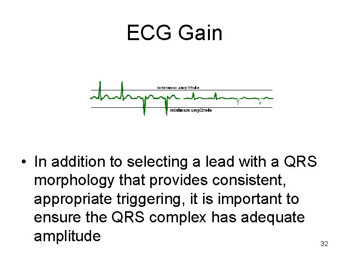
ECG Gain • In addition to selecting a lead with a QRS morphology that provides consistent, appropriate triggering, it is important to ensure the QRS complex has adequate amplitude 32
Triggering on the Arterial Pressure Waveform • Arterial pressure provides another signal to the IABP to determine where the cardiac cycle begins and ends • It is used when the ECG has too much interference from patient movement or poor lead connection 33
CPR • AP trigger is the appropriate choice during CPR • Once chest compressions are started an arterial pressure waveform will be generated • Triggering on the AP will produce pumping in synchrony to chest compression and has been shown to assist with coronary and carotid perfusion 34
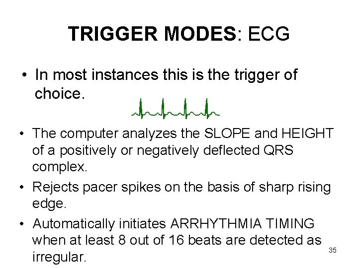
TRIGGER MODES: ECG • In most instances this is the trigger of choice. • The computer analyzes the SLOPE and HEIGHT of a positively or negatively deflected QRS complex. • Rejects pacer spikes on the basis of sharp rising edge. • Automatically initiates ARRHYTHMIA TIMING when at least 8 out of 16 beats are detected as 35 irregular.
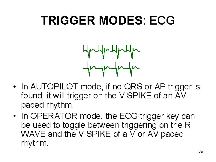
TRIGGER MODES: ECG • In AUTOPILOT mode, if no QRS or AP trigger is found, it will trigger on the V SPIKE of an AV paced rhythm. • In OPERATOR mode, the ECG trigger key can be used to toggle between triggering on the R WAVE and the V SPIKE of a V or AV paced rhythm. 36
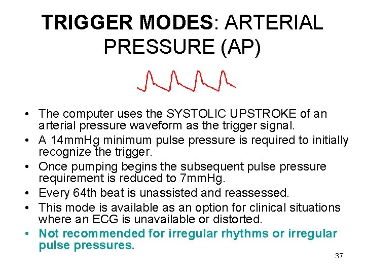
TRIGGER MODES: ARTERIAL PRESSURE (AP) • The computer uses the SYSTOLIC UPSTROKE of an arterial pressure waveform as the trigger signal. • A 14 mm. Hg minimum pulse pressure is required to initially recognize the trigger. • Once pumping begins the subsequent pulse pressure requirement is reduced to 7 mm. Hg. • Every 64 th beat is unassisted and reassessed. • This mode is available as an option for clinical situations where an ECG is unavailable or distorted. • Not recommended for irregular rhythms or irregular pulse pressures. 37
Gas Alarms/Balloon Pressure Waveform • During a cycle of inflation/deflation, helium is rapidly moved in and out of the balloon. • The environment within the balloon and the surrounding forces that affect balloon behavior all contribute to a predictable pattern of gas flow and pressure. • The gas pressure characteristics are converted into a waveform that is reflective of the behavior of the gas. 38
• This transduced waveform tells much about the interaction of the balloon within the patient's aorta. • Understanding the balloon pressure waveform is important for troubleshooting of the console as most alarms are based on the gas surveillance system. 39
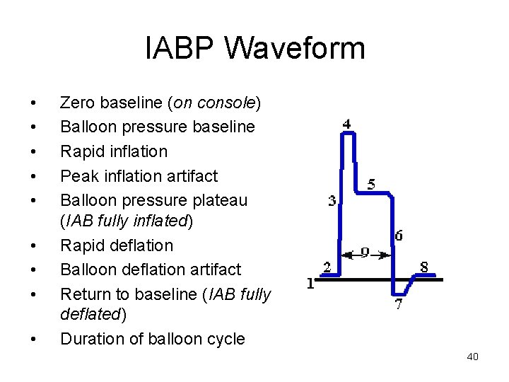
IABP Waveform • • • Zero baseline (on console) Balloon pressure baseline Rapid inflation Peak inflation artifact Balloon pressure plateau (IAB fully inflated) Rapid deflation Balloon deflation artifact Return to baseline (IAB fully deflated) Duration of balloon cycle 40
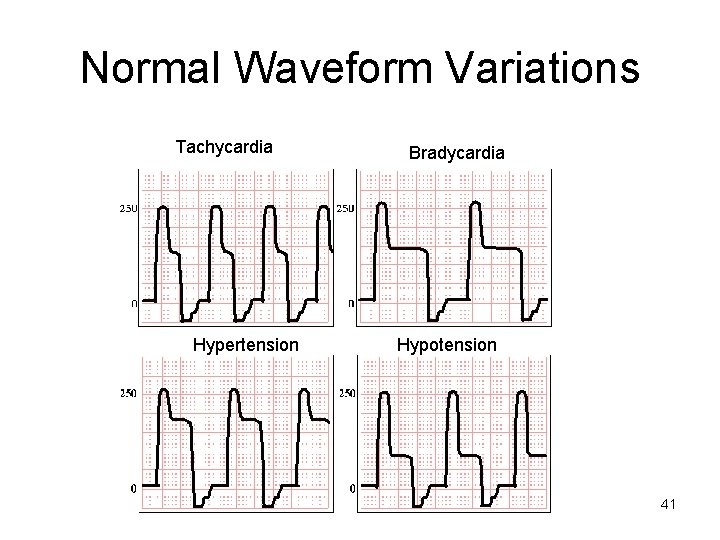
Normal Waveform Variations Tachycardia Hypertension Bradycardia Hypotension 41
Abnormal Waveform Variation: Wide Inflation and/or Deflation Artifact • Check for kinks, as they may trap gas in the IAB. • If water is present in the gas tubing, remove the condensation. – Pushing the helium through the water during inflation and deflation slows down gas transition. – If gas transition is prolonged too much, it can create alarms. 42
Abnormal Waveform Variation: Helium Loss / Gas Leakage Alarms • Observe for blood in the gas tubing. If even a slight amount is present, it may indicate a balloon rupture. – Do not resume pumping. – Notify physician immediately and prepare for IAB removal. • Check connections where gas tubing connects to IAB and to pump. – Secure if loose. 43
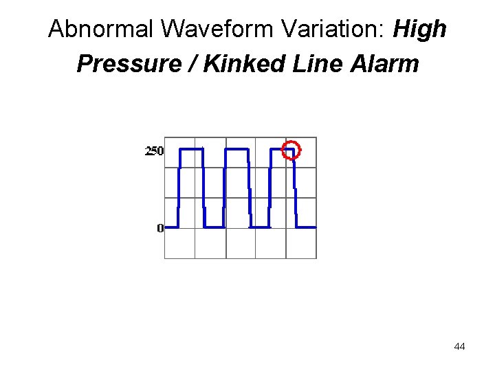
Abnormal Waveform Variation: High Pressure / Kinked Line Alarm 44
• • • Reposition patient. Keep affected leg straight. Use rolled towel under hip to hyperextend hip. Apply slight traction to the catheter if kinking at the insertion site or in the artery is suspected. The distal portion of the IAB may be in the sheath if a long introducer sheath was used. Pull sheath back until IAB bladder has exited the sheath. Introducer sheath may be kinked which in turn is kinking the balloon. Suspect this particularly if placement of the sheath was difficult. Pull sheath back or rotate sheath a partial turn. 45
• Check placement of the balloon; it may be too high or too low. • IAB may be partially wrapped if alarm occurs shortly after insertion. Take steps to facilitate unwrapping (consult IAB manufacturer). • The balloon may be too large for the patient. Reduce the helium volume the balloon is inflated with. (It is recommended to not reduce the volume below 2/3 of maximum. For example, do not decrease volume in a 40 cc IAB below 27 cc) 46
Abnormal Waveform Variation: High Baseline / Fill Pressure • • • Check for intermittent obstruction of gas lumen. Check for overfill of system. This condition may occur during ascent in air transport since gas expands as you go up in altitude. Reset the alarm and restart pumping. The volume will be adjusted automatically for current barometric pressure. 47
Complications of IABP • The following patients are at the greatest risk of developing complications associated with IABP: • Females, diabetics, smokers, obese patients • Patients with PVD, HTN, high SVR, shock 48
Complications of IABP • Aortic wall dissection, rupture or local vascular injury – Care as indicated • Emboli: thrombus, plaque or air – Care as indicated – Treatment of an air embolism is as follows: 100% oxygen, intubate, Trendelenberg in left lateral decubitus position, fluid resuscitation, etc. 49
Complications of IABP • IABP Rupture: • COFFEE GROUNDS seen in the drive line is a precursor to a rupture (make sure they are on the inside and not the outside of the tubing) – NOTIFY PHYSICIAN!!!!! – IF THERE IS A FLAGRANT RUPURE OF THE IABP CLAMP THE GAS LINE!!!!! 50
Complications of IABP • Infection – Check catheter insertion site often – STRICT ASEPTIC TECHNIQUE – Restrict movement while IABP in place 51
Complications of IABP • Obstruction • Malposition – Too high – obstruction of left subclavian and carotids • CHECK LEFT RADIAL ARTERY PULSE – Too low – obstruction of renal and mesenteric arteries • MONITOR URINE OUTPUT 52
Complications of IABP • Compromised circulation due to catheter • Ischemia – Routine nursing care and monitoring • Compartment syndrome – Rare complication seen in the LE, usually related to infection – Monitor calf circumference 53
Complications of IABP • Hematologic – ALL PATIENTS typed & crossmatched!!! • Bleeding – REMOVE THE DRESSING!!! – PUT ON STERILE GLOVES!!! – HOLD PRESSURE!!! • Thrombocytopenia – Routine monitoring 54
Weaning of IABP • Timing of weaning – Patient should be stable for 24 -48 hours • Decreasing inotropic support • Decreasing pump ratio – From 1: 1 to 1: 2 or 1: 3 • Decrease augmentation • Monitor patient closely – If patient becomes unstable, weaning should be immediately discontinued Weaning should only be attempted on order of a physician. 55
IAB Removal • • Discontinue heparin six hours prior Check platelets and coagulation factors Deflate the balloon Apply manual pressure above and below IABP insertion site • Remove and alternate pressure to expel any clots • Apply constant pressure to the insertion site for a minimum of 30 minutes • Check distal pulses frequently IAB should only be pulled by a physician or the cardiac P. A. 56

Thank you for your attention 57
- Slides: 57