Intestinal Protozoa CLASSIFICATION OF PARASITES PROTOZOA HELMINTHS Unicellular


 Giardia trophozoites (light microscope) can not")
 can survive in the environment and resist")


 Water, food Flies can act as vector.")


 SYMPTOMATIC EXTRAINTESTINAL INFECTED")
, molecular, and")
: diloxanide")


")


- Slides: 34
Intestinal Protozoa
CLASSIFICATION OF PARASITES PROTOZOA HELMINTHS Unicellular Single cell for all functions Multicellular Specialized cells 1: Amoebae: move by pseudopodia 2: Flagellates: move by flagella 3: Ciliates: move by cilia 4: Apicomplexa (Sporozoa) tissue parasites Round worms (Nematodes): - elongated, cylindrical, unsegmented. Flat worms: - Trematodes: leaf-like, unsegmented. - Cestodes: tape-like, segmented.
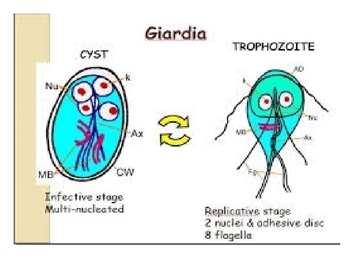
Flagellates: Giardia lamblia Giardia trophozoites ( electron microscopy) Giardia trophozoites (light microscope) can not survive in the environment, can not resist gastric acidity, diagnostic stage
Trichrome stain trophozoites Giardia cyst (light microscope) can survive in the environment and resist the gastric acidity, infective and diagnostic stage.
• Giardia lamblia a protozoan parasite capable of causing sporadic or epidemic diarrheal illness. Giardiasis is an important cause of waterborne and foodborne disease, daycare center outbreaks, and illness in international travelers. Giardiasis is especially common in areas with poor sanitary conditions and limited water-treatment facilities. Water is a major source of giardiasis transmission.
Giardia lamblia Life cycle
Giardia lamblia: Life cycle Giardia species have two forms: cysts & trophozoites. Cysts are the infectious stage of the parasite; they are excreted in stool. Following cyst ingestion, excystation occurs in the small intestine with release of trophozoites. Trophozoites are pear-shaped, bi-nucleate, multi-flagellated parasite forms capable of division by binary fission. Following cyst ingestion, infections have an incubation of a week or more before symptoms of acute giardiasis may develop. Trophozoites are localize to the small intestine, trophozoite attachment to the mucosal surface of the duodenum and jejunum, although the trophozoite does not invade the mucosal epithelium. Clinical manifestation: It is mainly asymptomatic infection occurs in both children and adults, and asymptomatic cyst & trophozoits shedding can last six months or more, however, If symptoms occur will be as diarrhea, malaise, abdominal cramps, flatulence, weight loss & vomiting. Complications: In a small number of patients, persistent infection is associated with development of malabsorption and weight loss, Chronic giardiasis may affect growth and development in children.
Giardia trophozoites in tissue section
Giardiasis: Laboratory diagnosis • Stools examination – Microscopy for cysts or trophozoits – Detection of Giardia antigens in stools • Examination of duodenal contents: trophozoites Chemotherapy • Drug of choice: Metronidazole
Intestinal Amoebae
Entaemoeba histolytica 500 million people are infected. 100, 000 deaths per year. It is a waterborne infection. There are 6 species of Entamoeba: 1. E. histolytica Amoebae that are pathogenic & invasive 2. E. dispar. The non-pathogenic, non invasive form 3. E. hartmanni 4. E. coli 5. E. gingivalis 6. E. polecki E. histolytica and E. dispar can’t be distinguished by microscopic observation
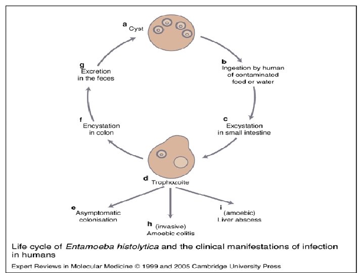
Entamoeba histolytica Amebiasis occurs worldwide; the prevalence is increased in developing countries because of poor socioeconomic conditions and sanitation levels. The parasite exists in two forms: a cyst stage (the infective form) and a trophozoite stage which causes invasive disease. The cysts pass through the stomach to the small intestine, where they excyst to form trophozoites. The trophozoites can invade and penetrate the mucous barrier of the colon, causing tissue destruction colitis and increased intestinal secretion and can thereby ultimately lead to bloody diarrhea.
Clinical manifestation: The majority of Entamoeba infections are asymptomatic some have Symptoms which range from mild diarrhea to severe amebic dysentery (abdominal pain, bloody diarrhea and mucus in stools) & fulminant amebic colitis. Weight loss occurs in about half of patients, and fever can occur. Fulminant colitis with bowel necrosis leading to perforation, and peritonitis has been observed in approximately 0. 5 percent of cases; associated mortality rate is more than 40 percent.
Entamoeba histolytica
Entamoeba histolytica Trophozoite: vegetative stage, must encyst to survive in the environment. It is a fragile structure. Cyst: infective stage. Resist the harsh conditions of the environment.
E. histolytica cyst E. histolytica trophozoite
Entamoeba histolytica Mode of infection (fecal-oral route) Water, food Flies can act as vector. Can be sexually transmitted person to person contacts Not a zoonosis The infective dose can be as little as 1 cyst (very virulent). The incubation period can be from few days to few weeks depending on the infective dose Cysts can survive for weeks at appropriate temperature and humidity.
Entamoeba histolytica PATHOLOGY Intsetinal amoebiasis : Remarkable and unique ability to produce enzymes that lyses host tissues. Lesions are found mainly in the colon. They may heal. Or it may cause serious complications: • Perforation of the colon. • Amoeboma: Granulomatous mass obstructing the bowel • Blood invasion; Amoebic liver abscess, lung, brain • Direct extension
PATHOLOGY: Intsetinal amoebiasis
PATHOLOGY: Intsetinal amoebiasis Complications
PATHOLOGY: Intsetinal amoebiasis Entamoeba histolytica E. Histolytica in mucosa. Numerous trophozoites can be seen with ingested erythrocytes
PATHOLOGY: Extra-intestinal amoebiasis : LUNG LIVER BRAIN
A 30 -year-old male experienced diarrhea for two weeks with fever of 39° C, nausea, vomiting, malaise and right upper abdominal pain. Physical examination revealed hepatomegaly 6 cm below the right costal margin. CT scan showed a single hypodense mass in the rigth lobe of 7. 8 x 5. 2 cm, round, with well defined borders. Serology was positive for Enamoeba histolytica at 1/512. Amebic liver abscess was diagnosed.
THE CLINICAL OUTCOMES OF INFECTION WITH Entamoeba histolytica INTESTINAL (Amoebic dysentery) SYMPTOMATIC EXTRAINTESTINAL INFECTED ASYMPTOMASTIC CARRIER
Laboratory Diagnosis of Amoebiasis • Diagnostic techniques include microscopy, antigen detection (serology), molecular, and colonoscopy with histological examination. – Stools examination (Microscopy): • Wet mount (cysts and trophozoites) • Concentration methods (only cysts) – Serology antigen detection ( mainly for invasive infections): IHA, ELISA. – Molecular — Detection of parasitic DNA or RNA in feces via probes can also be used to diagnose amebic infection and to differentiate between the different strains. • Extra-intestinal: – – – Serology: IHA , ELISA Surgical aspirate (not done as a diagnostic procedure due to risk of extension) trophozoite Sigmoidoscopy and/or colonoscopy and taking biopsy: trophozoite.
Main Drugs for Treatment of Amoebiasis • Intestinal : – Asympromatic (cysts only): diloxanide furoate (Furamide) – Symptomatic (cysts and trophozoites): metronidazole • Extra-intestinal: – Metronidazole
Cryptosporidium Parvum
Cryptosporidium • is an intracellular protozoan parasite that is associated with self-limited diarrhea in normal immunocompetent hosts • and severe debilitating diarrhea with weight loss and malabsorption in HIV-infected patients. The diagnosis of cryptosporidiosis is generally based upon microscopy since Cryptosporidium species cannot be cultivated in vitro. • Transmission of cryptosporidiosis occurs via spread from an infected person or animal, by fecal oral rout. Contaminated environment, such as food or water source.
Cryptosporidium Diagnosis Modified acid-fast stain ZN. safranin IMMUONOFLOURECENT (IF)
Cryptosporidium Diagnosis From stool . The diagnosis of cryptosporidiosis is made by finding oocysts in fecal smears when using modified acid –fast stain (ZN) And by Antigen detection by using ELIZA, IF. From duodenal aspirates, bile secretions & biopsy specimens from affected gastrointestinal tissue also we can do polymerase chain reaction (PCR), or enzyme immunoassays (ELIZA) & IF.
Cryptosporidiosis Treatment • Self-limited in immunocompetent patients • In AIDS patients: paromomycin