Interval History a The patient was admitted to


- Slides: 33

Interval History a. The patient was admitted to respiratory isolation to rule out active infection with Mycobacterium Tuberculosis b. Acid-fast bacilli culture and smear were negative c. The patient improved on empiric NSAIDs and was discharged home d. The patient returned to her baseline exercise tolerance and was asymptomatic within 1 week

Interval History a. Repeat CT imaging showed resolution of effusions, but persistent nodules, concerning for metastatic disease of unknown primary b. PET Scan and Abdominal CT scans did not show evidence of extrapulmonary malignancy c. The patient returned two months later for a thorascopy with lung wedge resection of a characteristic nodule

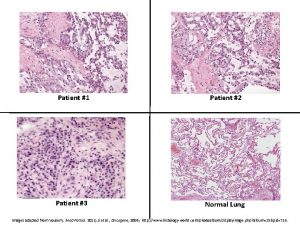
4 x

10 x

20 x

40 x

CD 1 a

S 100

Final Pathological Diagnosis PULMONARY LANGERHANS’- CELL HYSTIOCYTOSIS

PLCH: Introduction a. Histiocytosis encompasses a group of diverse disorders with the common primary event of the accumulation and infiltration of monocytes, macrophages, and dendritic cells in the affected tissues b. Langerhans Cells are a dendritic cell subtype and part of the monocytemacrophage lineage derived from bone marrow involved in antigen presentation in the tracheobronchial tree

Classification of Histiocytosis a. Single-organ involvement a. Lung (>85% of lung involvement occurs in isolation) b. Bone c. Skin d. Pituitary e. Lymph Nodes f. Thyroid, Liver, Spleen, Brain b. Multisystem Disease a. Multiorgan b. Multiorgan c. Multiorgan disease with lung involvement disease without lung involvement histiocytic disorder

Historical Terms a. Hystiocytosis X b. Eosinophilic Granuloma c. Letter-Siwe disease a. A rare systemic aggressive disease seen in adults d. Hand-Schüller-Christian disease a. Triad of exopthalmos, central diabetes insipidus, and bone lesions

Langerhans Cells a. Discovered by Paul Langerhans in 1868 b. The hallmark ultrastructural feature called the Birbeck Granule discovered in 1961 c. The CD 1 a cell surface antigen distinguises LC from other histiocytes

Epidemiology a. Precise incidence and prevalence is hard to define in this disease a. 1200 new cases per year b. 0. 5 -5. 4 cases / million b. 5% of lung-biopsy specimens in patients with ILD result in PLCH c. No known genetic susceptibility d. Mainly in caucasians. Male to female ratio is changing over the decades… e. >90% of PLCH patients are smokers

Proposed Pathogenesis of PLCH Vassallo R et al. N Engl J Med 2000; 342: 1969 -1978

Reactive vs. Neoplastic? a. Spontaneous remission b. Abscence of chromosomal abnormalities c. Overall good prognosis in majority of cases a. Monoclonal proliferation in extrapulmonary tissue b. Infiltration of aberrent cells into normal tissue c. Response to chemotherapy and possible fatal outcome in more severe cases

Histopathological Features a. Proliferation of Lagerhans Cells along the small airways serves as the nidus of cellular/fibrotic nodules from 5 mm to 1. 5 in size. Eosinophils may be present b. In severe disease, nodules may interconnect and cavitate to produce distinctive honeycomb-like structures c. Given that most patients are smokers, concominant COPD and ILD 2/2 respiratory bronchiolitis is often present

Clinical Presentation a. Cough (50 -70%) b. Dyspnea (30 -50%) c. Fever, weight loss, diaphoresis (2030%) d. Asymptomatic (25%) e. Chest Pain (10%)

Clinical Presentation a. Pneumothorax (10 -20%) b. Extrapulmonary disease (15%) c. Pulmonary hypertension d. Respiratory failure e. Secondary malignancy • Physical Exam and Laboratory findings are variable

Chest Radiography a. Symmetrical micronodular and Interstitial infiltration predominantly in the middle and upper lobes b. Increased lung volumes c. Rare: alveolar infiltrates, hilar LAD, pleural effusion


Tissue Diagnosis a. Bronchoalveolar Lavage b. Transbronchial Biopsy c. Open vs. Thorascopic Lung Biopsy


Management a. Smoking Cessation b. Corticosteroids c. Chemotherapy a. Vinblastine, MTX, Cyclophosphamide, Etoposide b. 2 -chlorodeoxyadenosine d. Immune modulation: Etanercept e. Pleurodesis of pneumothoraces f. Serial TTE and PFTs to monitor progression

Prognosis a. For a majority of patients, the disease regresses with smoking cessation b. It is not known to predict those who tend to progress, although age, prolonged constitutional symptoms, extrapulmonary involvement, abnormal PFTs are markers of poor outcome

Back to our case… a. This patient has baseline respiratory insufficiency 2/2 PLCH and COPD, but presented with an acute respiratory illness not characteristic of these diagnoses b. She endorsed chills, dyspnea, and chest pain. There was radiographic evidence of pleuropericarditis which symptomatically and radiographically improved within 1 -2 weeks on NSAIDs

Dfdx of pleuropericarditis a. Viral / Acute idiopathic b. Drug-induced c. Collagen vascular: Sarcoid, RF, Lupus d. Tuberculosis e. Malignancy f. Infarction pericarditis g. Uremia h. Atypical infections: fungal

Follow-up a. Pleural fluid was negative for Acid-Fast, Bacterial or Fungal organisms b. HIV Negative c. The patient continues to struggle with smoking cessation and reports baseline shortness of breath and cough d. The patient reports an increase in smoking because of the anxiety of “having cancer” e. Steroids have not been offerred due to the relatively mild course of her disease

Dfdx of pleuropericarditis a. Viral / Acute idiopathic b. Drug-induced c. Collagen vascular: Sarcoid, RF, Lupus d. Tuberculosis e. Malignancy f. Infarction pericarditis g. Uremia h. Atypical infections: fungal

Dfdx of pleuropericarditis a. Viral / Acute Idiopathic b. Drug-induced c. Collagen vascular: Sarcoid, RF, Lupus d. Tuberculosis e. Malignancy f. Infarction pericarditis g. Uremia h. Atypical infections: fungal

Final Diagnoses a. Pulmonary Langerhans’-Cell Histiocytosis, b. Acute Viral Plueropericarditis • • • Active Tobacco Abuse Coronary Artery Disease COPD Essential HTN Anxiety / Dysthymia

CPC 9. 12. 08 Flowsheet HTN Age CAD Chronic respiratory insufficiency, cough and exercise intolerance Active Tobacco Abuse PLCH COPD Diminished epithelial defenses and mucociliary elevator Viral respiratory pathogen? Pleuropericarditis Acute self-limited Increased cough, Acute phase reactants: dyspnea and Subjective chills Platelets, Ferritin, ESR atypical chest pain Chronic illness: Anemia of chronic disease and low albumin Dysthymia/Anxiety

Thank You! a. Dr. Martin Blaser b. Dr. Anthony Grieco c. Dr. Elvio Ardilles d. Dr. Jonathon Ralston e. Dr. Kristin Remus f. Dr. James Tsay g. Dr. Christina Yoon
 A newly admitted patient was found wandering
 Admission procedure in fundamentals of nursing
 Nada c g berjarak
 Primary vs secondary reinforcers
 Variable interval
 Facts admitted need not be proved
 Patient 2 patient
 Interval history sample
 History of patient safety
 Ros in history taking
 Sacred seven mc
 Opqrsta
 History taking respiratory system
 History taking of patient
 Hình ảnh bộ gõ cơ thể búng tay
 Slidetodoc
 Bổ thể
 Tỉ lệ cơ thể trẻ em
 Gấu đi như thế nào
 Thang điểm glasgow
 Chúa sống lại
 Các môn thể thao bắt đầu bằng tiếng chạy
 Thế nào là hệ số cao nhất
 Các châu lục và đại dương trên thế giới
 Công thức tiính động năng
 Trời xanh đây là của chúng ta thể thơ
 Mật thư anh em như thể tay chân
 Làm thế nào để 102-1=99
 Phản ứng thế ankan
 Các châu lục và đại dương trên thế giới
 Thơ thất ngôn tứ tuyệt đường luật
 Quá trình desamine hóa có thể tạo ra
 Một số thể thơ truyền thống
 Bàn tay mà dây bẩn


