
Intertwistingbend process in DNA condensation Prof Dr Xizeng
Intertwisting-bend process in DNA condensation Prof. Dr. Xizeng FENG Nankai University 1
Outline Ⅰ. Experiment—Phenomenon--Model • 1. DNA Toroids and Microstructure • 2. DNA Toroid Size and Mechanisms of Formation Ⅱ. Microstructure– Biofunction -- Model • 3. Cationic-Complex Induced DNA Condensates • 4. Questions and some thinks…… 2
Ⅰ. Experiment—Phenomenon--Model 1. DNA Toroids and Microstructure • The DNA condensates have served as models of high density packing in biological systems, in virus, and have found applications in the Non-viral gene delivery. 3
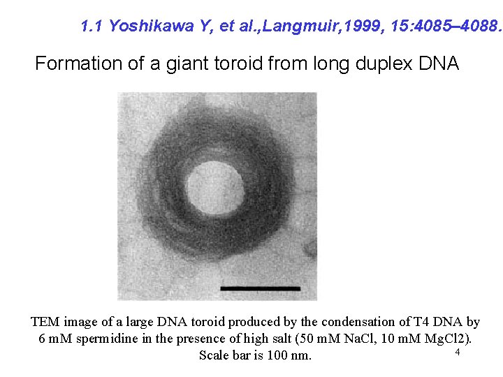
1. 1 Yoshikawa Y, et al. , Langmuir, 1999, 15: 4085– 4088. Formation of a giant toroid from long duplex DNA TEM image of a large DNA toroid produced by the condensation of T 4 DNA by 6 m. M spermidine in the presence of high salt (50 m. M Na. Cl, 10 m. M Mg. Cl 2). 4 Scale bar is 100 nm.
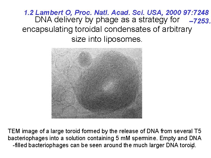
1. 2 Lambert O, Proc. Natl. Acad. Sci. USA, 2000 97: 7248 DNA delivery by phage as a strategy for – 7253. encapsulating toroidal condensates of arbitrary size into liposomes. TEM image of a large toroid formed by the release of DNA from several T 5 bacteriophages into a solution containing 5 m. M spermine. Empty and DNA -filled bacteriophages can be seen around the much larger DNA toroid. 5
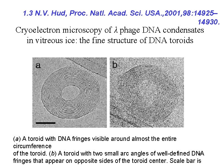
1. 3 N. V. Hud, Proc. Natl. Acad. Sci. USA. , 2001, 98: 14925– 14930. Cryoelectron microscopy of λ phage DNA condensates in vitreous ice: the fine structure of DNA toroids (a) A toroid with DNA fringes visible around almost the entire circumference of the toroid. (b) A toroid with two small arc angles of well-defined DNA 6 fringes that appear on opposite sides of the toroid center. Scale bar is
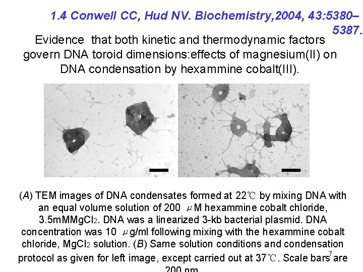
1. 4 Conwell CC, Hud NV. Biochemistry, 2004, 43: 5380– 5387. Evidence that both kinetic and thermodynamic factors govern DNA toroid dimensions: effects of magnesium(II) on DNA condensation by hexammine cobalt(III). (A) TEM images of DNA condensates formed at 22℃ by mixing DNA with an equal volume solution of 200 μM hexammine cobalt chloride, 3. 5 m. MMg. Cl 2. DNA was a linearized 3 -kb bacterial plasmid. DNA concentration was 10 μg/ml following mixing with the hexammine cobalt chloride, Mg. Cl 2 solution. (B) Same solution conditions and condensation protocol as given for left image, except carried out at 37℃. Scale bars 7 are
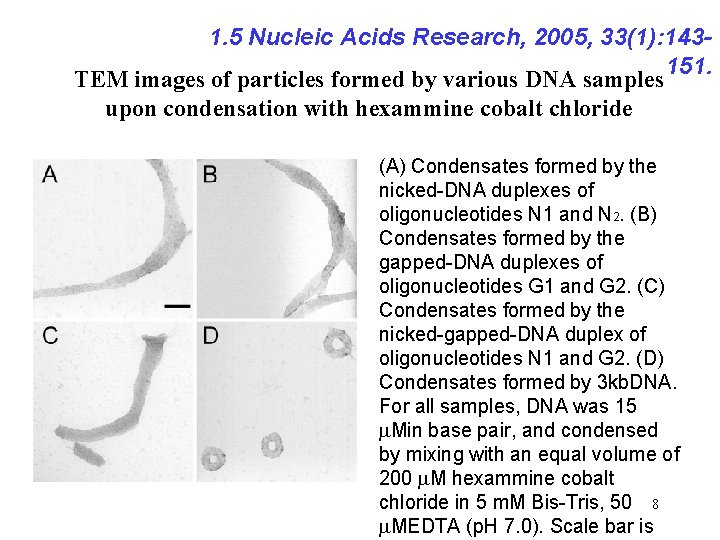
1. 5 Nucleic Acids Research, 2005, 33(1): 143151. TEM images of particles formed by various DNA samples upon condensation with hexammine cobalt chloride (A) Condensates formed by the nicked-DNA duplexes of oligonucleotides N 1 and N 2. (B) Condensates formed by the gapped-DNA duplexes of oligonucleotides G 1 and G 2. (C) Condensates formed by the nicked-gapped-DNA duplex of oligonucleotides N 1 and G 2. (D) Condensates formed by 3 kb. DNA. For all samples, DNA was 15 m. Min base pair, and condensed by mixing with an equal volume of 200 m. M hexammine cobalt chloride in 5 m. M Bis-Tris, 50 8 m. MEDTA (p. H 7. 0). Scale bar is
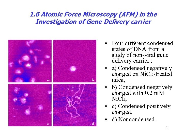
1. 6 Atomic Force Microscopy (AFM) in the Investigation of Gene Delivery carrier • Four different condensed states of DNA from a study of non-viral gene delivery carrier : • a) Condensed negatively charged on Ni. Cl 2 -treated mica, • b) Condensed negatively charged with 0. 2 m. M Ni. Cl 2, • c) Condensed positively charged, • d) Noncondensed. 9
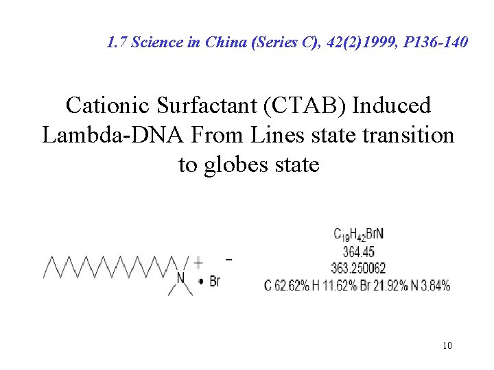
1. 7 Science in China (Series C), 42(2)1999, P 136 -140 Cationic Surfactant (CTAB) Induced Lambda-DNA From Lines state transition to globes state 10
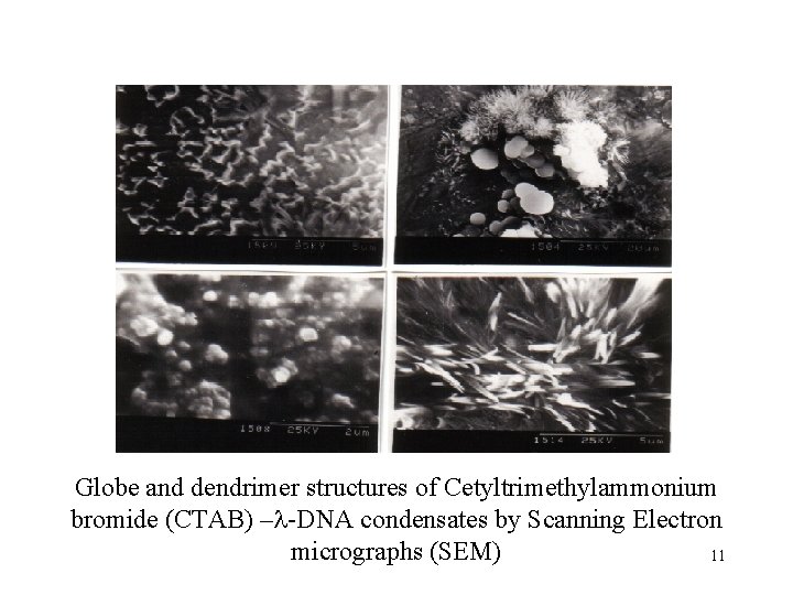
Globe and dendrimer structures of Cetyltrimethylammonium bromide (CTAB) –l-DNA condensates by Scanning Electron micrographs (SEM) 11
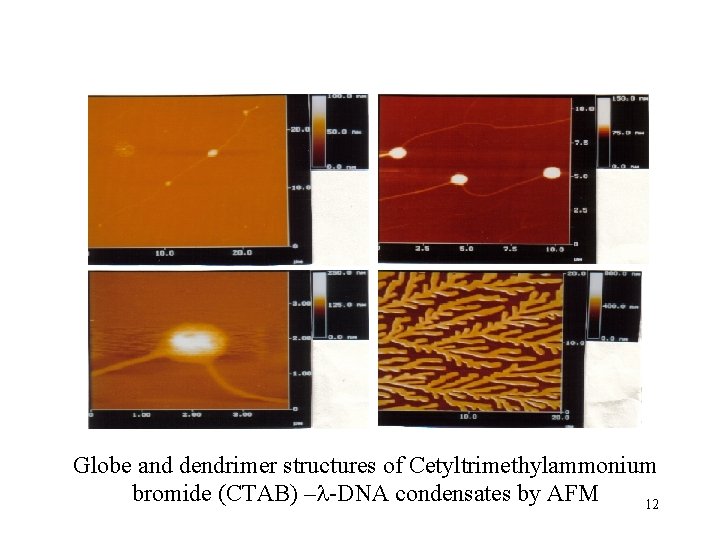
Globe and dendrimer structures of Cetyltrimethylammonium bromide (CTAB) –l-DNA condensates by AFM 12
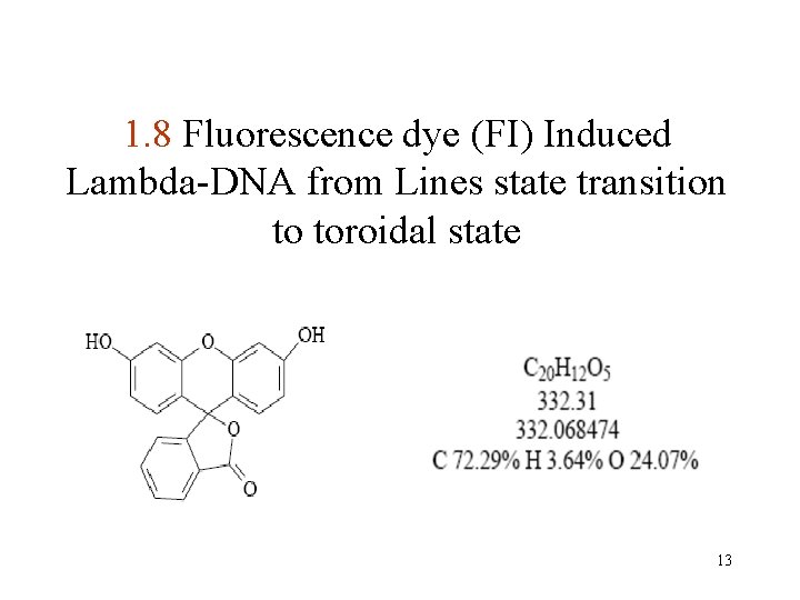
1. 8 Fluorescence dye (FI) Induced Lambda-DNA from Lines state transition to toroidal state 13
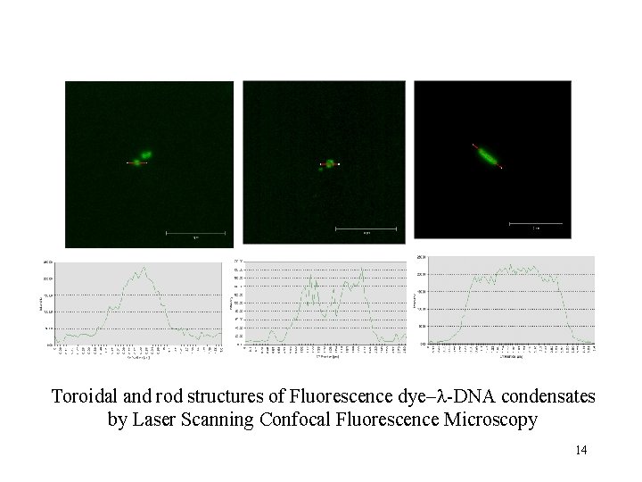
Toroidal and rod structures of Fluorescence dye–l-DNA condensates by Laser Scanning Confocal Fluorescence Microscopy 14

Toroidal and rod structures of Fluorescence dye–l-DNA condensates by Laser Scanning Confocal Fluorescence Microscopy 15
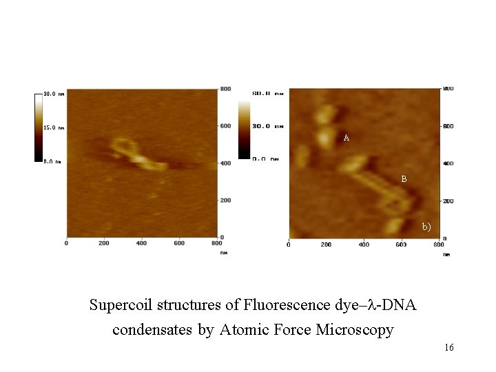
A B b) Supercoil structures of Fluorescence dye–l-DNA condensates by Atomic Force Microscopy 16

A A BB CC c) Toroidal structures of Fluorescence dye–l-DNA condensates by Atomic Force Microscopy d) 17
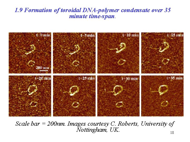
1. 9 Formation of toroidal DNA-polymer condensate over 35 minute time-span. Scale bar = 200 nm. Images courtesy C. Roberts, University of Nottingham, UK. 18
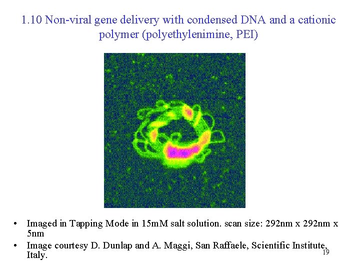
1. 10 Non-viral gene delivery with condensed DNA and a cationic polymer (polyethylenimine, PEI) • Imaged in Tapping Mode in 15 m. M salt solution. scan size: 292 nm x 5 nm • Image courtesy D. Dunlap and A. Maggi, San Raffaele, Scientific Institute, 19 Italy.
Ⅰ. Experiment—Phenomenon--Model 2. DNA Toroid Size and Mechanisms of Formation • A DNA toroid 100 nm in outside diameter with a 30 nm hole contains approximately 50 kbp of DNA. 20
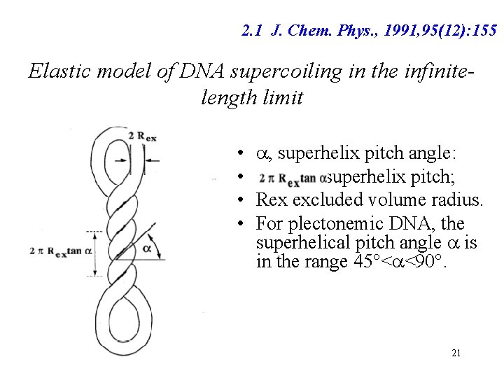
2. 1 J. Chem. Phys. , 1991, 95(12): 155 Elastic model of DNA supercoiling in the infinitelength limit • a, superhelix pitch angle: • superhelix pitch; • Rex excluded volume radius. • For plectonemic DNA, the superhelical pitch angle a is in the range 45°<a<90°. 21
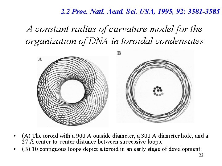
2. 2 Proc. Natl. Acad. Sci. USA, 1995, 92: 3581 -3585 A constant radius of curvature model for the organization of DNA in toroidal condensates • (A) The toroid with a 900 Å outside diameter, a 300 Å diameter hole, and a 27 Å center-to-center distance between successive loops. • (B) 10 contiguous loops depict a toroid in an early stage of development. 22
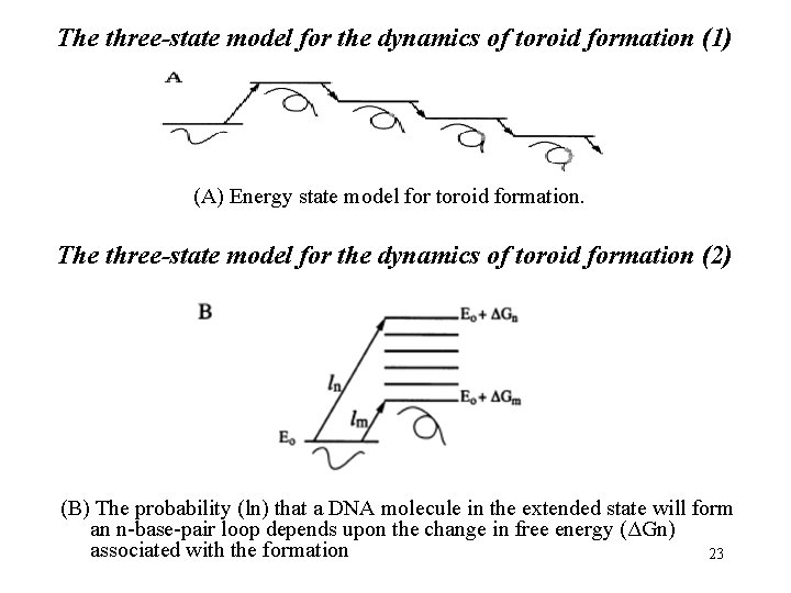
The three-state model for the dynamics of toroid formation (1) (A) Energy state model for toroid formation. The three-state model for the dynamics of toroid formation (2) (B) The probability (ln) that a DNA molecule in the extended state will form an n-base-pair loop depends upon the change in free energy (ΔGn) associated with the formation 23
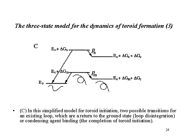
The three-state model for the dynamics of toroid formation (3) • (C) In this simplified model for toroid initiation, two possible transitions for an existing loop, which are a return to the ground state (loop disintegration) or condensing agent binding (the completion of toroid initiation). 24
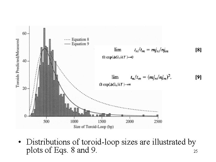
• Distributions of toroid-loop sizes are illustrated by 25 plots of Eqs. 8 and 9.
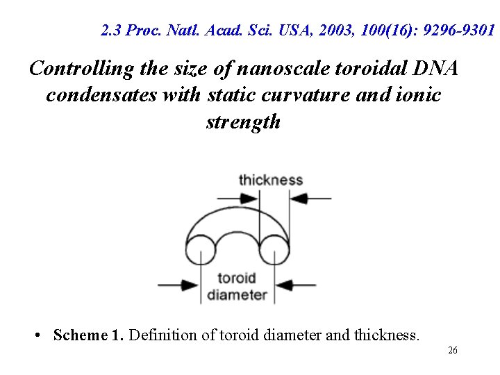
2. 3 Proc. Natl. Acad. Sci. USA, 2003, 100(16): 9296 -9301 Controlling the size of nanoscale toroidal DNA condensates with static curvature and ionic strength • Scheme 1. Definition of toroid diameter and thickness. 26
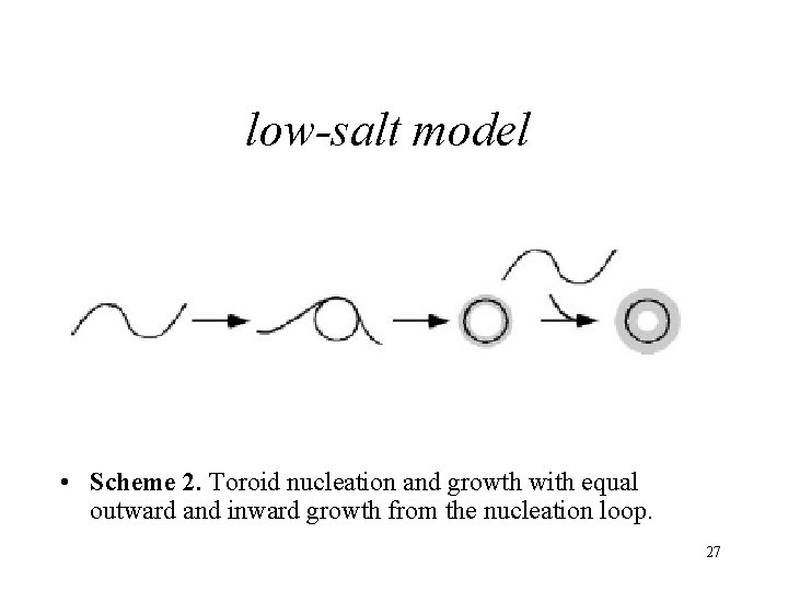
low-salt model • Scheme 2. Toroid nucleation and growth with equal outward and inward growth from the nucleation loop. 27

Transmission electron micrographs of toroids produced by the condensation of DNA with hexammine cobalt (III). • (A) Atract 60 (linear 3, 681 -bp DNA) condensed in the lowsalt buffer (0. 53 TE: 5 m. MTris 0. 5 m. MEDTA). • (B) 3 kb. DNA (linear 2, 961 -bp plasmid) condensed in the lowsalt buffer. 28
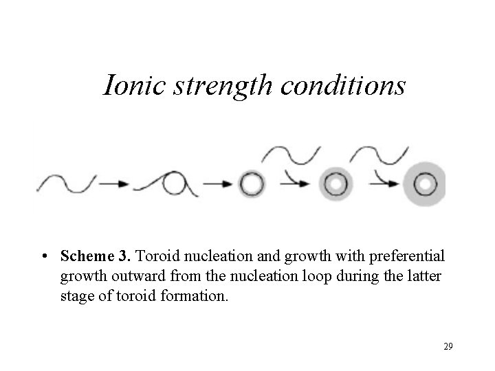
Ionic strength conditions • Scheme 3. Toroid nucleation and growth with preferential growth outward from the nucleation loop during the latter stage of toroid formation. 29

Transmission electron micrographs of toroids produced by the condensation of DNA with hexammine cobalt (III). • (C)3 kb. DNA condensed in 2. 5 m. M Na. Cl, 0. 53 TE. • (D) 3 kb. DNA condensed in 1. 75 m. M Mg. Cl 2 0. 53 TE. 30
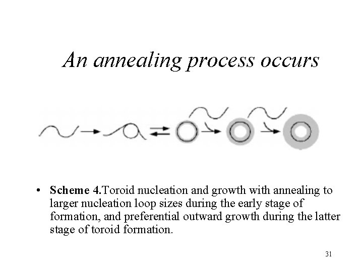
An annealing process occurs • Scheme 4. Toroid nucleation and growth with annealing to larger nucleation loop sizes during the early stage of formation, and preferential outward growth during the latter stage of toroid formation. 31

Transmission electron micrographs of toroids produced by the condensation of DNA with hexammine cobalt (III). • (E) 3 kb. DNA condensed in 3. 75 m. M Na. Cly 0. 53 TE. • (F) 3 kb. DNA condensed in 2. 5 m. M Mg. Cl 2 y 0. 53 TE. • All samples were 8. 5 mgyml in DNA and 100 m. M hexammine cobalt chloride. (Scale bar: 100 nm. ) 32
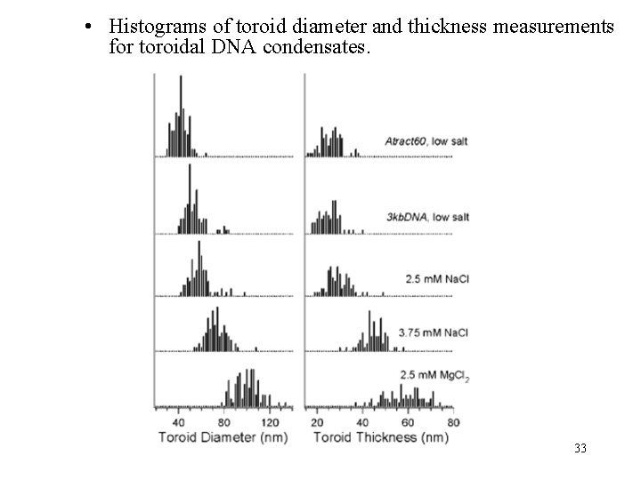
• Histograms of toroid diameter and thickness measurements for toroidal DNA condensates. 33
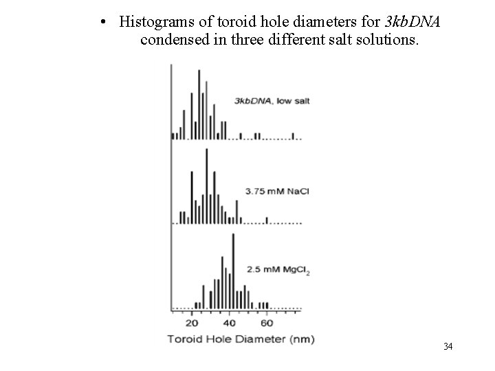
• Histograms of toroid hole diameters for 3 kb. DNA condensed in three different salt solutions. 34

2. 4 Eur. phys. Lett. , 2004, 67 (3): 418– 424. Twist-bend instability for toroidal DNA condensates Fig. 1 – Illustration of the flow field on a toroidal condensate which features additional twist. The slenderness of the torus is ξ = 1. 5. 35
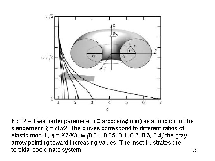
Fig. 2 – Twist order parameter τ ≡ arccos(nϕ, min) as a function of the slenderness ξ = r 1/r 2. The curves correspond to different ratios of elastic moduli, η = K 2/K 3 ∈ {0. 01, 0. 05, 0. 1, 0. 2, 0. 3, 0. 4}, the gray arrow pointing toward increasing values. The inset illustrates the toroidal coordinate system. 36
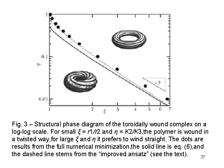
Fig. 3 – Structural phase diagram of the toroidally wound complex on a log-log scale. For small ξ = r 1/r 2 and η = K 2/K 3, the polymer is wound in a twisted way, for large ξ and η it prefers to wind straight. The dots are results from the full numerical minimization, the solid line is eq. (6), and the dashed line stems from the “improved ansatz” (see the text). 37
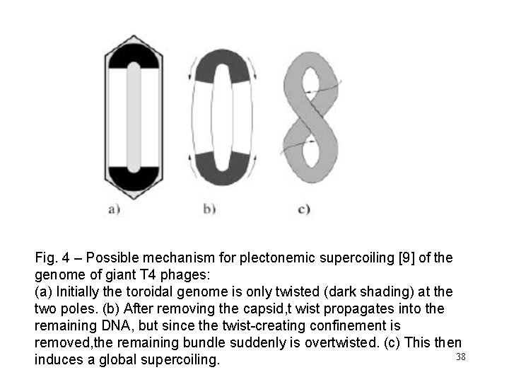
Fig. 4 – Possible mechanism for plectonemic supercoiling [9] of the genome of giant T 4 phages: (a) Initially the toroidal genome is only twisted (dark shading) at the two poles. (b) After removing the capsid, t wist propagates into the remaining DNA, but since the twist-creating confinement is removed, the remaining bundle suddenly is overtwisted. (c) This then 38 induces a global supercoiling.

• DNA Condensation by Multivalent Cations, Curr. Op. Struct. Biol. , 1996, 6: 334. • Deformation of toroidal DNA condensates under surface stress, Eur. phys. Lett. , 1996, 33 (5): 353 -358. • DNA Aggregation Induced by Polyamines and Cobalthexamine J Biol. Chem. , 1996, 271, (10): p 5656– 5662. • Toroidal Condensates of Semiflexible Polymers in Poor Solvents: Adsorption, Stretching, and Compression, Biophysical Journal, 2001, 80: 161– 168. 39
• A director-field model of DNA packaging in viral capsids, Journal of the Mechanics and Physics of Solids, 2003, 51: 1815 – 1847. • Cryoelectron microscopy of phage DNA condensates in vitreous ice: The fine structure of DNA toroids, PNAS, 2001, 98(26): 14925. • Spontaneous condensation in DNA-polystyrene-bpoly(l-lysine) polyelectrolyte block copolymer mixtures, Eur. Phys. J. E. , 2006, 20, 1 -6. 40
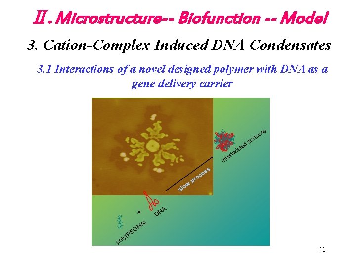
Ⅱ. Microstructure-- Biofunction -- Model 3. Cation-Complex Induced DNA Condensates 3. 1 Interactions of a novel designed polymer with DNA as a gene delivery carrier 41
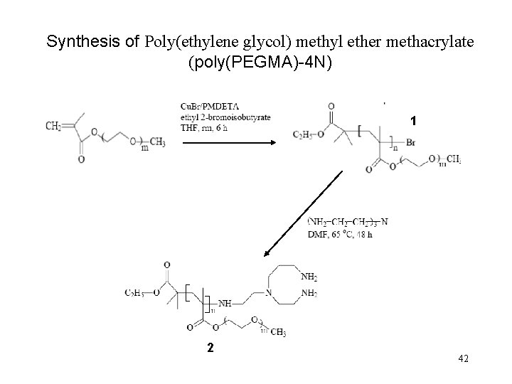
Synthesis of Poly(ethylene glycol) methyl ether methacrylate (poly(PEGMA)-4 N) 1 2 42
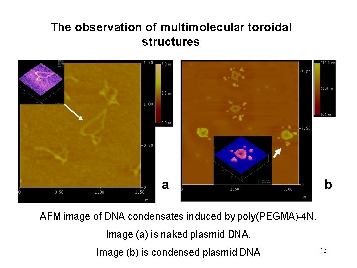
The observation of multimolecular toroidal structures AFM image of DNA condensates induced by poly(PEGMA)-4 N. Image (a) is naked plasmid DNA. Image (b) is condensed plasmid DNA 43
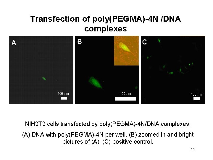
Transfection of poly(PEGMA)-4 N /DNA complexes NIH 3 T 3 cells transfected by poly(PEGMA)-4 N/DNA complexes. (A) DNA with poly(PEGMA)-4 N per well. (B) zoomed in and bright pictures of (A). (C) positive control. 44
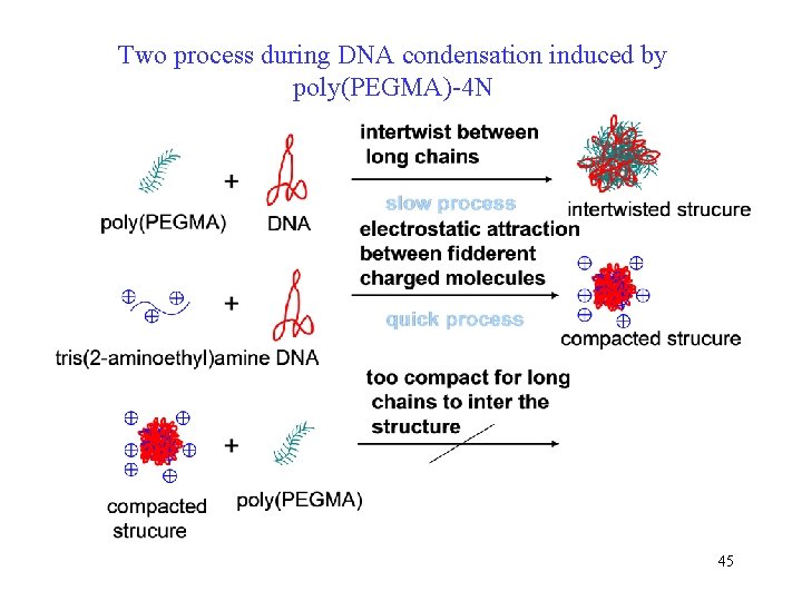
Two process during DNA condensation induced by poly(PEGMA)-4 N 45
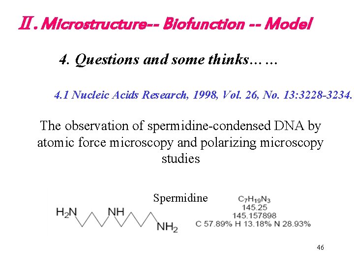
Ⅱ. Microstructure-- Biofunction -- Model 4. Questions and some thinks…… 4. 1 Nucleic Acids Research, 1998, Vol. 26, No. 13: 3228 -3234. The observation of spermidine-condensed DNA by atomic force microscopy and polarizing microscopy studies Spermidine 46
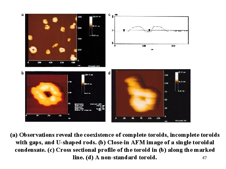
(a) Observations reveal the coexistence of complete toroids, incomplete toroids with gaps, and U-shaped rods. (b) Close-in AFM image of a single toroidal condensate. (c) Cross sectional profile of the toroid in (b) along the marked line. (d) A non-standard toroid. 47
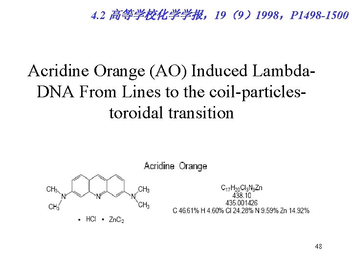
4. 2 高等学校化学学报,19(9)1998,P 1498 -1500 Acridine Orange (AO) Induced Lambda. DNA From Lines to the coil-particlestoroidal transition 48
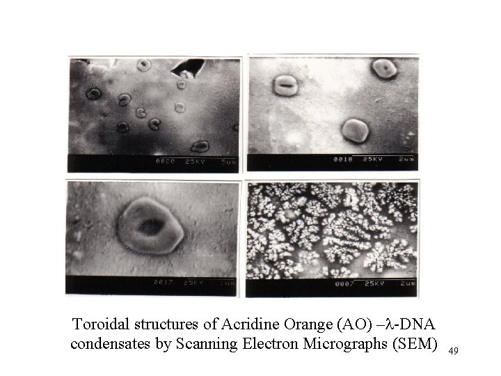
Toroidal structures of Acridine Orange (AO) –l-DNA condensates by Scanning Electron Micrographs (SEM) 49
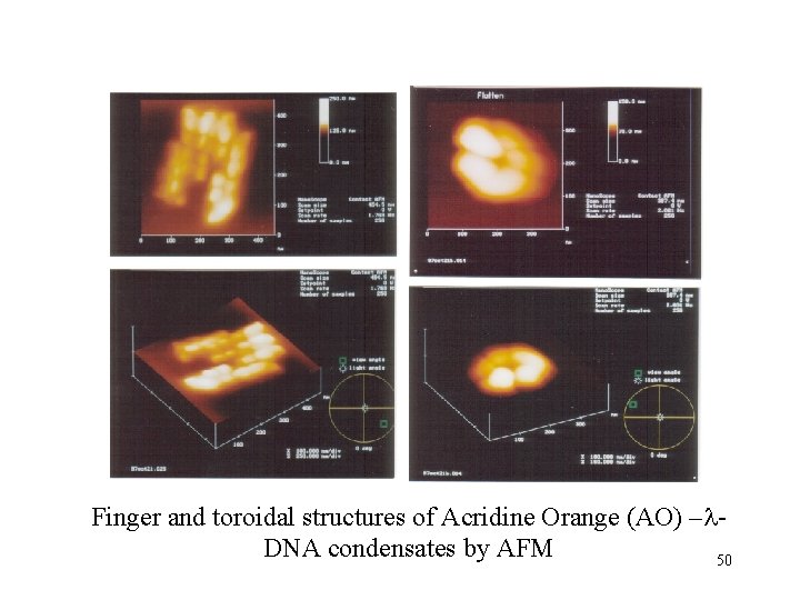
Finger and toroidal structures of Acridine Orange (AO) –l. DNA condensates by AFM 50
- Slides: 50