Interstitial syndrome Groundglass attenuation Miliary and nodular images


 •")


")


 • Viral infections •")


- Slides: 51
Interstitial syndrome Ground-glass attenuation Miliary and nodular images linear images Etienne Leroy Terquem – Pierre L’Her SPI / ISP Soutien Pneumologique International / International Support for Pulmonology
Acinus and primary lobule 1 Terminal bronchiol; 2, 3, 4. respiratory bronchioli (BR 1, BR 2, BR 3); 5. alveolar canal; 6. alveola sac; 7. alveola. There are 14 divisions between the trachea and terminal bronchiol Lobule: morphological unit. Dimension: 10 to 25 mm. It is composed of 3 to 5 acini (functional unit) (7. 5 mm); 30 to 50 primary lobules (0. 5 to 1 mm). 1 et 1'. centrolobular Bronchiol and artery; 2. terminal bronchiol and artery; 3. respiratory bronchiol; 4. canal; 5. sac; 6. alveolar; 7. perilobular vena and lymphatic vessels.
• The different parts of intersticial tissue: Under-pleural tissue – Intra-lobular tissue – Peri-lobular tissue – Under-pleural tissue Axial tissue Peri-lobular tissue Intra-lobular tissue – Axial or peri-broncho vascular tissue (according to Dr. Bernadac)
• The different parts of interstitial tissue: Under-pleural tissue – Intra-lobular Axial tissue (according to Pr. Bernadac ) Peri-lobular tissue Intra-lobular tissue
Intra-lobular interstitial tissue images: -Ground glass attenuation -Miliary: micronodules <3 mm -Nodules: between 3 and 7 mm -Macronodules > 7 mm
Ground glass attenuation Normal chest X ray Easy to recognise on scanner, it is more difficult on CXR
Ground glass attenuation Main etiologies: • Cardiac failure (initial phase before alveolar oedema) • Viral or atypical bacterial infections • Lyphoma, haemopatologic malignancies…. • Pneumocystosis
Pneumocystosis: one of the main opportunistic lung diseases in AIDS cases
Ground glass attenuation: pneumocystosis
Man, HIV+ severe dyspnea, normal auscultation , Sa 02 86 % It is a pneumocystosis. CXR features in acute phase: § Ground glass attenuation and or alveolar pictures § Diffuse and bilateral picture § No retraction, no systematisation
2 1 3 Improvment after 3 weeks of treatment with cotrimoxazole.
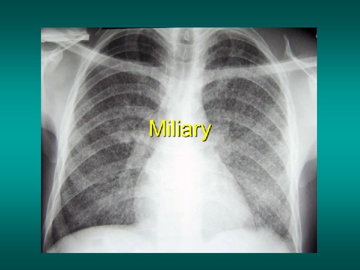
Miliary: diffuse micronodules < 3 mm Normal CXR Easy to recognise on scanner, it is more difficult on CXR
It can be difficult to distinguish miliary from ground glass attenuation…
miliary Ground glass attenuation
Miliary is sometimes barely visible
And sometimes the diagnosis is obvious…
AFB+ in bronchial aspiration. Note the assymetry of the miliary M, 68 y African, t° 40°C, weight loss, dyspnea, miliary left predominant, AFB negative in sputum
M, 25 years old Non-productive cough T° 39°C Dyspnea with effort AFB – in sputum Miliary. Improvement with anti-TB treatment
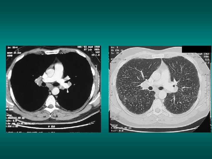
Man, 30 years old, non-smoker, dyspnea and cough progressively increasing. 2 chest x-ray with 2 -month interval. AFB - In countries with high incidence, TB is the most probable diagnosis, But it could be also a sarcoïdosis
Man, 53 years old , refugee from mauritania. Cough and worsening condition. AFB neg. . Headache (notice the unequal size of nodules which is unusual in TB miliary)
Previous case: carcinomatous miliary with metastatic adenopathies, bone and brain metastasis
Woman, 20 y HIV+ Cough, dyspnea, t° 38° 5 C Miliary AFB - BAL: Histoplasmosis
Diagnostic of miliary : • Requires an excellent quality CXR and very attentive analysis by the physician Negatoscope • Pictures often barely visible, in contrast with the importance of general signs (asthenia, dyspnea, fever, weight loss) in case of TB • AFB in sputum is usually negative The primary diagnosis is TB Main differential diagnosis (miliary and nodules<7 mm) are: – fungal infection, particularly in cases of AIDS, (histoplasmosis, – – cryptococcosis, . . . ) sarcoidosis (incidence in your countries ? ) carcinomatosis miliary pneumoconiosis (probably frequent in your countries / Mines) auto-immune affection, haemotologic malignancies, immunoallergic pneumopathy…
In countries with high incidence of TB and HIV, If X-ray shows miliary pattern - First diagnostic suspect is TB - If not clinical response after 3 weeks of treatment, in PLHIV, consider adding anti-fungal treatment - BUT DO NOT INTERRUPT TB TREATMENT! - Among PLHIV, If severe clinical picture with X-ray ground glass attenuation picture and hypoxemia , consider PCP and start treatment
Nodules
Main etiologies of diffused nodules>7 mm • Tuberculosis • Pulmonary metastasis Less frequent etiologies: § silicosis § sarcoidosis § lymphoma § fungal infection § multiple abcesses by septic emboly § hydatid cyst § bronchiolo-alveolar cancer and multiple bronchial cancers § vascularitis, Wegener, auti-immune disease, histiocytosis…
Man, 55 years old Antecedent of left pleural effusion Hemoptoic sputum AFB+
Notice the excavation. This explains why this patient is AFB positive
Woman 55 years old, cough and dyspnea with effort. Tobacco >1 pack/ day
Bronchial adenocarcinoma with lung metastis
Bronchial carcinoma with carcinomatous miliary
Lung metastasis ( uterus leiomyosarcoma)
Man, 65 years old, Asthenia, no respiratory symptoms except light cough. Antecedent of rectal cancer treated by surgery…
Chest radio 6 months later
Balloon release: pulmonary metastasis of rectum cancer
Silicosis in an older mineworker
Silicosis in an older mineworker
• The different parts of intesticial tissue: Under-pleural tissue – Under-pleural tissue Axial tissue Peri-lobular tissue Intra-lobular tissue ( according to Pr. Bernadac )
The pathology of under-pleural tissue is characterized by linear images called Kerley A or B lines. Kerley B lines Kerley A lines
Kerley lines: main etiologies • Cardiac failure (B kerley lines) • Viral infections • Less frequent: - carcinomatous lymphangitis - Fibrosis, regardless of the etiology (toxic, auto-immune, allergic, idiopathic. . ) - pneumoconiosis - haemopathy -…
• The different parts of intersticial tissue: Under-pleural tissue – Peri-lobular tissue Axial tissue Peri-lobular tissue Intra-lobular tissue ( according to Pr. Bernadac )
The increasing thickness of the peri-lobular septa and their intersection produce reticular opacities which line polyedric spaces: « Meshed net » aspect. The main etiology is pulmonary fibrosis, regardless of the etiology ( idiopathic, toxic, autoimmun…)
Idiopathic fibrosis
Idiopathic fibrosis
• The different parts of intersticial tissue: Under-pleural tissue Axial or peribronchovascular tissue Axial tissue (according to Dr. Bernadac ) Peri-lobular tissue Intra-lobular tissue
The pathology of peribronchovascular tissue is characterised by linear opacities from the hilus to the periphery of the lungs. Main etiologies are: -Carcinomatous lymphangitis -Fibrosis regardless of the etiology (idiopathic, autoimmune, allergic, toxic) -Sarcoidosis -Viral and fungal infection Carcinomatous lymphangitis The association of nodular and linear images is highly suggestive of carcinomatous lymphangitis
Man, 60 years old. Dyspnea with effort, poor condition and jaundice . The association of nodular and linear images suggests in this context a carcinomatous lymphangitis (pancreatic origin)
… Previous case
Conclusions • The Interstitial pathology is complicated • The etiologies are numerous • The intrication with alveolar pictures is frequent (cardiac failure, TB, infections. . ) • The interpretation requires a very good quality chest radiography (especially for miliary) • CT scan is much more performant but not always easily accessible