Interpretation of Histological sections Lecturer Farah E Ismaeel


 like nucleic acids (BLUE-VIOLET) �Using")


- Slides: 13
Interpretation of Histological sections Lecturer Farah E. Ismaeel
Preparation Of histological slides
Fixation �Small pieces of tissue are placed in solutions of chemicals �Preserve by cross-linking proteins and inactivating degradative enzymes �Physical & chemical fixation (formaldehyde 37%)
Dehydration �The tissue is transferred through a series of increasingly concentrated alcohol solutions, ending in 100%, which removes all water Ethanol 70% Ethanol 80% Ethanol 99%
Clearing �Alcohol is removed in xylene or other solvents in which both alcohol and paraffin are miscible.
Infiltration & embedding �The tissue is then placed in melted paraffin until it becomes completely infiltrated with this substance. • The paraffin-infiltrated tissue is placed in a small mold with melted paraffin and allowed to harden
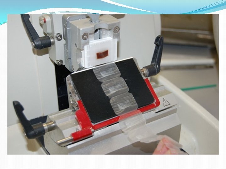
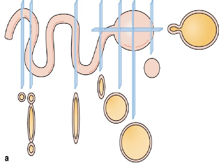
Sectioning �Microtome used for sectioning paraffinembedded tissues for light microscopy � 1 and 10 μm thick sections put on glass slides
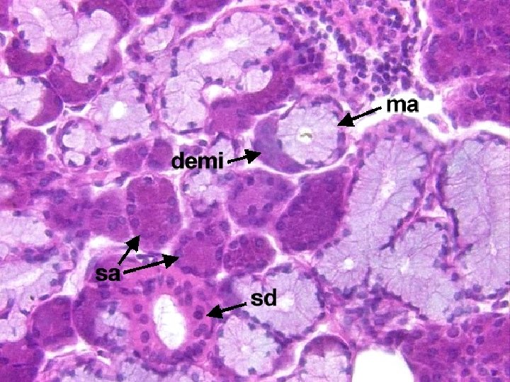
Staining �Using basic dyes to stain acidic components (basophilic) like nucleic acids (BLUE-VIOLET) �Using acidic dyes to stain basic components (acidiophilic) like proteins with amino groups PINK • Basic dyes that stain basophilic components: toluidine blue, methylene blue & Hematoxyline • Acidic dyes stain the acidophilic components: orang G & Eosin • Hemotoxylin & Eosin is the most used stain
Hematoxylin only Eosin only H&E
Thank you for listening