Interhospital Conference v Past history no underlying disease


 § Finding: Irregular mass at left tonsillar fossa, invaded")


 v. A 2. 1 x 2. 2 cm hypermatabolic")
, likely")
 v. Bone lesion at skull is corresponding with")


 v. Section show fragments of bony")


 v. Normal attenuation of muscles and")


,")


 § Infectious mononucleosis v")
- Slides: 57
Interhospital Conference
v. Past history : no underlying disease no history of drug allergy v. Family history : no malignancy
Physical Examination v. GA: A Thai boy, good consciousness, well co-operative v. HEENT: pink conjunctiva, anicteric sclera § Irregular mass below left tonsillar fossa, mass invaded posterior pillar and posterior pharyngeal wall, invade valleculae, but abutted epiglottis v. LN : impalpable v. Heart: regular, normal S 1 S 2, no murmur v. Lungs: clear both lungs v. Abd: normoactive, no hepatosplenomegaly
Investigation v. CBC Hb 11. 6 g/dl, Hct 35. 7%, WBC 5, 680 cells. mm 3 (N 59. 9%, L 32. 9%, M 4. 2%, E 2. 5%, B 0. 5%), Plt 200, 000 cells/mm 3 v. Blood chemistry § BUN 12 mg/dl, Cr 0. 53 mg/dl § Electrolyte: Na 138 mmol/L, K 4. 7 mmol/L, Cl 99 mmol/L, CO 2 28 mmol/L § TB 0. 23 mg/dl, DB 0. 02 mg/dl, SGOT 19 mg/dl, SGPT 22 mg/dl, ALP 194 U/L
PROBLEM LISTS
INVESTIGATION
Tonsillar Biopsy
Management v. Intraoral biopsy (28/04/52) § Finding: Irregular mass at left tonsillar fossa, invaded posterior pillar, posterior pharyngeal wall, valleculae, abutted epiglottis § Operation: partial excision about 50% by electrocautery
Pathological Report
Pathology Report v. Section of mucosa show vascular lesion infiltrating in underlying stroma. There are lined by plump endothelial cells witch have round to spindle nuclei, vascular chromatin, distinct nuclei and moderate amount of eosinophilic cytoplasm. Mitoses are frequently seen. Intervening stroma reveals hyalinization. Hemorrhage and many chronic and acute inflammatory cells infiltrate are observed. Covering mucosa display focal ulcer with fibrinous exudate and acute inflammatory cells infiltrate. Pseudoepitheliomatous hyperplasia is occationally noted. v. Hemangioendothelioma
Progression v. ENT consult Tumor conference v. Tumor conference suggest MRI กอน plan management เพมเตม
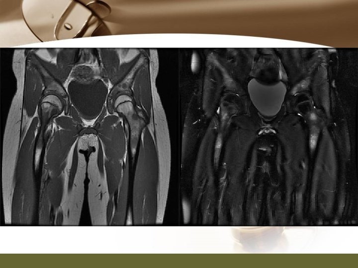
PET/CT and MRI
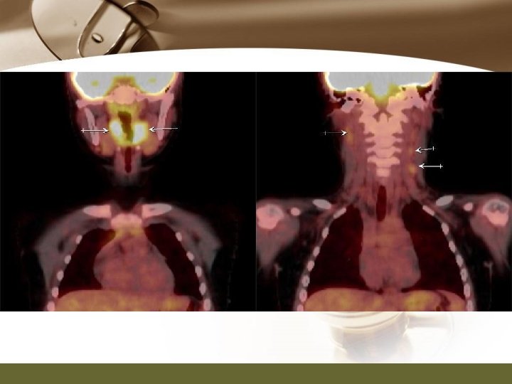
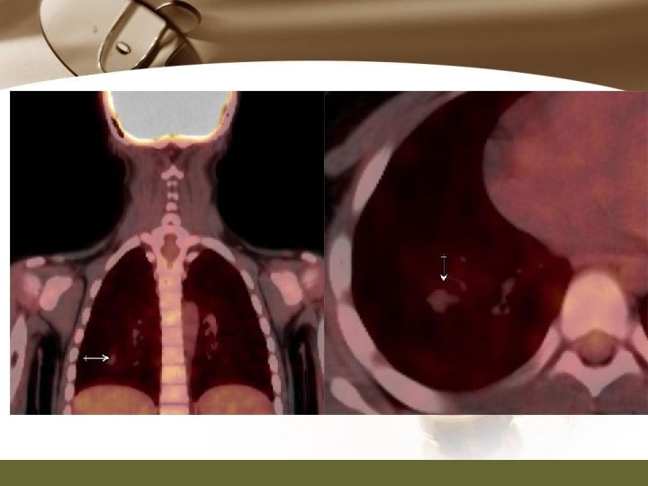
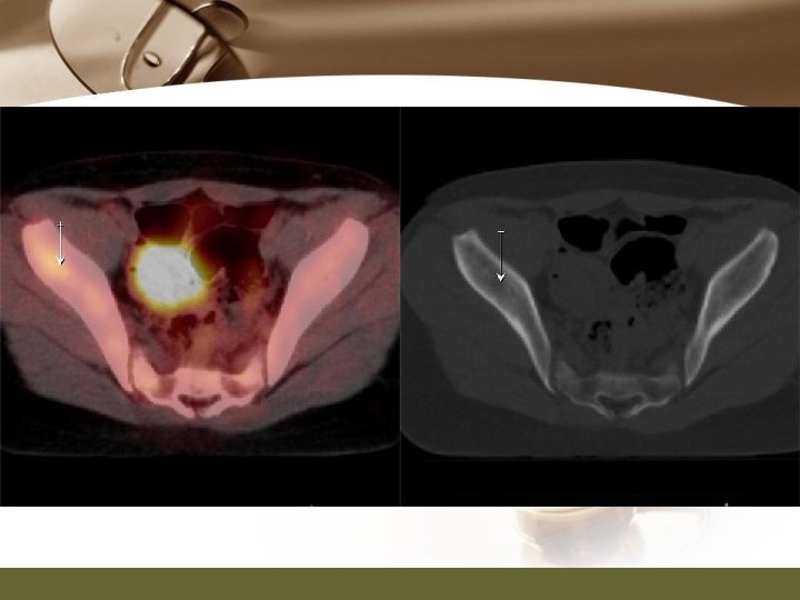
PET/CT Report v. PET/CT (03/07/52) v. A 2. 1 x 2. 2 cm hypermatabolic irregular rim enhancing mass with central hypodensity at left palatine tonsillar fossa, consistent with history of hamangioendothelioma. This is possible residual disease v. Mild focal bulging medical contour of right palatine tonsil without definite abnormal enhancing area, showing homogeneous FDG accumulation, small focal lesion cannot be excluded. Tissue diagnosis is recommended.
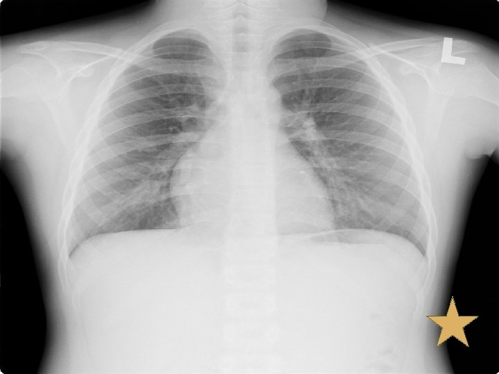
PET/CT Report v. Multiple hypermetabolic bilateral cervical lymph nodes (more on the left), likely nodal metastases. v. Multiple pulmonary nodules, probably pulmonary metastases. v. Multiple small hypermetabolic poorly osteolytic and nonosteolytic lesion, probably inhomogeneous marrow activity in child or foci of marrow infiltration. Correlation with other imaging modality such as bone scan is recommended
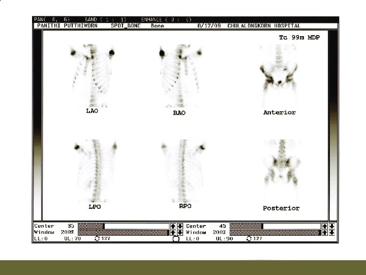
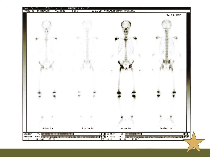
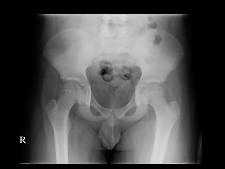
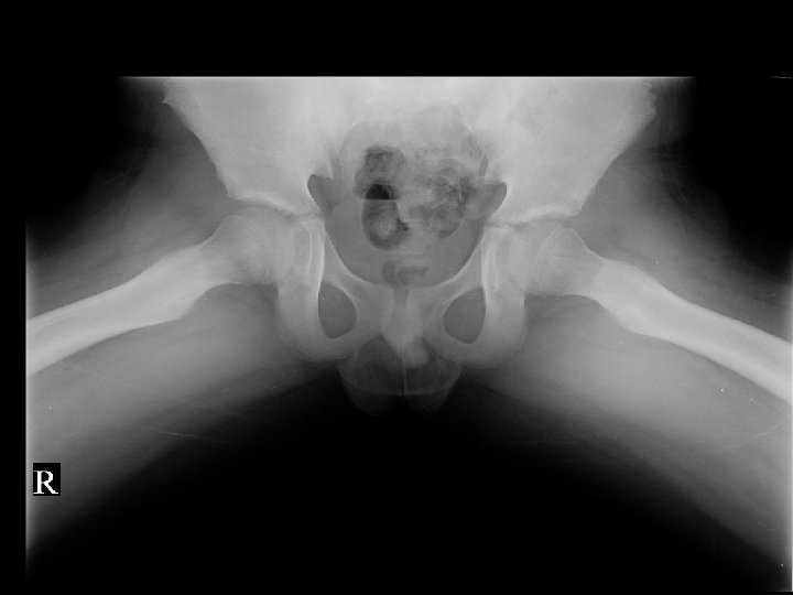
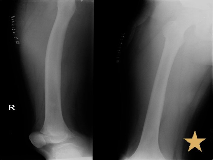
Bone Scan v. Bone scan (17/08/52) v. Bone lesion at skull is corresponding with multiple geographic lytic lesions without sclerotic rim and some blastic lesions in diploic space of bilateral parietal bone, likely bone metastases. Bone lesion at C 2 vertebral body, pelvic rim, right acetabulum, head, proximal, mid shaft and distal right femur, likely due to bone metastases as correlated with lytic lesion seen on PET/CT.
X-Ray
Management v. Due to PET/CT found multiple small hypermetabolic poorly osteolytic and non-osteolytic lesion, suspected bone metastases consult orthopaedics 23/7/52 Excision Bone at Right iliac, right proximal tumor
Pathological report of Bone Biopsy
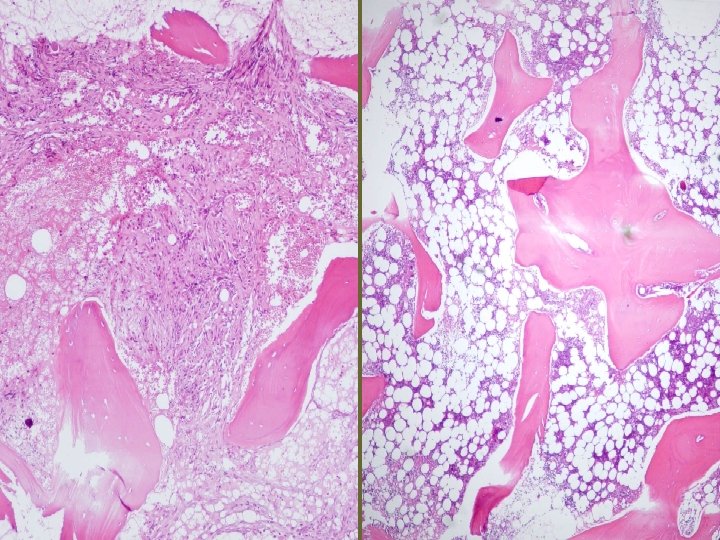
Pathology Report v. Right iliac bone biopsy (23/07/52) v. Section show fragments of bony tissue and marrow element. Few pieces of bone reveal proliferation of blood vessels with occasionally lined by round to spindleshaped cells. Cells have round nuclei, fine chromatin, visible nucleoli v. Immunohistochemistry: epithelioid hemangioendothelioma
Pathology Report v. Right proximal femur : no definite vascular tumor
v. Impression : Gorham disease
Progression v 03/08/52 Start Radiotherapy at tonsils and lymph node 70 Gy v 06/08/52 รบ consult at OPD ผปวยมปญหาเรองปวดทขาขางขวาต งแตตนขาถงปลายขา ไมมปวดบรเวณอน § CBC: Hb 8. 8 g/dl , WBC 11, 800 cells/mm 3 (N 78. 3 %, L 14. 9 %, M 3. 6% , E 2. 9% , B 0. 3% ), Platelet 122, 000 cells/mm 3 § LD-PRC 300 ml IV drip in 3 hour
Progression v 7/8/52 CBC: Hb 11 g/dl , WBC 9, 850 /mm 3 (N 66 %, L 21 %, M 3% , E 7% , AL 3% ), Platelet 104, 000 /mm 3 PTT 36. 8 sec [30. 4] PT 15. 4 sec [13. 4] Fibrinogen 641 mg/dl D-dimer 0. 2 mcg/dl [< 0. 3] v 10/08/52 OPD Follow up ผปวยยงมอาการปวดขาไมดขน Bonefos (800) 2 cap oral OD เชา
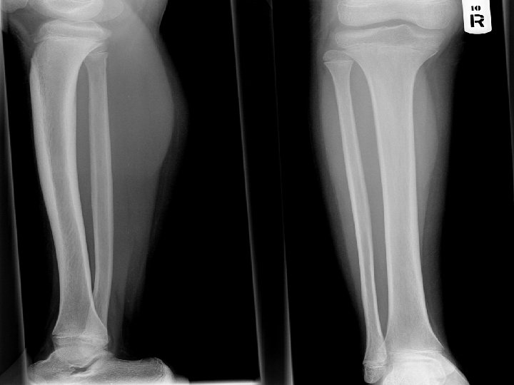
Progression v 20/08/52 OPD Follow up, ปวดขามากโดยเฉพาะทบรเวณนอง admit for further investigation § § § , ซดลง Investigation : plain X-ray leg [right], นด U/S right leg Consult pain: MO IV prn for pain Blood component as needed Continue Bonefos Continue Radiation
Progression Follow up Lab § CBC: Hb 10. 1 g/dl , WBC 4, 360 cells/mm 3 (N 76%, L 12%, M 5%, E 6%, AL 1%), Plt 62, 000 cells/mm 3 § Fibrinogen = 4. 66 G/L (1. 7 -4. 0) § D-dimer Vidas =3, 322 ng/ml (<500)
Ultrasound v. Ultrasonography of the right calf (26/08/52) v. Normal attenuation of muscles and subcutaneous tissue of right calf. No fluid or space taking lesion within right calf is observed. The color Doppler ultrasound show on evidence of hypervascularity or abnormal vascular formation within right calf.
Progression 25/8/52 Start systemic treatment: Vinblastion, Prednisolone vหลง start systemic treatment ได 2 wk, อาการปวดลดลง และปวดหางมากขน v. Continue systemic treatment และสามารถcontrol painไดดวยยากน v. D/C 15/9/52 then F/U as OPD case
Gorham’s Disease Dipak at el, Clinical Medicine & Research, Volume 3, Number 2: 65 -74
v. A rare disorder characterized by proliferation of vascular channels that results in destruction and resorption of osseous matrix. v. There have been fewer than 150 cases reported in the literature.
v. Etiology of Gorham’s disease remains poorly understood v. The pathological process is the replacement of normal bone by an aggressively expanding but non -neoplastic vascular tissue similar to a hemangioma or lymphangioma. Wildly proliferating neovascular tissue causes massive bone loss.
v. No evidence of a malignant, neuropathic, or infectious component involved in the causation of this disorder.
Clinical presentation v. The clinical presentation of Gorham’s disease is variable and depends on the site of involvement. v. Gorham’s disease may complain of dull aching pain or insidious onset of progressive weakness. In some cases, pathologic fracture often leads to its discovery.
Clinical presentation v. Gorham’s disease can involve men or women and any age group v. Although most cases are discovered before the age of 40 years. v. No familial predisposition has been found. v. The process may affect the appendicular or the axial skeleton. The shoulder and the pelvis are the most common sites.
Treatment v. The medical treatment for Gorham’s disease includes radiation therapy, anti-osteoclastic medications (bisphonates), and alpha-2 b interferon. v. Surgical treatment options include resection of the lesion and reconstruction using bone grafts and/or prostheses.
Treatment v. Radiation therapy are used for management of patients who have large, symptomatic lesions with long-standing, disabling functional instability. v. Definitive radiation therapy in moderate doses (4045 Gy in 2 Gy fractions) appears to result in a good clinical outcome with few long-term complications.
Treatment v. In general, no single treatment modality has proven effective in arresting the disease. v. The prognosis for patients with Gorham’s disease is generally good unless vital structures are involved.
CBC : Hb 12. 6 g%, WBC 6, 000 /mm 3 [N 56%, L 28 %, atypical L 13%, Mo 3 %] platelet 349, 000 /mm 3 PT 13. 1 sec [11. 3], PTT 38. 9 sec [33. 3] Impression : Kasabach Meritt syndrome Treatment กนยายน 2541 - พฤศจกายน 2542 - Prednisolone + Interferon alpha ตลาคม 2542 - มกราคม 2544 - Vincristine clinical improve, platelet count and coagulogramปกต
v พฤษภาคม 2552 ผปวยเรมมปนสแดงขนมาใหมและม เลอดออกไมหยดบรเวณ lesion รวมกบมเลอดออกตามไรฟน จงมาโรงพยาบาล ระหวาง admission ผปวยมปญหา hemothorax both lungs, compression fracture at T 12 and multiple osteolytic lesion at spine and rib Treatment vincristine weekly [total 7 doses] vinblastine INF alfa 2 b [ 45 doses]
THANK YOU FOR YOUR ATTENTION
A case report of epithelioid hemangioendothelioma metastasizing to the tonsil v. A 40 -year-old man admitted for right throat pain vhe underwent radical surgery. v. Epithelioid hemangioendothelioma was first diagnosed v. Lung specimens at open biopsy 4 years earlier showed the same histological features indicating he had epithelioid hemangioendothelioma lesion since that time. v. We assumed this epithelioid hemangioendothelioma had originated in the lung and metastasized to the right tonsil Nippon Jibiinkoka Gakkai Kaiho. 2002 Sep; 105(9): 937 -40.
v. Epithelioid hemangioendothelioma is an extremely rare, difficult-to-diagnose vascular tumor mainly originating from the lung or liver. v. Primary tumors in the head and neck are very rare Nippon Jibiinkoka Gakkai Kaiho. 2002 Sep; 105(9): 937 -40.
Differential Diagnosis for Tonsillar hypertrophy v Infectious Disorders (Specific Agent) § Infectious mononucleosis v Infected organ, Abscesses § Pharyngitis § Adenoiditis, acute § Tonsillitis/exudative, acute § Tonsillitis, chronic v Neoplastic Disorders § Tonsil lymphosarcoma § Tonsil, Lymphoepithelioma v Metabolic, Storage Disorders § Tangier's disease v Hereditary, Familial, Genetic Disorders § Lipodystrophy, generalized v Reference to Organ System § Adenoid hypertrophy § Tonsillar hypertrophy syndrome