Interactive Grand Rounds Blair Lonsberry MS OD MEd

Interactive Grand Rounds Blair Lonsberry, MS, OD, MEd. , FAAO Professor of Optometry Pacific University College of Optometry blonsberry@pacificu. edu 1

Disclosures • I have received honoraria from the following companies: – Alcon – Carl Zeiss Meditec – Optovue – Shire – Sun

CASE 1

Case History • 38 black male, complaining that the vision in his right eye is blurry. – Got the current Rx 3 weeks previously, and started out good but in last couple of days OD vision has become blurry • Medical Hx: no current health concerns and no medications

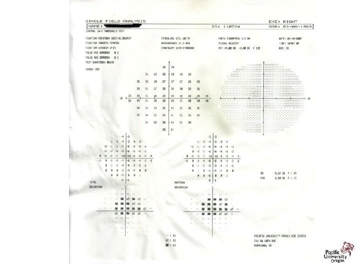
Entrance Skills • • • Va’s: OD: 20/25, OS: 20/20 Pupils: PERRL CVF: full to finger count EOM’s: FROM Amsler: central metamorphopsia OD HVF: 10 -2 (see VF)

Which of the following OCT’s goes with this patient? 1 2 3 4
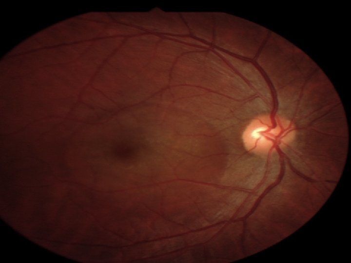

Central Serous Retinopathy • an exudative chorioretinopathy characterized by an exudative neurosensory retinal detachment with or without an associated detachment of the retinal pigment epithelium (RPE) • Patients experience blurry vision, metamorphopsia and micropsia • individuals between 20 and 50 years of age

Central Serous Retinopathy • incidence in men vs women is approximately 6 to 1 • associated with stress and stress hormones (ie, corticosteroids and epinephrine); • individuals with a "type A personality" who are under stress • recurrence in the ipsilateral eye is approximately 30% and CSR in the fellow eye was 32%

Central Serous Retinopathy

Central Serous Retinopathy • 80% to 90% of cases resolve spontaneously within 3 months • Treatment options: – include laser photocoagulation, – "safety-enhanced" PDT, – Acetazolamide reduced the time for subjective and objective CSR resolution, but it had no effect on final VA or recurrence rate. Most patients in the experimental group in that study had side effects from the acetazolamide, including paresthesias, nervousness, and gastric upset

Central Serous Retinopathy • Treatment options: – Topical NSAIDs: • Conflicting reports • Michael Singer, MD, from Medical Center Ophthalmology in San Antonio reported an increase in resolution time by 50% • PRADEEP VENKATESH, MD reports that NSAIDS treatment could possibly slow down or cause a rebound CSR

CASE 2

Case • 55 yr white female complains of fluctuating vision – Worse at near – Spends 8 -10 hours/day on the computer • Medical Hx: – Hypertension for 10 years – Joint pain • Medications: – HCTZ for HTN – Celebrex for her joint pain
: OD: 20/25, OS: 20/25 • PERRL • EOM’s: FROM")
Exam Data • VA (corrected): OD: 20/25, OS: 20/25 • PERRL • EOM’s: FROM • CVF: FTFC • SLE: – TBUT 5 sec OD, OS – Positive Na. Fl staining and Lissamine green staining of conj and cornea – Decreased tear prism

Additional Testing/Questions • Schirmer: < 5 mm of wetting in 5 minutes OD, OS • RF and ANA: normal for patients age • SS-A: 2. 0 (normal < 1. 0), SS-B: 1. 9 (normal <1. 0) • Additional symptoms reported: – Patient experiences dry mouth and taking Salagen • Diagnosis: Sjogren’s Syndrome

Differential Diagnosis of Dry Eye

Signs and Symptoms of Dry Eye Signs: – Ocular Surface Damage • Corneal Staining (Fluorescein and/or Rose Bengal) • Conjunctival Staining (Lissamine Green ) – Decreased Tear Quantity • Schirmer Score • Phenol Red Thread Test • Tear Meniscus Height – Decreased Tear Quality • Tear Break Up Time (TBUT) • Tear Osmolarity Symptoms: – – – Grittiness Burning Irritation Stringy discharge Blurring of vision Ocular Surface Disease Index (OSDI)

Inflamma. Dry • Point of care testing to measure MMP-9 levels – MMP-9 is an inflammatory biomarker found to be elevated in patients with dry eye • Marketed by RPS
 – Recommended")
Treatment • We initiated: – Omega-3 supplements (3 -4 grams per day) – Recommended warm compresses and lid washes qhs – Testosterone cream 3% applied to upper lid bid • Patient had significant improvement in symptoms with the use of the topical testosterone cream. – However, she was still symptomatic at the end of the day and she still had significant staining on her cornea and conjunctiva – Initiated FML tid for 1 month, restasis bid after 2 weeks • 2 months later patient reported further improvement in her symptoms • No conjunctival staining was noted and only slight SPK • Schirmer values improved to OD: 9 mm, OS: 10 mm

Role of Androgens? • Recent studies have suggested that androgen deficiency may be the main cause of the meibomian gland dysfunction, tear-film instability and evaporative dry eye seen in Sjogren patients • Transdermal testosterone 3% promotes increased tear production and meibomian gland secretion, thereby reducing dry eye symptoms (Dr. Charles Connor). • Progesterone 0. 05%/Testosterone 0. 05% Ophthalmic Solution BID (available from Leiter’s Pharmacy)

SJOGREN’S SYNDROME: OLD/NEW CLASSIFICATION • Old: – 1 o Sjogrens: occurs when sicca complex manifests by itself • no systemic disease present – 2 o Sjogrens: occurs in association with collagen vascular disease such as • RA and SLE • significant ocular/systemic manifestations • New: – The diagnosis of SS should be given to all who fulfill the new criteria while also diagnosing any concurrent organ-specific or multiorgan autoimmune diseases, without distinguishing as primary or secondary.
 was funded by")
Diagnosis: New Criteria • • Sjogren’s International Collaborative Clinical Alliance (SICCA) was funded by the National Institutes of Health to develop new classification criteria for SS New diagnostic criteria requires at least 2 of the following 3: – 1) positive serum anti-SSA and/or anti-SSB or (positive rheumatoid factor and antinuclear antibody titer >1: 320), – 2) ocular staining score >3, or – 3) presence of focal lymphocytic sialadenitis with a focus score >1 focus/4 mm 2 in labial salivary gland biopsy samples
 • The ocular surface score (OSS) is the sum of:")
Ocular Surface Score (OSS) • The ocular surface score (OSS) is the sum of: – 0 -6 score for fluorescein staining of the cornea and – 0 -3 score for lissamine green staining of both the nasal and temporal bulbar conjunctiva, – yielding a total score ranging from 0 -12.

Antibodies to SS-A and SS-B • Sjogren’s syndrome A and B • Typically tested by ELISA and immunoblot • Associated Conditions: – Uncommon in the normal population and in patients with rheumatic diseases other than Sjogren’s syndrome and SLE – Present in 75% of patients with “primary” Sjogren’s but only 10 -15% of patients with RA and secondary Sjogren’s

Antibodies to SS-A and SS-B • Indications: – Should be measured in patients with a clinical suspicion of Sjogren’s or SLE • Interpretation: – Presence of AB’s is a strong argument for the diagnosis of Sjogren’s Syndrome in a patient with sicca syndrome

Sjogren’s Ocular and Systemic • Recently published article comments: – One or more extraglandular ocular manifestations (e. g. corneal scarring/melts, uveitis) were present in 35% of patients – 13% had vision threatening findings – Approximately 55% with a vision threatening ocular finding did not have an established diagnosis of primary SS at the time of presentation Ocular and Systemic Morbidity in a Longitudinal Cohort of Sjögren’s Syndrome. Ophthalmology 2014

Sjogren’s Ocular and Systemic • Recently published article comments: – all patients had dry eye symptoms for approximately 10. 4 years before presentation – 42% of the patients had systemic manifestations resulting from primary SS – SS has been shown to be an independent risk factor for the development of non. Hodgkin’ s lymphoma. Ocular and Systemic Morbidity in a Longitudinal Cohort of Sjögren’s Syndrome. Ophthalmology 2014

Sjogren’s Ocular and Systemic • Authors recommendation: – primary SS is associated with vision- and lifethreatening complications – presence of SS needs to be explored in patients with clinically significant dry eye because dry eye precedes the occurrence of the systemic manifestations Ocular and Systemic Morbidity in a Longitudinal Cohort of Sjögren’s Syndrome. Ophthalmology 2014

Dry Eye Summit • Held in December 2014 – Combination of optometrists, an ophthalmologist and industry • Goal: – to find a way to encourage optometrists to look for, diagnose and manage dry eye in their patients – Come to a consensus on the minimum: • 3 questions that should be asked to identify dry eye patients • 3 diagnostic tests • 3 initial treatments 31

REV. as of March 13, 2015 Consensus on Baseline Management 1. For all patients: A. Ocular lubrication B. Lid hygiene C. Nutrition 2. Topical anti-inflammatories Recommendations from the Dry Eye Summit 2014

Dry Eye and Lid Disease? • It is estimated that 67 -75% of patients who have dry eye have some form of lid disease – it is often the most overlooked cause for dry eye symptoms • Important to address the lids in any treatment plans for patients with dry eye

Treatment of MGD At Home Therapy – Warm compresses – Eyelid Scrubs – Self expression Low Compliance In-Office Therapy – Manual Expression – Off-Label Pharmacotherapy – Oral tetracycline/doxycycline -systemic side effects – Topical Antibiotics – erythro. , tobra. – Topical Steroids – dexamethasone -antibiotic resistance, poor gland penetrance -risk of cataract, glaucoma, poor gland penetrance 37

LIPIFLOW® THERMAL PULSATION SYSTEM The Lid Warmer: Comprised of a precision heater, eye insulation & vaulted shape The Eye Cup: Comprised of an inflatable bladder & rigid eye cup 38 Lipi. Flow Auto Console pictured (inset) is not approved for Caution: Investigational device. The use in the U. S. Limited by United States law to investigational use.

QUICKIE

Lid Nevi • Lid nevi: – congenital or acquired – occur in the anterior lamella of the eyelid and can be visualized at the eyelid margin. • The congenital eyelid nevus is a special category with implications for malignant transformation. • With time, slow increased pigmentation and slight enlargement can occur. • An acquired nevus generally becomes apparent between the ages of 5 and 10 years as a small, flat, lightly pigmented

Congenital Nevus • The nevus is generally well circumscribed and not associated with ulceration. • The congenital nevus of the eyelids may present as a "kissing nevus" in which the melanocytes are present symmetrically on the upper and lower eyelids. – Presumably this nevus was present prior to eyelid separation

Congenital Nevus • Most nevi of the skin are not considered to be at increased risk of malignancy. – However, the large congenital melanocytic nevus appears to have an increased risk of malignant transformation of 4. 6% during a 30 year period

Acquired Lid Nevi • Acquired nevi are classified as: – junctional (involving the basal epidermis/dermis junction), typically flat in appearance – intradermal (involving only the dermis), tend to be dome shaped or pedunculated – compound (involving both dermis and epidermis) tend to be dome shaped

CHRPE vs Nevus 44

Nevi Trivia • 31% of choroidal nevi show slight enlargement over time without the transformation to a melanoma (Ophthalmology 2011) • The prevalence of choroidal nevi in the white U. S. population ranges from 4. 6% to 7. 9% – If it is assumed that all choroidal melanomas arise from preexisting nevi, then the published data suggest a low rate (1/8845) of malignant transformation of a choroidal nevus in the U. S. white population. (Ophthalmology 2005) • Choroidal melanoma risk for metastasis, ranging from 16% to 53% (at 5 years of follow-up) depending on the size of the tumor at the time of diagnosis. (Arch Ophthalmol 1992)

TFSOM—“To Find Small Ocular Melanoma” Thickness: lesions >2 mm Fluid: any subretinal fluid (suggestive of serous retinal detachment) Symptoms: photopsia, vision loss Orange pigment overlying the lesion Margin touching optic nerve head (<3 mm) • None of these factors = 3% risk of a nevus converting to melanoma in five years. One of these factors = 8% risk of conversion in five years. Two or more factors = 50% risk of conversion in five years. For any changes noted during the course of follow-up, refer the patient to a retinal practice or an ocular oncology service.

TFSOM-UHHD: “To Find Small Ocular Melanoma Using Helpful Hints Daily” Thickness: lesions >2 mm • Choroidal nevi showing no features should be initially Fluid: subretinal fluid monitored twice yearly and followed up annually Symptoms: photopsia, • 1 or 2 features should be vision loss monitored every 4 to 6 Orange pigment months. overlying the lesion • Nevi with 3 or more features Margin touching optic should be evaluated at an experienced center for nerve head (<3 mm) management alternatives and Ultrasound Hollowness possible treatment owing to the high risk of ultimate Halo absence growth Drusen absence

From: Enhanced Depth Imaging Optical Coherence Tomography of Small Choroidal Melanoma: Comparison With Choroidal Nevus Arch Ophthalmol. 2012; 130(7): 850 -856. doi: 10. 1001/archophthalmol. 2012. 1135 Figure Legend: Date of download: 10/9/2014 Copyright © 2014 American Medical Association. All rights reserved.

From: Enhanced Depth Imaging Optical Coherence Tomography of Small Choroidal Melanoma: Comparison With Choroidal Nevus Arch Ophthalmol. 2012; 130(7): 850 -856. doi: 10. 1001/archophthalmol. 2012. 1135 Figure Legend: Date of download: 10/9/2014 Copyright © 2014 American Medical Association. All rights reserved.

Case • 65 yr old white male – Notices spot in vision in his left eye – Diabetes for 15 years • Vision: 20/20 (6/6) and 20/40 (6/12 ) • Dilated exam: – Large lesion noted in left eye (not noted in exam 6 months previously – See photo and B-scan

Ocular Tumors Astrocytic Hamartoma Retinoblastoma Amelanotic Melanoma Metastatic Choroidal Tumor

Choroidal Melanoma Metastases • 80 to 90% of metastases from uveal melanoma occurred in the liver, less common sites being the skin and lung. – Gragoudas ES, Seddon JM, Egan KM, et al. Longterm results of proton beam irradiated uveal melanomas. Ophthalmology. 1987; 94: 349– 53.

Melanoma and Mortality • Tumor Size: – 5 -year mortality after enucleation: • 16% for small melanoma, • 32% for medium melanoma, and • 53% for large melanoma. – the prognostic importance of tumor size: • each 1 -mm increase in melanoma thickness adds approximately 5% increased risk for metastatic disease at 10 years • Tumor genetics: – Chromosome monosomy 3 (apprx 50% of patients) • 50% of them develop metastasis within 5 years of diagnosis • 70% mortality within 4 years of ocular treatment • one of the most important independent risk factors of poor survival

CASE 3

Case: Gonzalez • 33 HF presents with a painful, red right eye – Started a couple of days ago, deep boring pain – Has tried Visine but hasn’t helped the redness • PMHx: patient reports she has been diagnosed with rheumatoid arthritis 3 years ago – Takes Celebrex for the joint pain – Patient reports she occasionally gets a skin rash when she is outdoors in the sun • POHx: unremarkable • PMHx: mother has rheumatoid arthritis
 OD, – 6/6 (20/20) OS •")
Case: Gonzalez • VA: – 6/7. 5 (20/30) OD, – 6/6 (20/20) OS • • • Pupils: PERRL –APD VF: FTFC OH EOM’s: FROM OU BP: 130/85 mm Hg RAS SLE: see picture – 2+ cells, mild flare • IOP’s: 16, 16 mm HG • DFE: see fundus photo

Etiologies of Cotton Wool Spots Vascular Occlusive Disease Hypertension Ocular Ischemic Syndrome Autoimmune Disease e. g. SLE Hyperviscosity syndromes Trauma Pre-eclampsia Radiation Retinopathy Toxic e. g. interferon Neoplastic e. g. leukemia Anterior Ischemic Syndrome Infectious e. g. HIV

Patient Update • Patient was worked up for lupus and diagnosed with lupus. • Patient was already taking Celebrex which was not effective in treating the scleritis she presented with – upon referral to rheumatology it was discovered that she had several organs already being affected by the lupus – she was put on immunosuppressive agents to treat the systemic and ocular manifestations • Patient was taken off of Celebrex and put on plaquenil (hydroxychloroquine) 400 mg po qd

Treatment and Management: Antimalarials • hydroxychloroquine more common and less toxic than more effective chorloquine • usual dose is 200 -400 mg/d @night with onset of action after a period of 2 -4 months • has mild DMARD effect, does not slow radiographic progression and has relatively slow onset of action, useful with other DMARD’s 28

Treatment and Management: Antimalarial Ocular Complications • Have affinity for pigmented structures such as iris, choroid and RPE • Toxic affect on the RPE and photoreceptors leading to rod and cone loss. • Have slow excretion rate out of body with toxicity and functional loss continuing to occur despite drug discontinuation.

Question Which of the following depicts a retina undergoing hydroxychloroquine toxicity? ARMD Macular Hole OHS Bull’s Eye Maculopathy

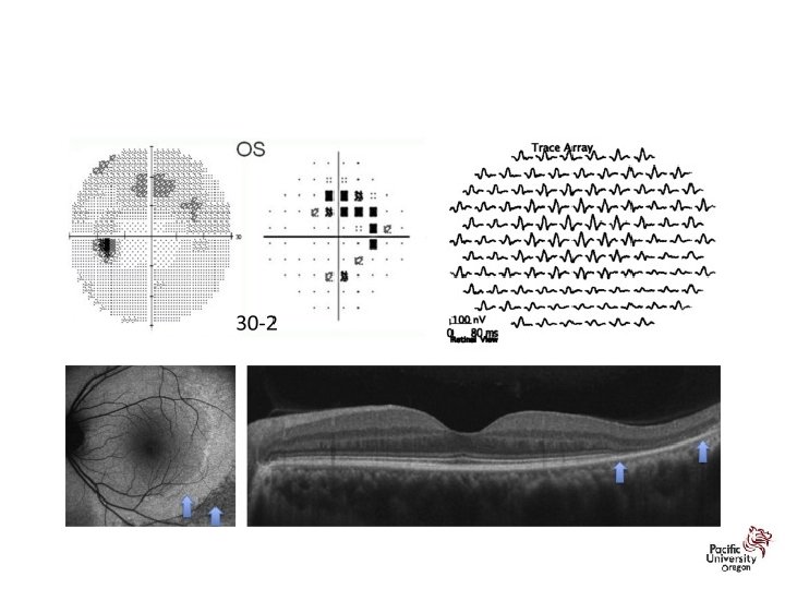
Question Which OCT goes with a patient undergoing hydroxychloroquine toxicity? 1 2 3 4

Treatment and Management: Antimalarial Ocular Complications • Toxicity can lead to whorl keratopathy, “bulls eye” maculopathy, retinal vessel attenuation, and optic disc pallor. • Early stages of maculopathy are seen as mild stippling or mottling and reversible loss of foveal light reflex • “Classic” maculopathy is in form of a “bulls eye” and is seen in later stages of toxicity – this is an irreversible damage to the retina despite discontinuation of medication

Treatment and Management: Antimalarials 29 Bulls Eye Maculopathy Whorl Keratopathy

Revised Recommendations on Screening for Hydroxychloroquine Retinopathy • 2002 recommendations for screening were published by Ophthalmology • Revised recommendations on screening published in Ophthalmology 2011; 118: 415 -42 – Significant changes in light of new data on the prevalence of retinal toxicity and sensitivity of new diagnostic techniques – Risk of toxicity after years of use is higher than previously believed • Risk of toxicity approaches 1% for patients who exceed 5 years of exposure

“New” New Recommendations • Recommendations on Screening for Chloroquine and Hydroxychloroquine Retinopathy – Ophthalmology 2016; 123: 1386 -1394 – Released March 2016 from American Academy of Ophthalmology – revised in light of new information about the prevalence of toxicity, risk factors, fundus distribution, and effectiveness of screening tools.

2016 Recommendations • maximum daily HCQ use of 5. 0 mg/kg real weight, which correlates better with risk than ideal weight. • risk of toxicity is dependent on daily dose and duration of use. – at recommended doses: • risk of toxicity up to 5 years is under 1% • up to 10 years is under 2% • rises to almost 20% after 20 years. However, even after 20 years, a patient without toxicity has only a 4% risk of converting in the subsequent year.

2016 Recommendations • High dose and long duration of use are the most significant risks. – Other major factors are concomitant renal disease, or use of tamoxifen • A baseline fundus examination should be performed to rule out preexisting maculopathy. • Begin annual screening after 5 years for patients on acceptable doses and without major risk factors.

2016 Recommendations • primary screening tests are automated visual fields plus spectral-domain optical coherence tomography (SD OCT) • most patients of Asian descent will show initial damage in a more peripheral extramacular distribution near the arcades (require a 24 -2 as opposed to 10 -2 and OCT scans need to be analyzed further out)

Revised Recommendations on Screening for Retinopathy • Parafoveal loss of visual sensitivity may appear before changes are seen on fundus evaluation • Many instances where retinopathy was unrecognized for years as field changes were dismissed as “non-specific” until the damage was severe • 10 -2 VF should always be repeated promptly when central or parafoveal changes are observed to determine if they are repeatable • Advanced toxicity shows well-developed paracentral scotoma

Paracentral Scotomas

Normal Retina: VF/OCT/ERG TD-OCT Outer Nuclear Layer PIL SD-OCT Rodriguez-Padilla, J. A. et al. Arch Ophthalmol 2007; 125: 775780. PIL=PR Integrity Line Copyright restrictions may apply.

Mild Maculopathy Normal Foveal Peak Paracentral Scotomas Thinned Outer Nuclear Layer PIL Rodriguez-Padilla, J. A. et al. Arch Ophthalmol 2007; 125: 775780. Copyright restrictions may apply.

Bull’s Eye Maculopathy Flattened Foveal Peak Dense Para/Central Defects RPE Atrophy Remnant of PIL Rodriguez-Padilla, J. A. et al. Arch Ophthalmol 2007; 125: 775780. Copyright restrictions may apply.

Major Risk Factors

Screening Recommendations

QUICKIE

Pre-Malignant Eyelid Lesions: Keratoacanthoma • solitary, rapidly growing nodule on sun exposed areas • umbilicated with a distinctive crater filled with keratin • Lesion develops over weeks and undergoes spontaneous involution within 6 mo to leave an atrophic scar • Complete excision is recommended as there are invasive variants

Pre-Malignant Eyelid Lesions: Actinic Keratosis • Also known as solar or senile keratosis • Most common pre-malignant skin lesion • Develops on sun-exposed areas and commonly affect the face, hands and scalp (less commonly the eyelids) – Predominately white males • Development of SCC in untreated lesions as high as 20% • Management is surgical excision or cryotherapy (following biopsy)
 • Most common malignant lesion of the")
Malignant Eyelid Lesions: Basal Cell Carcinoma (BCC) • Most common malignant lesion of the lids (85 -90% of all malignant epi eyelid tumors) • 50 -60% affect the lower lid followed by medial canthus 25 -30% and upper lid 15% • Metastases is rare but local invasion is common and can be very destructive
 • Much less common than BCC on")
Malignant Eyelid Lesions: Squamous Cell Carcinoma (SCC) • Much less common than BCC on the eyelid but has much higher potential for metastatic spread • Typically affects elderly, fairskinned and usually found on the lower lid • Lesions have a high tendency towards ulceration and tend to affect lid margin and medial canthus

Malignant Eyelid Lesions: Malignant Melanoma • account for about 1% of all eyelid malignancies • Incidence been increasing and it causes about 2/3 of all tumor related deaths from cutaneous cancers

Malignant Eyelid Lesions: Malignant Melanoma • Risk factors: – congenital and dysplastic nevi, – changing cutaneous moles, – excessive sun exposure – family history, – age greater than 20 and white. • History of severe sunburns rather than cumulative actinic exposure thought to be a major risk factor

Malignant Eyelid Lesions: Malignant Melanoma • Prognosis and metastatic potential are linked to the depth of invasion and thickness of the tumor • Treatment is wide surgical excision confirmed with histological monitoring

CASE 5

30 YR WM • Patient calls from his PCP office asking if we can see him today because he has had red/painful eyes for over a week and has not resolved • Medical history: – Past week has been experiencing painful urination and discharge – New sexual partner apprx 10 days ago, who also had developed a red eye – Chlamydia and gonorrhea testing were negative – Has tested positive for HSV 2 but no current flare up 87

30 YO WM • Medications: – In the past week patient: • • • 2 courses of azythromycin (1 gram each) Injection of rocephin Injection of penicillin G Currently taking doxycycline 100 mg bid Valtrex 1 gram 3 times per day for 7 days (d/c 1 day ago) Was on Vigamox qid for 7 days (d/c 1 day ago) • VA: 6/7. 5 (20/25) OD, OS • Entrance skills unremarkable though some pain on eye movement 88

30 YO WM • SLE: – 2+ injection conjunctival both eyes – 1 -2+ lid edema – Mixed papillary and follicular response – 1 -2+ diffuse SPK (no staining noted above infiltrates) – No cells or flare noted 89

30 YO WM • Adeno. Plus: – Performed on the right eye (patient felt that was the worst eye) – Negative 90

30 YO WM • Started patient on the miracle drop – Tobradex 4 times per day and scheduled patient to come back the next day • 1 day f/u – Patient was feeling better – Less redness and much reduced photophobia and discomfort – No improvement on painful urination or discharge and is now seeing blood in his urine – Continue tobradex 4 times per day and RTC in 4 days for f/u with dilation and told to contact PCP to update on the blood in the urine 91

30 YO WM • 4 day f/u: – Patient says his eyes are doing great and that all of his urogenital problems abruptly stopped on Saturday – Discussion with PCP: Kidney stone What did we learn from this? 92
- Slides: 89