Intellectual Functions of the Brain Learning and Memory

Intellectual Functions of the Brain, Learning and Memory Dr. Ejlal Abu-El-Rub , Pharm. D, Ph. D Department of Basic Medical Sciences Yarmouk University
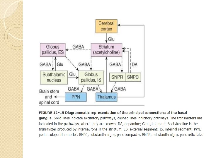

Parkinson's disease is a common disorder characterized by tremor, rigidity, and bradykinesia. This disease is caused by loss of neurons in the pars compacta of the substantia nigra. Consequently, the striatum suffers a severe loss of dopamine. Neurons of the locus caeruleus and the raphe nuclei, as well as other monoaminergic nuclei, are also lost. The loss of dopamine diminishes the activity of the direct pathway and increases the activity of the indirect pathway (The net effect is an increase in the activity of neurons in the internal segment of the globus pallidus. This results in greater inhibition of neurons in the ventral anterior and ventral lateral nuclei of the thalamus (VA and VL) and less pronounced activation of the motor cortical areas. The consequence is slowed movement (bradykinesia)

In Huntington's disease, which results from a genetic defect that involves an autosomal dominant gene. This defect leads to the loss of GABAergic and cholinergic neurons of the striatum (and also degeneration of the cerebral cortex, with resultant dementia). Loss of inhibition of the external globus pallidus presumably leads to diminished activity of neurons in the subthalamic nucleus and the excitation of neurons of the internal segment of the globus pallidus would be reduced. This will disinhibit neurons in the VA and VL nuclei. The resulting enhancement of activity in neurons in the motor areas of the cerebral cortex may help explain the choreiform movements of Huntington's disease.

Physiologic Anatomy of the Cerebral Cortex

�The functional part of the cerebral cortex is a thin layer of neurons covering the surface of all the convolutions of the cerebrum. �Three types of neurons: 1. Granular (stellate) 2. Fusiform 3. Pyramidal
 neurons: �Short axons �Function as interneurons �May be excitatory (glutamate) or")
1. Granular (stellate) neurons: �Short axons �Function as interneurons �May be excitatory (glutamate) or inhibitory (GABA). �Found in the sensory areas of the cortex �Found in association areas between sensory and motor areas

1. Fusiform and 2. Pyramidal neurons �Give rise to almost all output fibers from the cortex �Pyramidal are larger and more numerous than fusiform �Pyramidal - source of nerve fibers that go all the way to the spinal cord.

� Most incoming specific sensory signals from the body terminate in cortical layer IV. � Most of the output signals leave the cortex through neurons located in layers V and VI � The very large fibers to the brain stem and cord arise generally in layer V � Fibers to the thalamus arise in layer VI � Layers I, II, and III perform most of the intracortical association functions.

Anatomical and Functional Relations of the Cerebral Cortex to the Thalamus and Other Lower Centers Thalamic excitation of the cortex is necessary for almost all cortical activity. Areas of the cerebral cortex that connect with specific portions of the thalamu

Functions of Specific Cortical Areas

Functions of Specific Cortical Areas Functional areas of the human cerebral cortex as determined by electrical stimulation during neurological surgery or examination.

Association areas Areas that receive and analyze signals simultaneousl y from multiple regions of both the motor and sensory cortices, as well as from subcortical structures.

Association areas 1. Parieto-occipitotemporal association area • Analysis of the spatial coordinates of the body • Wernicke’s Area - important for language comprehension • Angular gyrus area - needed for reading • Area for naming objects

Association areas 2. Pre-frontal association area Ø 3. Broca’s area-neural circuitary for word formation Limbic association area Ø behavior, emotions, and motivation

Area for Recognition of Faces -Located in the Temporal lobe, -Prosopagnosia

Comprehensive Interpretative Function of the Posterior Superior Temporal Lobe - “Wernicke’s Area” (a General Interpretative Area) The somatic, visual, and auditory association areas all meet one another in the posterior part of the superior

Comprehensive Interpretative Function of the Wernicke’s Area �This area is especially highly developed in the dominant side of the brain [the left side in almost all right-handed people]. �It plays the greatest single role in intelligence. �Also known as the general interpretative area, the gnostic area, the knowing area, the tertiary association area. �Activation of this area can call forth complicated memory patterns that involve more than one sensory modality even though most of the individual memories may be stored elsewhere.

Angular Gyrus - Interpretation of Visual Information �Is the most inferior portion of the posterior parietal lobe, lying immediately behind Wernicke’s area. �Its destruction causes dyslexia (word blindness) the person may be able to see words and even know that they are words but not be able to interpret their meanings.

Concept of the dominant hemisphere �The general interpretative functions of Wernicke’s area and the angular gyrus, as well as the functions of the speech and motor control areas, are usually much more highly developed in one cerebral hemisphere than in the other. This hemisphere is called the dominant hemisphere. �In about 95 percent of all people, the left

Concept of the dominant hemisphere �Because the left posterior temporal lobe at birth is usually slightly larger than the right, the left side normally begins to be used to a greater extent than the right. �Mind tends to direct one’s attention to the better developed region the rate of learning in the cerebral hemisphere that gains the first start increases rapidly.

Concept of the dominant hemisphere �The premotor speech area (Broca’s area; responsible formation of the words) is also almost always dominant on the left side of the brain. �The motor areas for controlling hands are also dominant in the left side of the brain in 9 of 10 persons. �Temporal lobe and angular gyrus and motor areas are more developed in the left side, but they receive sensory information and control motor activity in both hemispheres.

Role of Language in the Function of Wernicke’s Area and in Intellectual Functions �Majority of sensory experience is converted into its language equivalent before being stored in the memory areas of the brain and before being processed for other intellectual purposes. �The sensory area for interpretation of language is Wernicke’s area – also associated with both primary and secondary hearing areas of the temporal lobes later in life,

Functions of the parieto-occipitotemporal cortex in the nondominant hemisphere �Nondominant hemisphere is important for: ◦ understanding and interpreting music, ◦ nonverbal visual experiences, ◦ spatial relations between person and surrounding ◦ the significance of “body language” and intonations of people’s voices ◦ many somatic experiences related to use of the limbs and hands.

Higher Intellectual Functions of the Prefrontal Association Areas �Individuals that underwent pre-frontal lobotomy developed: 1. Decreased aggressiveness and inappropriate social responses Ø from loss of the ventral parts of the frontal lobes (part of limbic association cortex behavior control). 2. Inability to progress toward goals or to carry through sequential thoughts.

Higher Intellectual Functions of the Prefrontal Association Areas 3. Impaired elaboration of thought, prognostication, and performance of higher intellectual functions Ø “working memory” = ability to keep track of many bits of information simultaneously and to cause recall of this information promptly as it is needed for subsequent thoughts.

Function of the Corpus Callosum and Anterior Commissure to Transfer Thoughts, Memories, Training and Other Information Between the Two Cerebral Hemispheres

Cutting the Corpus Callosum: � Blocks transfer of information from the dominant hemisphere to the motor cortex on the opposite side � Prevents transfer of somatic and visual info from the right to left hemisphere � Person would have two entirely separate conscious portions of the brain.

Thoughts, Consciousness and Memory

�A thought results from a “pattern” of stimulation of many parts of the nervous system at the same time, probably involving most importantly the cerebral cortex, thalamus, limbic system, and upper reticular formation of the brain stem holistic theory of thoughts. �Consciousness can be described as continuing stream of awareness of either the

Memory - Roles of Synaptic Facilitation and Synaptic Inhibition �Memories are stored in the brain by changing the basic sensitivity of synaptic transmission between neurons as a result of previous neural activity. �New or facilitated pathways are called memory traces can be selectively activated.

Memory - Roles of Synaptic Facilitation and Synaptic Inhibition Positive and Negative Memory - “Sensitization” or “Habituation” of Synaptic Transmission �The brain has the capability to learn to ignore information that is of no consequence. �This results from inhibition of the synaptic pathways for this type of information; the resulting effect is called habituation negative memory for information that causes important consequences. �It results from facilitation of the synaptic pathways, and the process is called memory sensitization. �Positive

Classification of memory 1. Declarative memory - memory of the various details of integrated thought (i. e. memory of surroundings, time relationships, causes of experiences, meaning of an experience). 2. Skill memory - associated with motor activities based on previous learning (i. e. hitting a tennis ball).

� Short-Term Memory Lasts for a few seconds to a few minutes at a time, but lasting only as long as the person continues to think about the numbers or facts. May be caused by: 1. Continual neural activity resulting from nerve signals that travel around a temporary memory trace in a circuit of reverberating neurons; or 2. Presynaptic facilitation or inhibition.

�Intermediate Long-Term Memory Lasts for many minutes or even weeks. ü Lost unless memory traces are activated enough to become more permanent. ü ü Result from chemical and/or physical changes in the presynaptic terminal or postsynaptic neuronal

Molecular Mechanisms of Intermediate Memory a. Mechanism of habituation – progressive closure of calcium channels in sensory terminals less transmitter released. b. Mechanism of facilitation – Stimulation of facilitator presynaptic terminal serotonin release activation of adenyl cyclase c. AMP activity

� Long-Term Memory Structural changes in synapses during the development of long-term memory 1. Increase in vesicle release site for secretion of transmitters 2. Increase in number of transmitter vesicles released 3. Increase in the number of presynaptic terminals 4. Changes in the structure of dendritic spines that permit transmission of stronger signals

� Number of neurons and their connectivities change during learning � Consolidation of Memory a. Rehearsal enhances the transference of short-term memory into long-term memory. b. New memories are codified during consolidation.

Consolidation of Memory �Roles of specific parts of the brain in the memory process: ◦ Hippocampus promotes storage of memories ◦ Hippocampi are not important in reflexive learning (skill learning).
- Slides: 39